. 2016 Oct;75(10):981-997.
doi: 10.1093/jnen/nlw075. Epub 2016 Sep 9.

Hippocampal Formation Maldevelopment and Sudden Unexpected Death across the Pediatric Age Spectrum

Affiliations

Hippocampal Formation Maldevelopment and Sudden Unexpected Death across the Pediatric Age Spectrum

Hannah C Kinney et al. J Neuropathol Exp Neurol. 2016 Oct.

Abstract

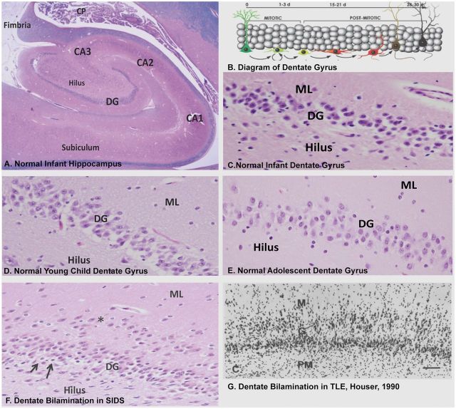
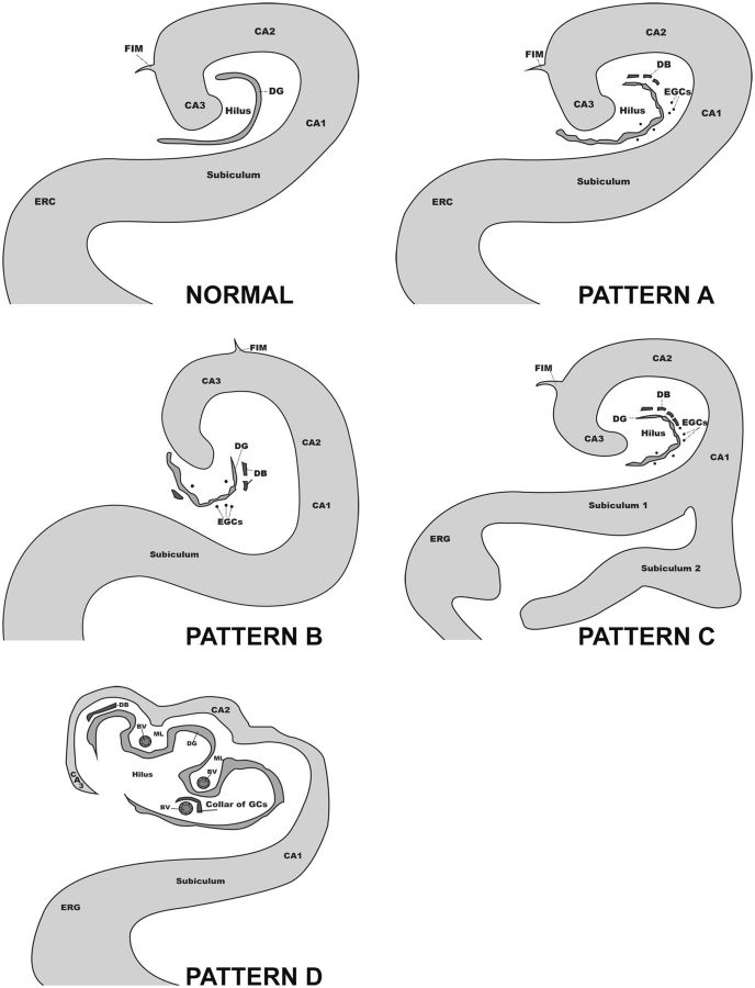
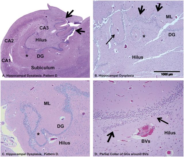
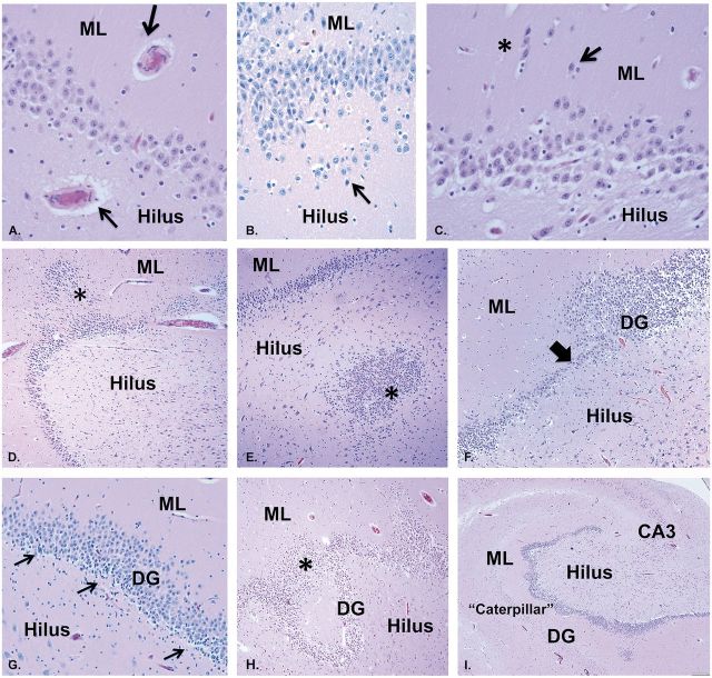
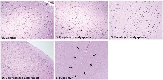
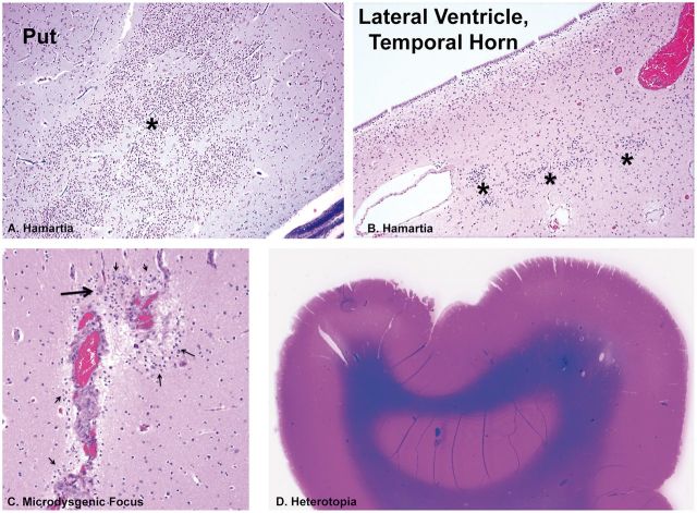
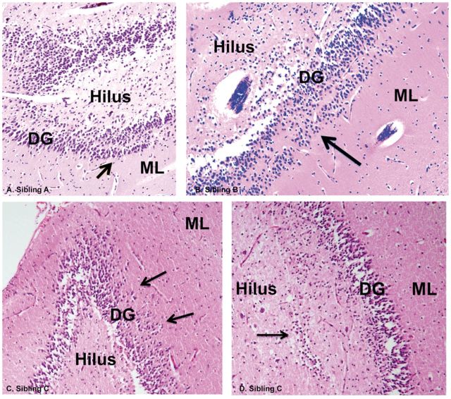
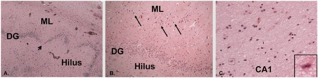
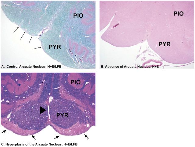
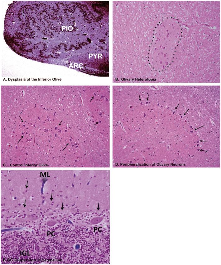
Sudden infant death syndrome (SIDS) and sudden unexplained death in childhood (SUDC) are defined as sudden death in a child remaining unexplained despite autopsy and death scene investigation. They are distinguished from each other by age criteria, i.e. with SIDS under 1 year and SUDC over 1 year. Our separate studies of SIDS and SUDC provide evidence of shared hippocampal abnormalities, specifically focal dentate bilamination, a lesion classically associated with temporal lobe epilepsy, across the 2 groups. In this study, we characterized the clinicopathologic features in a retrospective case series of 32 children with sudden death and hippocampal formation (HF) maldevelopment. The greatest frequency of deaths was between 3 weeks and 3 years (81%, 26/32). Dentate anomalies were found across the pediatric age spectrum, supporting a common vulnerability that defies the 1-year age cutoff between SIDS and SUDC. Twelve cases (38%) had seizures, including 7 only with febrile seizures. Subicular anomalies were found in cases over 1 year of age and were associated with increased risk of febrile seizures. Sudden death associated with HF maldevelopment reflects a complex interaction of intrinsic and extrinsic factors that lead to death at different pediatric ages, and may be analogous to sudden unexplained death in epilepsy.

Keywords: Dentate gyrus; Febrile seizures; Granule cell dispersion; Sudden infant death syndrome (SIDS); Sudden unexpected death in pediatrics with hippocampal formation maldevelopment (SUDP-HFM); Sudden unexplained death in childhood (SUDC); Sudden unexplained death in epilepsy (SUDEP).


Figures

FIGURE 1.
FIGURE 1.
The normal dentate gyrus compared with the abnormal focal DB in sudden unexpected/unexplained death in children and TLE. (A) Low-power view of the normal hippocampus of an infant demonstrating the interlocking relationship of the dentate gyrus and Ammon’s horn of the hippocampus proper, and the subiculum, the major outflow site, adjacent to CA1. (B) Diagram of the GC layer of the dentate gyrus showing the site of genesis of GCs in the subgranular layer from neuronal precursors (mitotic zone), and transition to differentiated GCs (postmitotic zone), with upward migration and elaboration of their dendritic arbor in the molecular layer of the dentate gyrus (from reference 60 Copyright (2006) National Academy of Sciences, U.S.A). Normal dentate gyrus of an infant (C) compared with that of a young child (D) and adolescent (E) . H&E-LFB, ×20. There is normally a single layer of packed GCs between the molecular layer and pleomorphic layer of the hilus, the 3 components of the dentate gyrus. (F) Focal DB of the GC layer in a SIDS infant with a row of GCs in 1 layer (asterisks) separated by a cell free zone from the main body of GCs. There are immature cells in the subgranular layer (arrows). Focal DB is seen in Patterns A–D. H&E-LFB, ×20. The displaced isolated layer, 1- to multiple-cells-thick, occurs at different points and with different lengths along the upper and/or lower blade of the dentate gyrus; the acellular layer of neuropil separates the 2 GC layers, and gliosis is not present. There is no gliosis or neuronal loss in Ammon’s horn, supporting the concept of a developmental lesion without major acquired changes. (G) GC dispersion in TLE, as first defined by Houser ( 13 ), with a widened GC layer and GCs dispersed in the molecular layer. Abbreviations: DG, dentate gyrus; GC, granule cell; ML, molecular layer; TLE, temporal lobe epilepsy.
FIGURE 2.
FIGURE 2.
Schematic drawings of 4 morphological patterns of developmental pathology in the hippocampus and subiculum of the cohort of 32 infants and children with sudden unexpected death.
FIGURE 3.
FIGURE 3.
Hippocampal asymmetry and/or malrotation in coronal slices of the brains (mid-thalamic level) of an infant (A) , young child (B) , and adolescent (C) . Hippocampal asymmetry is defined as a difference in the size and shape of the right and left hippocampi compared to each other, as noted in each of the 3 cases at different ages. Malrotation of the hippocampus proper is notable for a rounded and upright shape, exemplified in the brain of the infant bilaterally (A) , but more pronounced on the encircled hippocampus, and in the young child (B) , also distinct in the encircled hippocampus. The young child also has abnormal folding of the subiculum (Pattern C) (arrow), whereas the subiculum is not abnormal in the infant (A) or adolescent (C) . In the young child, the right hippocampus is abnormally formed as well as the left, and the right is smaller than the left (encircled), both without distinct landmarks. In this case (B) , the insular cortex is also asymmetric (asterisk on each side of insular cortex). In the adolescent (C) , the left and right superior temporal gyri are asymmetric (asterisk). The hippocampus on the left side (encircled) is larger than on the right side, and slanted downward and to the right. Abbreviations: CC, corpus callosum; In, insula; LGN, lateral geniculate nucleus; STG, superior temporal gyrus; Th, thalamus.
FIGURE 4.
FIGURE 4.
Abnormal folding of the subiculm (Pattern C) in the brains of 2 different young children of the cohort in whole mount sections of the HF stained with H& E/LFB. (A) Duplication of the subiculum is characterized by the “splitting” of CA1 of Ammon’s horn into 2 thick “branches” of subiculum (arrows, enclosed in dashed circles). Whole mount. (B) Abnormal subicular folding is characterized by an uneven thickness in its shape. Whole mount. Of note, the degree of myelination is age-appropriate in the intrinsic and extrinsic temporal lobe pathways. Abbreviations: CP, choroid plexus; DG, dentate gyrus; LGN, lateral geniculate nucleus; OT, optic tract.
FIGURE 5.
FIGURE 5.
Hippocampal dysplasia (Pattern D) demonstrated in the brains of 1 infant and 1 young child in the cohort. (A) Hippocampal dysplasia (Pattern D) in a SIDS infant with excessive convolutions of the GC layer of the dentate gyrus at the medial surface (arrows). A GC heterotopia is in the molecular layer (asterisk). H&E, Whole mount. (B) In a young child with SUDC, there is displacement of elements of the GC and molecular layer and blood vessels undulating towards and away from the center of the hilus (all arrows). In the molecular layer, there is a central blood vessel (long arrow) (Pattern D); there is also focal DB is present (short arrows). There is a blood vessel in the hilus with a partial collar of GCs towards the body of the DG (asterisk). H&E-LFB, ×40. (C) In a second SIDS infant, at 1 level there is a closed loop of GCs separate from the main body of the granular cell layer that becomes attached to the main body (asterisk) on deeper cuts, H&E, ×10. (D) A small artery and vein, side by side in the hilus, are partially surrounded by a collar of GCs (arrows) on the side of the main body of the DG, H&E, ×20. BVs, blood vessels; DG, dentate gyrus; GC, granule cell; ML, molecular layer.
FIGURE 6.
FIGURE 6.
Additional features in the abnormal dentate gyrus associated with DB in representative infants and young children with SUDP-HFM. A: Blood vessels with thick walls, shown here in the hilus and molecular layer (arrows), H&E, ×40. (B) Ectopic GCs in a row in the hilus below the subgranular layer (arrow), H&E, ×20. (C) Clusters (asterisk) and single (arrow) ectopic GCs in the molecular layer of the DG, H&E, ×40. (D) GC heterotopia in the molecular layer (asterisk), H&E, ×10. (E) GC heterotopia in the hilus (asterisk), H&E, ×10. (F) Focal lack of GCs (thick arrow), suggesting abnormal cell migration to this position, in an uneven GC layer with alternating areas of cell thickness of different degrees. H&E, ×10. (G) Immature cells in the subgranular layer of the dentate gyrus (arrows), characterized by dark nuclei and scant or rod shaped cytoplasm in clusters, H&E, ×20. (H) Marked GC dispersion with a “smear” of GCs bridging 2 convolutions (asterisk) of the abnormal GC layer, H&E ×10. (I) Marked undulation of the GC layer of the dentate gyrus mimicking the shape of a caterpillar in movement, H&E, ×2. DB, dentate bilamination; DG, dentate gyrus; ML, molecular layer; GC, granule cell.
FIGURE 7.
FIGURE 7.
Anomalies of development in the temporal and extra-temporal cerebral cortex in infants and children of the SUDP-HFM cohort. (A) Normal cerebral cortex of an infant for comparison, ×10. (B) FCD in a SIDS infant with vertical layering of neurons in rows of at least eight cells (arrows), H&E, ×10. (C) Higher power of vertical neuronal layering in FCD, Type I, ×20. (D) Disorganized cortex in a SIDS infant with an undulating molecular layer and absence of an organized laminar cortex directly underneath the molecular layer, ×10. (E) Fused gyrus between 2 cortical gyri (arrows), ×10. All are H&E.
FIGURE 8.
FIGURE 8.
Microdysgenetic features in extra-temporal lobe sites in infants and children of the SUDP-HM cohort. (A) Hamartia (asterisk) in the putamen of the basal ganglia, H&E, ×10. (B) Hamartia (asterisk) in the periventricular region of the lateral ventricle of the temporal horn. H&E, ×20. (C) Microdysgenetic focus with clusters of immature neurons (thin arrows) in the molecular layer of the cerebral cortex around thickened leptomeningeal vessels between 2 gyri, with a single mature neuron (thick arrow), H&E, ×20. (D) A heterotopia in the subcortical white matter of the frontal lobe. Whole mount, H&E-LFB. Put, putamen.
FIGURE 9.
FIGURE 9.
Familial DB. In 1 family, a 1-month-old boy (A) , 3-month-old girl (B) , and 3-week old girl (C) died suddenly and unexpectedly during a sleep period. In all 3 cases, DB (arrows) and GC dispersion were found upon microscopic examination in sibling A (A) , sibling B (B) , and sibling C (C) , H&E, ×40. In sibling C, there is a single layer of GCs deep in the hilus (arrow) underneath the subgranular gyrus (D) . DG, dentate gyrus; GC, granule cell; ML, molecular layer.
FIGURE 10.
FIGURE 10.
Temporal lobe resection for TLE in a young adult father of a young child with SUDP-HFM showed AHS. (A) Irregular dentate gyrus with focal depopulation of GCs (arrow), ×10. (B) Focal bilamination of the dentate gyrus with GCs in a separate layer from the main body (arrows). H&E, ×20. (C) Neuronal loss and gliosis in CA1; insert, reactive astrocyte with hypertrophic, eosinophilic cytoplasm, ×20. All are H&E. DG, dentate gyrus; ML, molecular layer.
FIGURE 11.
FIGURE 11.
Arcuate nucleus anomalies associated with SUDP-HFM. (A) Normal arcuate nucleus in clusters (long arrows) for comparison at ventral surface of the medulla within the rim of the pyramid, H&E./LFB, ×2. (B) Absence of clusters of arcuate nucleus along the ventral rim of the medullary surface. H&E, ×2. (C) Hyperplasia of the arcuate nucleus (arrows) extending along the entire ventral rim of the medullary surface associated with fusion of the pyramids. There is a perivascular space entrapped between the 2 fused pyramids (arrowhead). PIO, principal inferior olive; PYR, pyramid.
FIGURE 12.
FIGURE 12.
Abnormal derivatives of the rhombic lip in the infant and children cases of the SUDP-HFM cohort. (A) Olivary dysplasia, exhibiting abnormal configuration of a hyperconvoluted principal inferior olive, with closely packed convolutions H&E, ×2. (B) Olivary heterotopia (misplaced clusters of olivary neurons) (dotted line) in the inferior cerebellar peduncle, H&E, ×20. (C) High-power view of normal inferior olive for comparison showing loosely packed separated neurons (arrows). H&E, ×20. (D) High-power view of olivary neurons (arrows) that are marginalized to the periphery of the convolutions of the nucleus (arrows). H&E, ×20. (E) A putative defect in migration of GCs from the external to internal granular cell layer in the cerebellum in infants and children in the SUDP-HFM cohort. Illustrated is dispersion of GCs (arrows) in the molecular layer of the cerebellar cortex, presumably having not reached the internal granular layer from its site of migration from the external granular cell layer, H&E, ×20. ARC, arcuate nucleus; GC, granule cell; IGL, internal granular layer; ML, molecular layer. PC, Purkinje cell. PIO, principal inferior olive; PYR, pyramid.

References

    1. Beckwith JB. Defining the sudden infant death syndrome . Arch Pediatr Adolesc Med 2003. ; 157 : 286 – 90 - PubMed
    1. Willinger M, James LS, Catz C. Defining the sudden infant death syndrome (SIDS): deliberations of an expert panel convened by the National Institute of Child Health and Human Development . Pediatr Pathol 1991. ; 11 : 677 – 84 - PubMed
    1. Krous HF, Chadwick AM, Crandall L , et al. . Sudden unexpected death in childhood: A report of 50 cases . Pediatr Dev Pathol 2005. ; 8 : 307 – 19 - PubMed
    1. Moon RY , Task Force on Sudden Infant Death Syndrome . SIDS and other sleep-related infant deaths: expansion of recommendations for a safe infant sleeping environment . Pediatrics 2011. ; 128 : 1030 – 9 - PubMed
    1. Rodriguez M, McMillan K, Crandall LA , et al. . Hippocampal asymmetry and sudden unexpected death in infancy: a case report . Forensic Med Pathol Sci 2012. ; 8 : 441 – 6 - PMC - PubMed