. 2016 Dec 1:69:733-43.
doi: 10.1016/j.msec.2016.07.060. Epub 2016 Jul 22.

Fabrication and characterization of carboxymethyl cellulose novel microparticles for bone tissue engineering

Affiliations

Fabrication and characterization of carboxymethyl cellulose novel microparticles for bone tissue engineering

Bipin Gaihre et al. Mater Sci Eng C Mater Biol Appl. .

Abstract

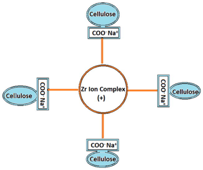
In this study we developed carboxymethyl cellulose (CMC) microparticles through ionic crosslinking with the aqueous ion complex of zirconium (Zr) and further complexing with chitosan (CS) and determined the physio-chemical and biological properties of these novel microparticles. In order to assess the role of Zr, microparticles were prepared in 5% and 10% (w/v) zirconium tetrachloride solution. Scanning electron microscopy (SEM) with energy dispersive X-ray spectrometer (EDS) results showed that Zr was uniformly distributed on the surface of the microparticles as a result of which uniform groovy surface was obtained. We found that Zr enhances the surface roughness of the microparticles and stability studies showed that it also increases the stability of microparticles in phosphate buffered saline. The crosslinking of anionic CMC with cationic Zr and CS was confirmed by Fourier transform infrared spectroscopy (FTIR) results. The response of murine pre-osteoblasts (OB-6) when cultured with microparticles was investigated. Live/dead cell assay showed that microparticles did not induce any cytotoxic effects as cells were attaching and proliferating on the well plate as well as along the surface of microparticles. In addition, SEM images showed that microparticles support the attachment of cells and they appeared to be directly interacting with the surface of microparticle. Within 10days of culture most of the top surface of microparticles was covered with a layer of cells indicating that they were proliferating well throughout the surface of microparticles. We observed that Zr enhances the cell attachment and proliferation as more cells were present on microparticles with 10% Zr. These promising results show the potential applications of CMC-Zr microparticles in bone tissue engineering.

Keywords: Carboxymethyl cellulose; Microparticles; Pre-osteoblasts; Surface roughness; Zirconium.


Figures

Figure 1
Figure 1
Ionic crosslinking interaction model for anionic CMC with cationic Zr ion complex
Figure 2
Figure 2
SEM image of P1 (a) and P2 (b) show spherical morphology of microparticle at low magnification. The rough surface of P1 (c) and P2 (d) can be seen at higher magnification.
Figure 3
Figure 3
Figure 3A. EDS elemental mapping of P1 (a-SEM image of microparticle, b-carbon, c-oxygen, d-zirconium) and P2 (A-SEM image of microparticle, B-carbon, C-oxygen, D-zirconium). Fig 3B: EDS spectra of P1 and P2 showing their elemental composition. Spectra on the right shows the intensity of Zr along the surface of P1 and P2.
Figure 3
Figure 3
Figure 3A. EDS elemental mapping of P1 (a-SEM image of microparticle, b-carbon, c-oxygen, d-zirconium) and P2 (A-SEM image of microparticle, B-carbon, C-oxygen, D-zirconium). Fig 3B: EDS spectra of P1 and P2 showing their elemental composition. Spectra on the right shows the intensity of Zr along the surface of P1 and P2.
Figure 4
Figure 4
XRD spectrum of microparticle showing amorphous state of Zr and CMC.
Figure 5
Figure 5
FTIR spectra of pure CMC and microparticles.
Figure 6
Figure 6
Variation in pH of PBS (pH 7.4) containing microparticles over 25 days (n=3). Stable neutral pH was maintained by both P1 and P2.
Figure 7
Figure 7
SEM images showing the stability of microparticles after 35 days of immersion in PBS (pH 7.4). The stability of P1 (a & b) was lower compared to P2 (c & d).
Figure 8
Figure 8
Figure 8A. Cells proliferating at the bottom of the wells without microparticles (a) with P1 (b) and with P2 (c) on day 10. Green fluorescence represents live and red dots represent dead cells (scale 100 μm). Figure 8B. Viable Cells (bright green spots) attached and proliferating on the surface of P1 on day 5 (a) and 10 (b) and on the surface of P2 on day 5 (c) and 10 (d). Figure 8C. Microparticles surface area covered with cells.
Figure 8
Figure 8
Figure 8A. Cells proliferating at the bottom of the wells without microparticles (a) with P1 (b) and with P2 (c) on day 10. Green fluorescence represents live and red dots represent dead cells (scale 100 μm). Figure 8B. Viable Cells (bright green spots) attached and proliferating on the surface of P1 on day 5 (a) and 10 (b) and on the surface of P2 on day 5 (c) and 10 (d). Figure 8C. Microparticles surface area covered with cells.
Figure 9
Figure 9
Figure 9A. SEM images of cells attached to P1 on day 5 (a & b) and on day 10 (c & d). Figure 9B. SEM images of cells attached to P2 on day 5 (a & b) and day 10 (c & d). Figure 9C. SEM images of cells proliferating along the cracks in P1 (a) and P2 (b) on day 10.
Figure 9
Figure 9
Figure 9A. SEM images of cells attached to P1 on day 5 (a & b) and on day 10 (c & d). Figure 9B. SEM images of cells attached to P2 on day 5 (a & b) and day 10 (c & d). Figure 9C. SEM images of cells proliferating along the cracks in P1 (a) and P2 (b) on day 10.
Figure 10
Figure 10
Figure 10A. Percent cumulative drug released from drug encapsulated P1 and P2. Figure 10B. Percent cumulative drug released from drug coated P1 and P2.
Figure 10
Figure 10
Figure 10A. Percent cumulative drug released from drug encapsulated P1 and P2. Figure 10B. Percent cumulative drug released from drug coated P1 and P2.

References

    1. Qiu XY, Hu SW. “Smart” Materials Based on Cellulose: A Review of the Preparations, Properties, and Applications. Materials. 2013;6:738–81. - PMC - PubMed
    1. Sannino A, Demitri C, Madaghiele M. Biodegradable Cellulose-based Hydrogels: Design and Applications. Materials. 2009:353–73.
    1. Jiang LY, Li YB, Xiong CD. Preparation and biological properties of a novel composite scaffold of nano-hydroxyapatite/chitosan/carboxymethyl cellulose for bone tissue engineering. J Biomed Sci. 2009:16. - PMC - PubMed
    1. Pasqui D, Torricelli P, De Cagna M, Fini M, Barbucci R. Carboxymethyl cellulose-hydroxyapatite hybrid hydrogel as a composite material for bone tissue engineering applications. J Biomed Mater Res A. 2014;102:1568–79. - PubMed
    1. Raucci MG, Alvarez-Perez MA, Demitri C, Giugliano D, De Benedictis V, Sannino A, et al. Effect of citric acid crosslinking cellulose-based hydrogels on osteogenic differentiation. Journal of Biomedical Materials Research Part A. 2015;103:2045–56. - PubMed

MeSH terms

LinkOut - more resources