. 2016 Sep 9;15(1):107.
doi: 10.1186/s12938-016-0231-9.

Patient-specific CFD simulation of intraventricular haemodynamics based on 3D ultrasound imaging

Affiliations

Patient-specific CFD simulation of intraventricular haemodynamics based on 3D ultrasound imaging

A M Bavo et al. Biomed Eng Online. .

Abstract

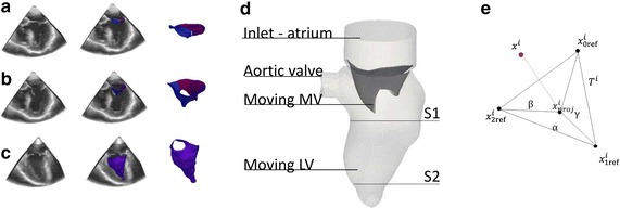
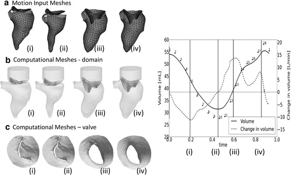
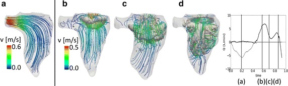
Background: The goal of this paper is to present a computational fluid dynamic (CFD) model with moving boundaries to study the intraventricular flows in a patient-specific framework. Starting from the segmentation of real-time transesophageal echocardiographic images, a CFD model including the complete left ventricle and the moving 3D mitral valve was realized. Their motion, known as a function of time from the segmented ultrasound images, was imposed as a boundary condition in an Arbitrary Lagrangian-Eulerian framework.

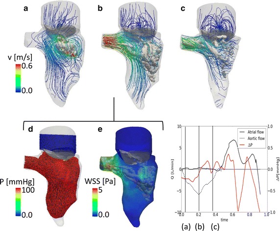
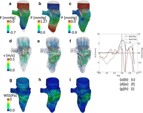
Results: The model allowed for a realistic description of the displacement of the structures of interest and for an effective analysis of the intraventricular flows throughout the cardiac cycle. The model provides detailed intraventricular flow features, and highlights the importance of the 3D valve apparatus for the vortex dynamics and apical flow.

Conclusions: The proposed method could describe the haemodynamics of the left ventricle during the cardiac cycle. The methodology might therefore be of particular importance in patient treatment planning to assess the impact of mitral valve treatment on intraventricular flow dynamics.

Keywords: CFD model with prescribed moving boundaries; Intraventricular flow; Patient-specific modeling; Real-time transesophageal ultrasound images.


Figures

Fig. 1
Fig. 1
From rt-TEE ultrasound images to segmented triangulated surfaces. a MV in closed configuration. b MV in open configuration. c LV during systole
Fig. 2
Fig. 2
Prescribed motion of the boundaries. Comparison between (a) the motion input meshes and (b, c) the computed meshes, complete domain and top view of the MV, in four time-points of the cardiac cycle
Fig. 3
Fig. 3
Systolic flow field. Streamlines and vortex structure (λ2) (a–c). Pressure and velocity vectors in the LV (d, e) on a section at peak of systole. f Flow curve and the intraventricular pressure difference (base–apex) during the cardiac cycle. The time-points used in a–e are indicated
Fig. 4
Fig. 4
Diastolic flow field. Pressure and velocity vectors in the LV (a–c) on a section. Streamlines and vortex structure (λ2) (d–f). WSS at the walls (g–i). j Flow curve and the intraventricular pressure difference (base–apex) during the cardiac cycle. The time-points used in a–i are indicated. Please note that the color scale for the pressure difference changes in each panel
Fig. 5
Fig. 5
Flow field for the model without the valve: vortex and velocity streamlines at peak systole (a) and in three time-points during diastole (b–d)

References

    1. Lopez-Perez A, Sebastian E, Ferrero JM. Three-dimensional cardiac computational modeling: methods, features and applications. Biomed Eng Online. 2015;14:35. doi: 10.1186/s12938-015-0033-5. - DOI - PMC - PubMed
    1. Baccani B, Domenichini F, Pedrizzetti G. Model and influence of mitral valve opening during the left ventricular filling. J Biomech. 2003;36:355–361. doi: 10.1016/S0021-9290(02)00420-7. - DOI - PubMed
    1. Shi JJ, Alenezy M, Smirnova IV, Bilgen M. Construction of a two-parameter empirical model of left ventricle wall motion using cardiac tagged magnetic resonance imaging data. Biomed Eng Online. 2012;11:79. doi: 10.1186/1475-925X-11-79. - DOI - PMC - PubMed
    1. Annerel S, Degroote J, Claessens T, Segers P, Verdonck P, Vierendeels J. The upstream boundary condition influences the leaflet opening dynamics in the numerical FSI simulation of an aortic BMHV. Int J Numer Method Biomed Eng. 2012;28:745–760. doi: 10.1002/cnm.2470. - DOI - PubMed
    1. Pedrizzetti G, Domenichini F, Tonti G. On the left ventricular vortex reversal after mitral valve replacement. Ann Biomed Eng. 2010;38(3):769–773. doi: 10.1007/s10439-010-9928-2. - DOI - PubMed

LinkOut - more resources