. 2017 Mar;28(3):761-768.
doi: 10.1681/ASN.2016040486. Epub 2016 Sep 9.

Hyperoxaluria Requires TNF Receptors to Initiate Crystal Adhesion and Kidney Stone Disease

Affiliations

Hyperoxaluria Requires TNF Receptors to Initiate Crystal Adhesion and Kidney Stone Disease

Shrikant R Mulay et al. J Am Soc Nephrol. 2017 Mar.

Abstract

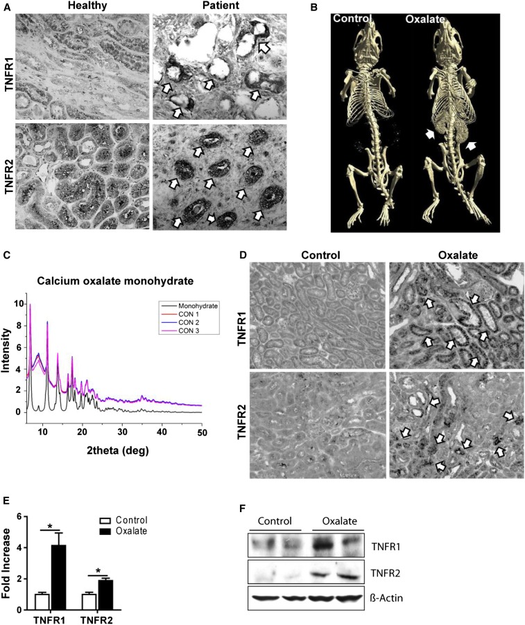
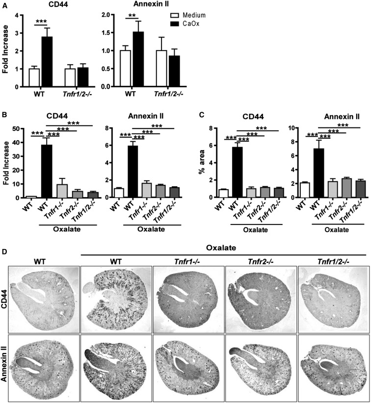
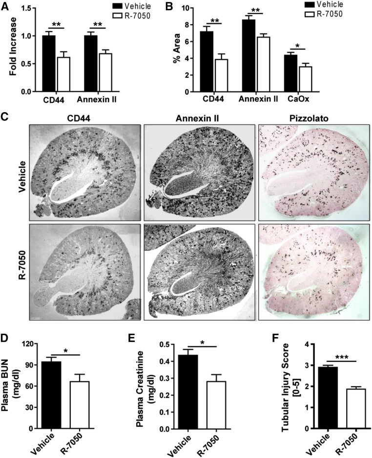
Intrarenal crystals trigger inflammation and renal cell necroptosis, processes that involve TNF receptor (TNFR) signaling. Here, we tested the hypothesis that TNFRs also have a direct role in tubular crystal deposition and progression of hyperoxaluria-related CKD. Immunohistochemical analysis revealed upregulated tubular expression of TNFR1 and TNFR2 in human and murine kidneys with calcium oxalate (CaOx) nephrocalcinosis-related CKD compared with controls. Western blot and mRNA expression analyses in mice yielded consistent data. When fed an oxalate-rich diet, wild-type mice developed progressive CKD, whereas Tnfr1-, Tnfr2-, and Tnfr1/2-deficient mice did not. Despite identical levels of hyperoxaluria, Tnfr1-, Tnfr2-, and Tnfr1/2-deficient mice also lacked the intrarenal CaOx deposition and tubular damage observed in wild-type mice. Inhibition of TNFR signaling prevented the induced expression of the crystal adhesion molecules, CD44 and annexin II, in tubular epithelial cells in vitro and in vivo, and treatment with the small molecule TNFR inhibitor R-7050 partially protected hyperoxaluric mice from nephrocalcinosis and CKD. We conclude that TNFR signaling is essential for CaOx crystal adhesion to the luminal membrane of renal tubules as a fundamental initiating mechanism of oxalate nephropathy. Furthermore, therapeutic blockade of TNFR might delay progressive forms of nephrocalcinosis in oxalate nephropathy, such as primary hyperoxaluria.

Keywords: Chronic inflammation; Hyperoxaluria; Kidney stone; pathology.


Figures

Figure 1.
Figure 1.
Humans and mice with hyperoxaluria express TNFR1 and TNFR2 in tubular epithelial cells. (A) Immunostaining for TNFR1 and TNFR2 in kidney tissue obtained from healthy donors, as well as from patients with hyperoxaluria-related CKD. Arrows indicate positivity for TNFR1 and TNFR2, respectively. (B–F) C57BL/6 male mice were fed either high control or high oxalate diet for 14 days. (B) Diagnostic imaging was conducted using computed tomography. Arrows indicate nephrocalcinosis. (C) The analysis of the mouse kidneys was performed by x-ray diffraction. Control (CON): CaOx nephropathy. (D) Immunostaining. Arrows indicate positivity for TNFR1 and TNFR2, respectively. Original magnification ×200. (E) Gene expression for TNFR1 and TNFR2 in kidney tissue obtained from these mice. (F) Protein expression of TNFR1 and TNFR2 was detected using Western blot. β-actin was employed as loading control. Data are means±SEM from six to seven mice in each group. *P<0.05 versus the control group.
Figure 2.
Figure 2.
Tnfr1-, Tnfr2-, and Tnfr1/2-deficient mice are protected from hyperoxaluria-related CKD and nephrocalcinosis. C57BL/6 wild type and Tnfr1-, Tnfr2-, and Tnfr1/2-deficient mice were fed either high oxalate or control diet for 14 days. (A) Plasma BUN and (B) plasma creatinine were measured. (C–F) Quantification of (C) tubular injury, (D) SMA+ area, (E) F4/80+ staining for macrophages, and (F) CD3+ cells per high power field. (G) Diagnostic imaging was performed using computed tomography. Arrows indicate the kidneys filled with CaOx crystals. (H) Pizzolato staining of kidney sections. Note that kidneys of wild-type mice, but not Tnfr1-, Tnfr2-, and Tnfr1/2-deficient mice fed with a high oxalate diet show CaOx monohydrate crystal deposition. Original magnification ×25. Data are means±SEM from six to seven mice in each group. ***P<0.001.
Figure 3.
Figure 3.
TNFRs are required to mediate CaOx crystal adhesion to tubular cells. (A) Primary isolated tubular epithelial cells from C57BL/6 and Tnfr1/2-deficient mice were stimulated with 300 µg/ml of CaOx monohydrate (CaOx) crystals for 6 hours and the gene expression for CD44 and annexin II were analyzed. Data are mean±SEM from three independent experiments. (B–D) C57BL/6 wild type and Tnfr1-, Tnfr2-, and Tnfr1/2-deficient mice were fed either high oxalate or control diet for 14 days. (B) Gene expression, (C) quantification, and (D) immunostaining for CD44 and annexin II in kidney tissue. Original magnification ×25. Data are mean±SEM from six to seven mice in each group. **P<0.01; ***P<0.001.
Figure 4.
Figure 4.
The TNFR antagonist R-7050 protects hyperoxaluric mice from nephrocalcinosis and subsequent CKD. C57BL/6 male mice were fed high oxalate diet for 14 days with or without R-7050 (12 mg/kg intraperitoneally, every alternate day) treatment. (A) Gene expression was analyzed for CD44 and annexin II. (B) Quantification and (C) immunostaining for CD44, annexin II, and Pizzolato staining for CaOx. Original magnification ×25. (D) Plasma BUN and (E) plasma creatinine were measured. (F) Quantification of tubular injury. Data are means±SEM from six to eight mice in each group. *P<0.05; **P<0.01 versus vehicle group.

Comment in

References

    1. Worcester EM, Coe FL: Clinical practice. Calcium kidney stones. N Engl J Med 363: 954–963, 2010 - PMC - PubMed
    1. Hoppe B: An update on primary hyperoxaluria. Nat Rev Nephrol 8: 467–475, 2012 - PubMed
    1. Evan AP, Lingeman JE, Coe FL, Parks JH, Bledsoe SB, Shao Y, Sommer AJ, Paterson RF, Kuo RL, Grynpas M: Randall’s plaque of patients with nephrolithiasis begins in basement membranes of thin loops of Henle. J Clin Invest 111: 607–616, 2003 - PMC - PubMed
    1. Bhasin B, Ürekli HM, Atta MG: Primary and secondary hyperoxaluria: Understanding the enigma. World J Nephrol 4: 235–244, 2015 - PMC - PubMed
    1. Aggarwal KP, Narula S, Kakkar M, Tandon C: Nephrolithiasis: molecular mechanism of renal stone formation and the critical role played by modulators. BioMed Res Int 2013: 292953, 2013 - PMC - PubMed

Substances

LinkOut - more resources