Review
. 2016 Dec;6(4):471-507.
doi: 10.1007/s13555-016-0141-6. Epub 2016 Sep 9.

Dermoscopy in General Dermatology: A Practical Overview

Affiliations
Review

Dermoscopy in General Dermatology: A Practical Overview

Enzo Errichetti et al. Dermatol Ther (Heidelb). 2016 Dec.

Abstract

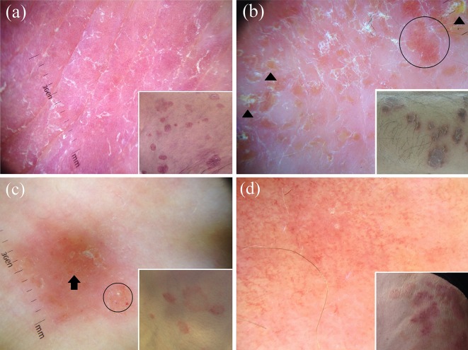
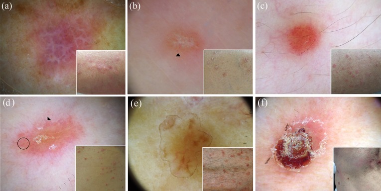
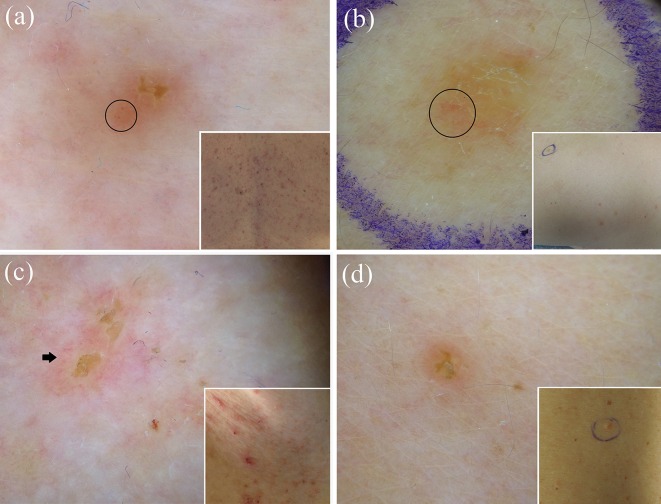
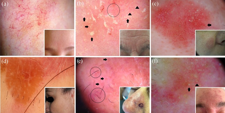
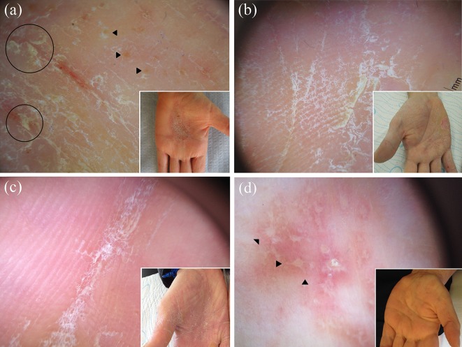
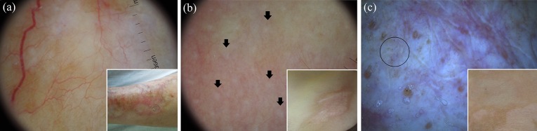
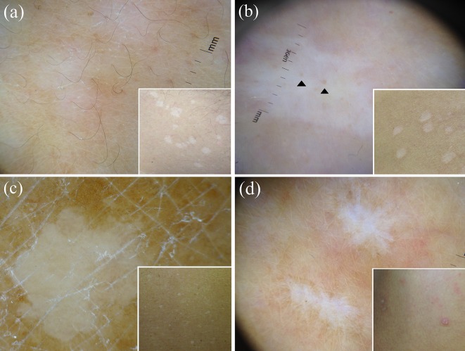
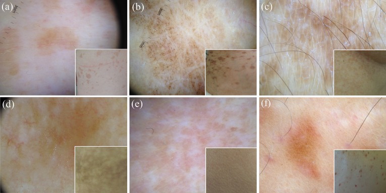
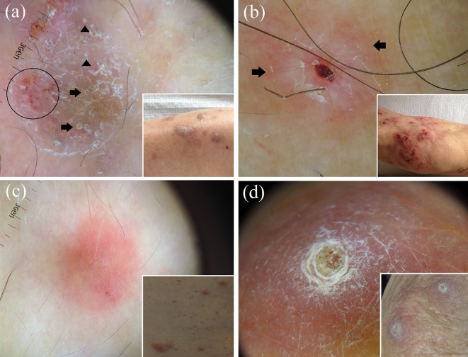
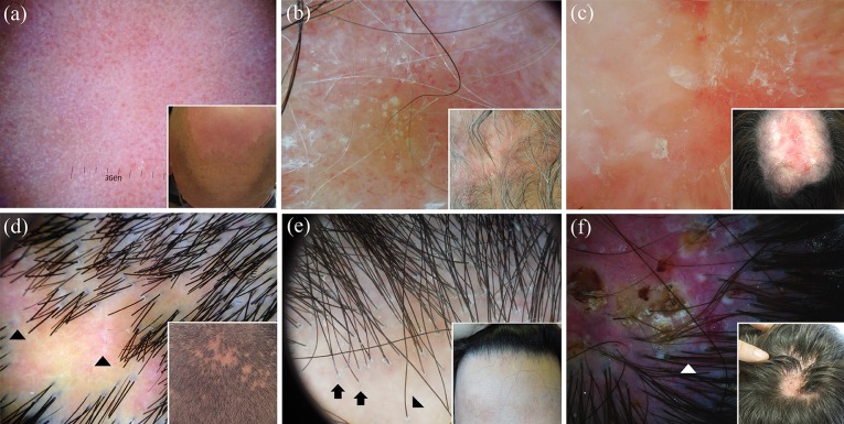
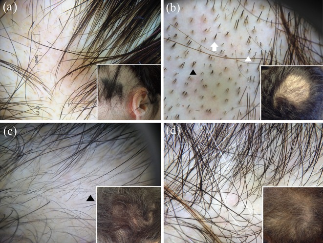
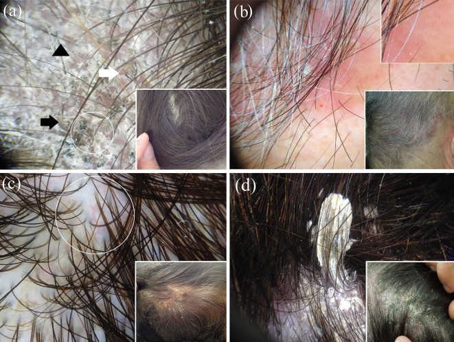
Over the last few years, dermoscopy has been shown to be a useful tool in assisting the noninvasive diagnosis of various general dermatological disorders. In this article, we sought to provide an up-to-date practical overview on the use of dermoscopy in general dermatology by analysing the dermoscopic differential diagnosis of relatively common dermatological disorders grouped according to their clinical presentation, i.e. dermatoses presenting with erythematous-desquamative patches/plaques (plaque psoriasis, eczematous dermatitis, pityriasis rosea, mycosis fungoides and subacute cutaneous lupus erythematosus), papulosquamous/papulokeratotic dermatoses (lichen planus, pityriasis rosea, papulosquamous sarcoidosis, guttate psoriasis, pityriasis lichenoides chronica, classical pityriasis rubra pilaris, porokeratosis, lymphomatoid papulosis, papulosquamous chronic GVHD, parakeratosis variegata, Grover disease, Darier disease and BRAF-inhibitor-induced acantholytic dyskeratosis), facial inflammatory skin diseases (rosacea, seborrheic dermatitis, discoid lupus erythematosus, sarcoidosis, cutaneous leishmaniasis, lupus vulgaris, granuloma faciale and demodicidosis), acquired keratodermas (chronic hand eczema, palmar psoriasis, keratoderma due to mycosis fungoides, keratoderma resulting from pityriasis rubra pilaris, tinea manuum, palmar lichen planus and aquagenic palmar keratoderma), sclero-atrophic dermatoses (necrobiosis lipoidica, morphea and cutaneous lichen sclerosus), hypopigmented macular diseases (extragenital guttate lichen sclerosus, achromic pityriasis versicolor, guttate vitiligo, idiopathic guttate hypomelanosis, progressive macular hypomelanosis and postinflammatory hypopigmentations), hyperpigmented maculopapular diseases (pityriasis versicolor, lichen planus pigmentosus, Gougerot-Carteaud syndrome, Dowling-Degos disease, erythema ab igne, macular amyloidosis, lichen amyloidosus, friction melanosis, terra firma-forme dermatosis, urticaria pigmentosa and telangiectasia macularis eruptiva perstans), itchy papulonodular dermatoses (hypertrophic lichen planus, prurigo nodularis, nodular scabies and acquired perforating dermatosis), erythrodermas (due to psoriasis, atopic dermatitis, mycosis fungoides, pityriasis rubra pilaris and scabies), noninfectious balanitis (Zoon's plasma cell balanitis, psoriatic balanitis, seborrheic dermatitis and non-specific balanitis) and erythroplasia of Queyrat, inflammatory cicatricial alopecias (scalp discoid lupus erythematosus, lichen planopilaris, frontal fibrosing alopecia and folliculitis decalvans), nonscarring alopecias (alopecia areata, trichotillomania, androgenetic alopecia and telogen effluvium) and scaling disorders of the scalp (tinea capitis, scalp psoriasis, seborrheic dermatitis and pityriasis amiantacea).

Keywords: Dermatitis; Dermatoscopy; Dermoscopy; Differential diagnosis; Inflammoscopy; Trichoscopy.


Figures

Fig. 1
Fig. 1
Dermoscopy of plaque psoriasis typically shows white scales and symmetrically and regularly distributed dotted vessels on a red background (a). The main dermoscopic criteria in eczematous dermatitis are dotted vessels (black circle) in a patchy distribution and yellow serocrusts (black arrowhead) (b). Both the herald patch and the secondary lesions of pityriasis rosea are dermoscopically characterised by peculiar peripheral whitish scales (“collarette” sign) as well as irregular or patchy dotted vessels (black circle); structureless orangish areas are also visible (black arrow) (c). Dermoscopic examination of mycosis fungoides reveals a combination of fine short, linear vessels with orange-yellowish patchy areas (d)
Fig. 2
Fig. 2
The dermoscopic analysis of classical lichen planus typically shows the Wickham striae over a purplish background (a). Dermoscopy of papulosquamous sarcoidosis shows the characteristic orange-yellowish background, in combination with in-focus fine linear vessels (black arrowhead); whitish lines and white scales are also evident in the centre (b). Guttate psoriasis lesions typically show a distinctive monomorphic dermoscopic picture, with dotted vessels distributed in a diffuse pattern (c). Dermoscopy of pityriasis lichenoides chronica frequently displays nondotted vessels, e.g. linear irregular vessels (black arrowhead), focally distributed dotted vessels (black circle) and orange-yellowish structureless areas (d). Dermoscopic examination of a case of disseminated superficial actinic porokeratosis displays the peculiar “cornoid lamella” at the periphery of the lesion (e). Dermoscopy of a necrotic lesion of lymphomatoid papulosis shows a central brown-grey structureless area (f)
Fig. 3
Fig. 3
Dermoscopy of Darier-like Grover disease displays a central branched polygonal brownish area surrounded by a thin whitish halo with peripheral dotted vessels (black circle) (a), while spongiotic Grover disease presents with whitish scaling over a reddish-yellowish background and irregular vessels (black circle) (b). Dermoscopic examination of Darier disease (c) and BRAF-inhibitor-induced acantholytic dyskeratosis (d) shows a pattern similar to that observed in Darier-like Grover disease, with a centrally located polygonal brownish area surrounded by a whitish halo and linear vessels (black arrow) in Darier disease (c) and a central branched polygonal brownish area surrounded by a thin whitish halo in the latter condition (d)
Fig. 4
Fig. 4
The main dermoscopic feature of rosacea is the presence of linear vessels, which are characteristically arranged in a polygonal network (a). The most typical dermoscopic finding of seborrheic dermatitis is represented by the presence of dotted vessels in a patchy distribution (black circle) and yellowish scales (black arrows); blurry linear branching vessels (black arrowheads) and whitish scales are also not uncommonly present (b). Dermoscopy of an intermediate-stage lesion of facial discoid lupus erythematosus reveals follicular white/yellowish rings/keratotic plugs, whitish scaling and blurred branching vessels (black arrow) over a reddish background (c). Dermoscopic examination of facial sarcoidosis displays a structureless orange-yellowish background with focussed linear vessels (d), while granuloma faciale features dilated follicular openings (black arrows) associated with linear/branching vessels (black circles) over a pinkish background (e). Dermoscopy of demodicidosis shows the so-called "Demodex tails", which are visualised as creamy/whitish gelatinous threads protruding out of follicular openings (black arrow), and “Demodex follicular openings”, which appear as round and coarse follicular openings containing light brown/greyish plugs surrounded by an erythematous halo (black arrowhead) (f)
Fig. 5
Fig. 5
Dermoscopy of chronic hand eczema typically reveals sparse whitish scales, yellowish scaling (black circles) and orangish dots/globules (black arrowheads), while palmar psoriasis and tinea manuum respectively display diffuse white scaling (b) and white scales mainly localised in the skin furrows (c). Dermoscopic examination of a case of palmar lichen planus shows roundish yellowish areas, some of which display peripheral projections in a star-like appearance (black arrowheads) over a purplish background (d)
Fig. 6
Fig. 6
Dermoscopic examination of an advanced lesion of necrobiosis lipoidica reveals elongated, branching and focussed serpentine vessels over a yellowish-orange/whitish background (a). Dermoscopy of morphea shows the typical fibrotic beams (black arrows) associated with linear branching vessels (b), while cutaneous lichen sclerosus displays several “comedo-like openings” (follicular keratotic plugs), whitish patches, dotted vessels (black circle) and delicate linear branching vessels (c)
Fig. 7
Fig. 7
Dermoscopy of achromic/hypochromic lesions of pityriasis versicolor usually shows a fairly demarcated white area with fine scales that are commonly localised in the skin furrows (a), while active lesions of guttate vitiligo typically display a well-demarcated, dense/glowing, often associated with perifollicular hyperpigmentation (black arrowheads) (b). Dermoscopic examination of idiopathic guttate hypomelanosis may show multiple small areas coalescing into irregular/polycyclic macules, with several white shades and both well- and ill-defined edges, surrounded by patchy hyperpigmented network (“cloudy sky-like” pattern) (c), whilst postinflammatory hypopigmentation often presents with some dermoscopic findings typical of the original lesions (in this case, the star-like arrangment typical of prurigo nodularis) (d)
Fig. 8
Fig. 8
Dermoscopy of hyperpigmented lesions of pityriasis versicolor often shows fine whitish scaling localised in the skin furrows associated with a diffuse brownish pigmentation (a). The most common dermoscopic finding of lichen planus pigmentosus is represented by fine/coarse, grey-blue/brown dots over a brownish background (b), while confluent and reticulated papillomatosis (Gougerot–Carteaud syndrome) displays fine whitish scaling and brownish, homogeneous, more or less defined, polygonal, flat globules separated by whitish/pale striae creating a cobblestone pattern (c). Dermoscopic examination of pigmented lesions of erythema ab igne may reveal diffuse brownish pigmentation with telangiectatic vessels/fine whitish scaling, while friction melanosis and urticaria pigmentosa typically display brownish structureless areas arranged in a reticular fashion (e) and a homogeneous light-brown blot with a pigment network (f), respectively
Fig. 9
Fig. 9
Dermoscopy of hypertrophic lichen planus shows a peculiar pattern characterised by a rippled surface with comedo-like structures filled with yellow keratinous plugs (black arrows) and/or round corneal structures (“corn pearls”) (black arrowheads); some irregular dotted vessels (black circle), scaling and central hyperpigmentation are also present in this picture (a). Dermoscopy of a prurigo nodularis lesion displays the typical “white starburst pattern”, with radially arranged whitish lines (black arrows) on a brownish and/or reddish background; central erosion and scales are also present in this case (b). Although dermoscopy of nodular scabies may often show the presence of mites (“hang glider sign”) and/or burrows (“jet with condensation trails”), it is not uncommon that the only detectable findings are nonspecific vascular features (mainly dotted vessels) (c). Dermoscopic examination of a case of reactive perforating collagenosis reveals the typical “three concentric areas” pattern, with a central round brownish-greenish/yellowish-brown structureless area, surrounded by a white keratotic collarette and an erythematous halo (d)
Fig. 10
Fig. 10
Dermoscopy of discoid lupus erythematosus of the scalp varies according to the disease stage: active lesions may be mainly characterised by red dots (a) or follicular keratotic plugs (quite large yellowish/whitish dots) and thick arborising vessels (b), while long-lasting lesions commonly display loss of follicular openings, white areas and thin vessels (c). The main dermoscopic hallmarks of active lichen planopilaris are perifollicular scales; characteristic (but not pathognomonic) white dots (fibrotic white dots) (black arrowheads) and a reddish background are also present in less active areas in this case (d). Dermoscopic examination of a case of frontal fibrosing alopecia reveals minor perifollicular scaling with an aflegmasic (ivory white to ivory beige) surrounding background; follicular openings with only one hair at the hair-bearing margin (black arrows) and lonely hair (black arrowhead) are also visible (e). Classic dermoscopic appearance of active folliculitis decalvans showing follicular pustules, yellow discharge, crusts and characteristic hair tufts that contain >10 hair shafts (white arrowhead); unspecific vessels and erythema are also evident in the picture (f)
Fig. 11
Fig. 11
Dermoscopic examination of a case of active alopecia areata shows black dots and micro-exclamation mark hairs; regular yellow dots are also evident (a), while dermoscopy of trichotillomania reveals a chaotic pattern of diverse findings related to hair fracturing, including (in this case) hairs broken at different lengths, black dots, flame-like hairs (white arrow), tulip-like hairs (short hairs with darker, tulip-shaped ends white arrowhead) and V-sign (two or more hairs emerging from one follicular unit that are broken at the same level black arrowhead) (b). Dermoscopy of androgenetic alopecia typically shows hair shaft thickness heterogeneity, a large number of follicular units with only one emerging hair shaft, and an increased proportion of thin and vellus hairs (>10% of the hairs); wavy hairs are also visible (black arrowhead) (c). The most indicative dermoscopic clue of telogen effluvium is the lack of features typical of other diseases; empty hair follicles and follicular units with only one hair are also evident in this case of chronic telogen effluvium (d)
Fig. 12
Fig. 12
Dermoscopic examination of a case of tinea capitis displays scaling and the peculiar “comma” hair (white arrow), “corkscrew” hair (white circle), “zigzag” hair (black arrow) and “Morse code” hair (black arrowhead) (a). Dermoscopy of scalp psoriasis reveals the typical dotted vessels (magnified in the upper-right box) and white scales; a haemorrhagic spot is also evident (b). Differently from psoriasis, scalp seborrhoeic dermatitis shows yellowish scales and the characteristic arborising vessels (white circle) (c). Dermoscopic examination of pityriasis amiantacea displays diffuse white scaling and the characteristic compact white keratotic material adhering to a tuft of hair (asbestos-like scale) (d)

References

    1. Errichetti E, Stinco G. The practical usefulness of dermoscopy in general dermatology. G Ital Dermatol Venereol. 2015;150:533–546. - PubMed
    1. Lallas A, Giacomel J, Argenziano G, et al. Dermoscopy in general dermatology: practical tips for the clinician. Br J Dermatol. 2014;170:514–526. - PubMed
    1. Lallas A, Zalaudek I, Argenziano G, et al. Dermoscopy in general dermatology. Dermatol Clin. 2013;31:679–694. - PubMed
    1. Zalaudek I, Argenziano G, Di Stefani A, et al. Dermoscopy in general dermatology. Dermatology. 2006;212:7–18. - PubMed
    1. Micali G, Lacarrubba F, Massimino D, Schwartz RA. Dermatoscopy: alternative uses in daily clinical practice. J Am Acad Dermatol. 2011;64:1135–1146. - PubMed