. 2016 Oct 28;291(44):23188-23198.
doi: 10.1074/jbc.M116.732743. Epub 2016 Sep 9.

Targeting Liver Fibrosis with a Cell-penetrating Protease-activated Receptor-2 (PAR2) Pepducin

Affiliations

Targeting Liver Fibrosis with a Cell-penetrating Protease-activated Receptor-2 (PAR2) Pepducin

Andrew M Shearer et al. J Biol Chem. .

Abstract

Chronic liver inflammation and fibrosis in nonalcoholic steatohepatitis can lead to cirrhosis and liver failure for which there are currently no approved treatments. Protease-activated receptor-2 (PAR2) is an emerging new target expressed on liver stellate cells and hepatocytes that regulates the response to liver injury and inflammation. Here, we identified a pepducin to block the deleterious actions of PAR2 in promoting liver fibrosis. Non-alcoholic fatty liver disease and early fibrosis were induced by the methionine-choline-deficient diet in mice. Fibrotic liver disease was induced by administering carbon tetrachloride for 8 weeks. Mice were treated with the pepducin PZ-235 either from onset of the experiment or after fibrosis was established. Hepatic fibrosis, collagen content, inflammatory cytokines, steatosis, triglycerides, and NAFLD activity score were assessed as primary outcome parameters depending on the model. The activity of the PAR2 pepducin on cultured stellate cell activation and hepatocyte reactive oxygen species production was evaluated. PZ-235 significantly suppressed liver fibrosis, collagen deposition, inflammatory cytokines, NAFLD activity score, steatosis, triglycerides, aspartate transaminase, alanine transaminase, and stellate cell proliferation by up to 50-100%. The PAR2 inhibitor afforded significant protective effects against hepatocellular necrosis and attenuated PAR2-mediated reactive oxygen species production in hepatocytes. PZ-235 was distributed to liver and other mouse tissues and was found to form a well structured α-helix that closely resembles the juxtamembrane helical region of the analogous TM6 and third intracellular region of the intact receptor that is critical for coupling to internal G proteins. The ability of PZ-235 to effectively suppress fibrosis, hepatocellular necrosis, reactive oxygen species production, steatosis, and inflammation indicates the potential for PAR2 pepducin inhibitors to be broadly efficacious in the treatment of liver fibrosis.

Keywords: G protein-coupled receptor (GPCR); PAR2; biotechnology; cell-penetrating peptide (CPP); fibrosis; pepducin; reactive oxygen species (ROS).


Figures

FIGURE 1.
FIGURE 1.
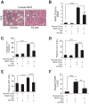
The PAR2 pepducin PZ-235 reduces hepatic steatosis and inflammation in the experimental MCD model in mice. C57BL/6 male mice were fed normal chow or an MCD diet for 3 weeks and treated daily with PZ-235 (5 mg/kg/day) or vehicle (Veh). A and B, representative 200× photomicrographs of H&E-stained liver sections from vehicle- and PZ-235-treated MCD mice. Liver histology was scored according to the NASH Clinical Research Network criteria to determine the NAS. Liver triglycerides (C), plasma ALT (D), and plasma ALT (E) were measured at the 3-week end point in the MCD mice. F, quantitative PCR of SYBR Green (mean ± S.E., n = 4–6) was determined by the ΔΔCT method where PAR2 mRNA was normalized to GAPDH and the lowest adjusted value in the control group. ****, p < 0.0001; ***, p < 0.001; **, p < 0.01 using the Holm-Sidak correction for multiple comparisons. Error bars represent S.E. gm, gram.
FIGURE 2.
FIGURE 2.
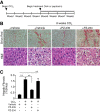
Suppression of CCl4-induced liver fibrosis with delayed PZ-235 treatment. A, timeline of treatment. C57BL/6 mice were treated twice weekly for 8 weeks with 1 μl of CCl4/g of body weight and PZ-235 (5 mg/kg/day), vehicle (Veh), or inactive control PAR2 pepducin PZ-234 (5 mg/kg/day) started at week 4. B, photomicrographs (200×) of Sirius red and H&E staining of liver sections from mice in the different treatment groups at the 8-week end point. C, quantification of percentage of Sirius red staining per 10 fields (mean ± S.E.). Vehicle (no CCl4), n = 5; CCl4 + vehicle, n = 8l CCl4 + PZ-235, n = 8; CCl4 + PZ-234, n = 3. **, p < 0.01. Error bars represent S.E.
FIGURE 3.
FIGURE 3.
PZ-235 suppresses liver expression of inflammatory cytokines in CCl4-treated mice receiving delayed pepducin treatment initiated at week 4. Livers were isolated from mice in Fig. 2 at week 8, and quantitative PCR by SYBR Green (mean ± S.E., n = 4–6) was determined by the ΔΔCT method where mRNA expression was normalized to GAPDH and the lowest adjusted value in the control group. #, p = 0.06; *, p < 0.05 using the Holm-Sidak correction for multiple comparisons. Error bars represent S.E. Veh, vehicle.
FIGURE 4.
FIGURE 4.
PZ-235 blocks PAR2-calcium signaling and activation of LX-2 stellate cells. A, stellate cell (LX-2) calcium mobilization in response to the PAR2 agonist SLIGRL (100 μm) is inhibited by PZ-235 (30 μm) but does not inhibit the PAR1 agonist thrombin (Thromb) (10 nm). B, ERK phosphorylation (pERK) induced by 30 μm SLIGRL or 1 nm trypsin in LX-2 cells is inhibited by 3 μm PZ-235. C, PZ-235 (1 μm) inhibits LX-2 cell viability induced by 10 nm trypsin (Tryp). 3-(4,5-Dimethylthiazol-2-yl)-2,5-diphenyltetrazolium bromide activity was determined after 96-h stimulation with 10 nm trypsin. D, secretion of MCP-1 into LX-2 medium in response to PAR2 stimulation by the agonists in A is inhibited by 3 μm PZ-235. *, p < 0.05; #, p = 0.06 based on analysis of variance. Error bars represent S.E. RFU, relative fluorescence units.
FIGURE 5.
FIGURE 5.
Suppression of CCl4-induced liver necrosis and fibrosis with concurrent PZ-235 treatment. A, timeline of treatment. C57BL/6 mice (n = 10) were treated twice weekly with 1 μl of CCl4/g of body weight and with PZ-235 (10 mg/kg/day) or vehicle (Veh) for the entire 8 weeks of the study. B, representative 200× photomicrographs of H&E and Picosirius red staining from the livers of the different treatment groups. C, quantification of nuclei number per 200× field (mean ± S.D. of 10 fields). D, quantification of percentage of Picosirius staining per 200× field (mean ± S.D. of 10 fields). **, p < 0.01; *, p < 0.05 using the Holm-Sidak correction for multiple comparisons. Error bars represent S.E.
FIGURE 6.
FIGURE 6.
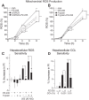
PZ-235 inhibits mitochondrial ROS production and enhances viability of hepatocytes. THLE2 hepatocytes were treated with vehicle (Veh) or 5 μm PZ-235 and then stimulated with LIGRLO (A) or trypsin (B) for the times indicated. Individual points represent the mean ± S.E. for that condition at a particular time point; n = 7–10 wells per point. C, serum-starved HepG2 cells were pretreated with 5 μm PZ-235 or vehicle before being stimulated with 10 nm trypsin for 2 h followed by 6-h treatment with 250 μm H2O2. Values shown are mean ± S.E. of the increase in PI from untreated controls; n = 4 for each condition. Comparisons between conditions and baseline were made by direct two-tailed t tests. D, serum-starved HepG2 cells were treated with 5 μm PZ-235 or vehicle, then exposed to CCl4 for 4 h, and analyzed as in C. Significance was determined using the Holm-Sidak correction for multiple comparison. ***, p < 0.001; **, p < 0.01; *, p < 0.05; #, p < 0.06. Error bars represent S.E.
FIGURE 7.
FIGURE 7.
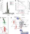
Liver residence and NMR structural determination of PZ-235. A, biodistribution of [1-14C]palmitate-PZ-235 to individual organs (50,000 cpm injected per mouse (n = 2) for each time point). Radioactive PZ-235 was mixed 1:2 with unlabeled PZ-235 (10 mg/kg final; 100 μl per injection). B, pharmacokinetics of s.c. and i.v. PZ-235 in CF-1 mice determined by LC-MS/MS from plasma samples withdrawn at 5-min–16-h time points (five to seven mice used for each dose). C, ensemble of 30 individual energy-minimized NMR-generated structures (green) of PZ-235 with the average structure (root mean square deviation, 0.78 ± 0.23 Å) shown in red. D, proposed mechanism of membrane insertion and flipping of the PAR2 pepducin and alignment with analogous i3 loop and TM6 regions of the off-state of PAR2. Error bars represent S.E. Hrt, heart; Kid, kidney; Spl, spleen; Panc, pancreas; Sm intest, small intestine.
FIGURE 8.
FIGURE 8.
Proposed mechanism of action of PAR2 in promoting liver fibrosis, necrosis, and steatosis in NASH. Procoagulant proteases such as factors VIIa and Xa are up-regulated in liver during the prothrombotic state that occurs during NASH progression. PAR2 is cleaved on the surface of stellate cells by these proteases to cause activation to a proliferative myofibroblast and enhanced secretion of collagen matrix and inflammatory cytokines, leading to liver fibrosis. Proteolytic cleavage of PAR2 on hepatocytes leads to steatosis, ROS production, and hepatocellular necrosis.

References

    1. LaBrecque D., Abbas A., Anania F., Ferenci P., Ghafoor-Khan A., Goh K.-L., Hamid S. S., Isakov V., Lizarzabal M., Mojica-Pernaranda M., Rivera-Ramos J. F., Sarin S., Stimac D., Thomson A. B., Umar M., et al. (2012) Nonalcoholic fatty liver disease and nonalcoholic steatohepatitis, in World Gastroenterology Organisation Global Guidelines (World Gastroenterology Organisation, ed) World Gastroenterology Organisation, Milwaukee, WI - PubMed
    1. Fujii H., and Kawada N. (2012) Inflammation and fibrogenesis in steatohepatitis. J Gastroenterol. 47, 215–225 - PubMed
    1. Machado M. V., and Diehl A. M. (2016) Pathogenesis of nonalcoholic steatohepatitis. Gastroenterology 150, 1769–1777 - PMC - PubMed
    1. Schuppan D., and Kim Y. O. (2013) Evolving therapies for liver fibrosis. J. Clin. Investig. 123, 1887–1901 - PMC - PubMed
    1. Wieser V., Adolph T. E., Enrich B., Kuliopulos A., Kaser A., Tilg H., and Kaneider N. C. (2016) Reversal of murine alcoholic steatohepatitis by pepducin-based functional blockade of interleukin-8 receptors. Gut 10.1136/gutjnl-2015-310344 - DOI - PMC - PubMed

Publication types