Review
. 2016 Nov;37(11):724-737.
doi: 10.1016/j.it.2016.08.010. Epub 2016 Sep 7.

Present Yourself! By MHC Class I and MHC Class II Molecules

Affiliations
Review

Present Yourself! By MHC Class I and MHC Class II Molecules

Kenneth L Rock et al. Trends Immunol. 2016 Nov.

Abstract

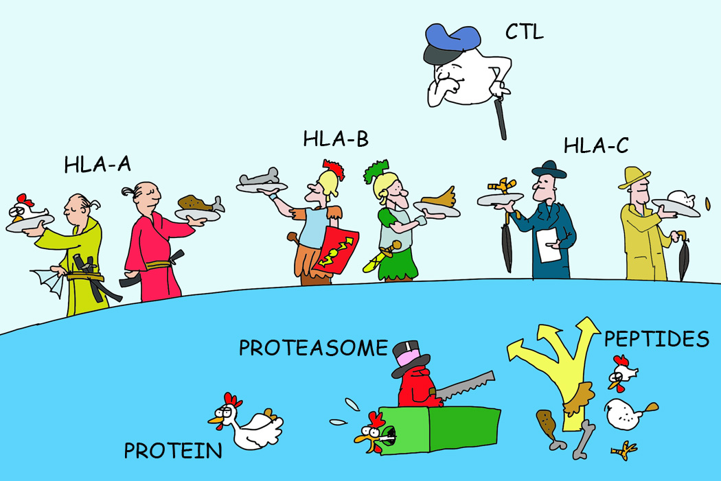
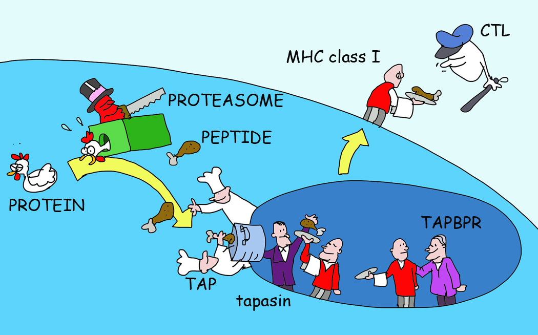
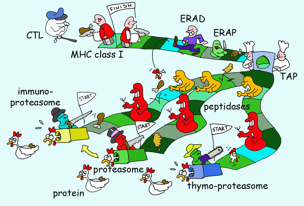
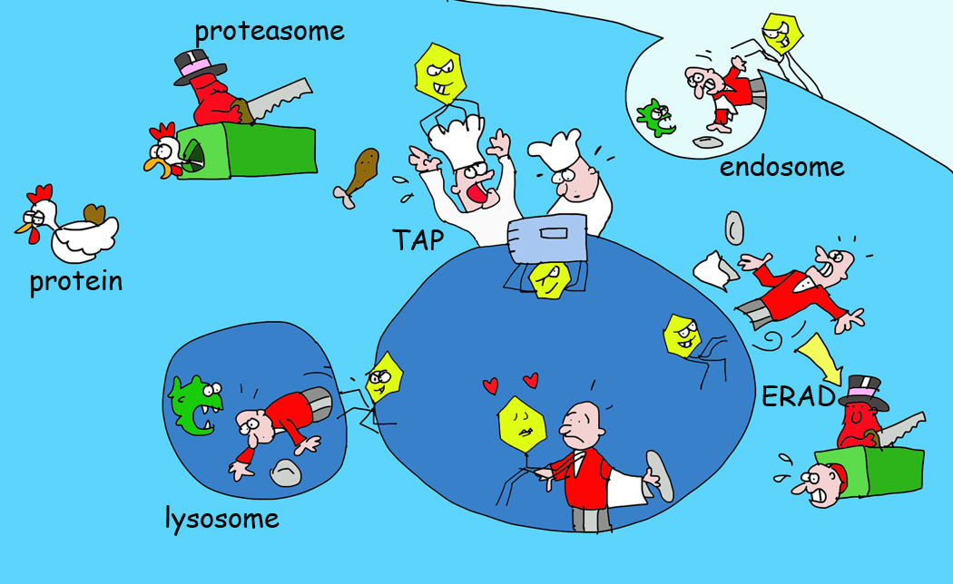
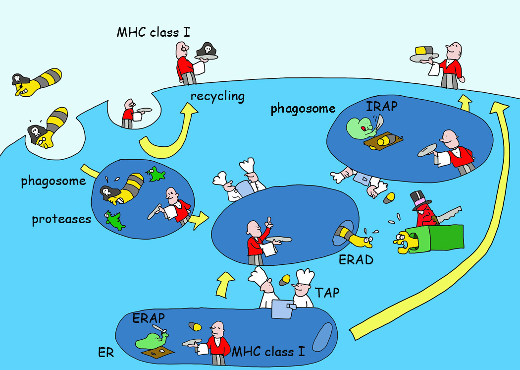
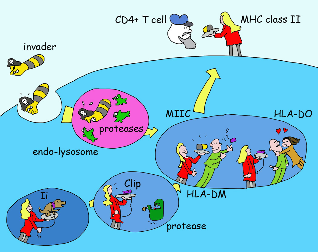
Since the discovery of MHC molecules, it has taken 40 years to arrive at a coherent picture of how MHC class I and MHC class II molecules really work. This is a story of the proteases and MHC-like chaperones that support the MHC class I and II molecules in presenting peptides to the immune system. We now understand that the MHC system shapes both the repertoire of presented peptides and the subsequent T cell response, with important implications ranging from transplant rejection to tumor immunotherapies. Here we present an illustrated review of the ins and outs of MHC class I and MHC class II antigen presentation.

Keywords: MHC class I; MHC class II; antigen presentation; autoimmune diseases; transplantation; tumor immunology.


Figures

Figure 1
Figure 1. Of MHC Locus and Allelic Products with Polymorphism
Most mammalian species express three different MHC I and three different MHC II molecules (shown here for the human MHC I HLA-A, HLA-B and HLA-C locus products). Since these are polymorphic and genetically encoded, a total of somewhere between 3 and 6 (depending on the differences between the inherited genes) alleles will be expressed on cells. These are polymorphic in the peptide-binding groove region of MHC molecules to present different peptides of a defined antigen.
Figure 2
Figure 2. A Simple Illustration of MHC I Antigen Presentation
Antigens are degraded by the proteasome to yield peptide fragments. These peptides are then translocated from the cytosol into the endoplasmic reticulum (ER) lumen where MHC I in waiting for peptides is retained by a series of chaperones including a dedicated chaperone tapasin in the peptide-loading complex. A second dedicated chaperone (TAPBPR) can further optimize the peptides in MHC I. Only MHC I with optimal peptides is allowed to leave the ER to present the peptide fragments at the cell surface to CD8+ T cells.
Figure 3
Figure 3. Survival of The Fittest for MHC I Presentation and The Many Proteasomes
There are three types of proteasomes with unique tissue expression. These proteasomes have an altered cleavage specificity yielding (in part) different degradation fragments. These fragments are released in the hostile environment of the cytosol where the majority of peptides will be destroyed by peptidases. Few peptides survive this massacre through translocation in the ER by transporter associated with antigen processing (TAP). Here they can be further trimmed by ER resident aminopeptidase (ERAP) or translocated back into the cytosol by the ER associated degradation (ERAD) system. Only few peptides survive the chaperone-mediated survival selection for low off-rate peptides for a defined MHC I allele and these are ultimately presented.
Figure 4
Figure 4. Various Viral Immune Evasion Strategies for MHC I Antigen Presentation
Pathogens have evolved different ways to obstruct processes selective in the antigen presentation pathway. Examples include viral proteins inhibiting peptide transport by TAP, retaining MHC I in the ER or recognizing MHC I in the ER for targeting these back into the cytosol for degradation by the proteasome (the ERAD system). Other viral proteins recognize MHC I at the cell surface for internalization and destruction in lysosomes.
Figure 5
Figure 5. Crossing Boundaries; Various Models of Cross-Presentation by MHC I
MHC I molecules can present exogenous antigens and antigens delivered in apoptotic bodies and other types of cell debris. For dendritic cells, this process can result in cross-priming of CD8+ T cells. MHC I can recycle through the endosomal pathway to acquire antigen fragments made by proteases such as insulin regulated aminopeptidase (IRAP)[129] for cross-presentation. Endosomes may also acquire TAP and other ER molecules that may help export antigens into the cytosol for proteasomal hydrolysis and the resulting peptides may be reimported into the endosomes for MHC I loading and recycling and/or be delivered in the normal antigen presentation pathway as shown in Figure 2.
Figure 6
Figure 6. A Simple Illustration of MHC II Antigen Presentation
MHC II proteins are made in the ER where they pair with a third chain, the invariant chain or Ii. Ii fills (through a peptide sequence called CLIP) the MHC II peptide-binding groove and allows efficient exit of MHC II from the ER. Ii also guides MHC II through the cells to a late endosomal compartment, MIIC. Ii is degraded by endosomal proteases, as are antigens taken up by endocytosis or phagocytosis. The CLIP segment is protected from destruction and exchanged for an antigenic peptide with the help of a dedicated chaperone called DM (HLA-DM in human). Another chaperone expressed in a few immune cell types (immature B cells, some DC forms). called DO (HLA-DO) can compete for DM binding to MHC II and thereby affect the peptide repertoire on MHC II that is ultimately presented at the cell surface to CD4+ T cells.

References

    1. Flajnik MF, Kasahara M. Origin and evolution of the adaptive immune system: genetic events and selective pressures. Nature reviews Genetics. 2010;11:47–59. - PMC - PubMed
    1. Brown JH, et al. Three-dimensional structure of the human class II histocompatibility antigen HLA-DR1. Nature. 1993;364:33–39. - PubMed
    1. Bjorkman PJ, et al. The foreign antigen binding site and T cell recognition regions of class I histocompatibility antigens. Nature. 1987;329:512–518. - PubMed
    1. Zhang P, et al. Crystal structure of the stress-inducible human heat shock protein 70 substrate-binding domain in complex with peptide substrate. PloS one. 2014;9:e103518. - PMC - PubMed
    1. Dong G, et al. Insights into MHC class I peptide loading from the structure of the tapasin-ERp57 thiol oxidoreductase heterodimer. Immunity. 2009;30:21–32. - PMC - PubMed

Publication types