Review
. 2017 Feb;41(1):108-113.
doi: 10.1016/j.jcjd.2016.07.002. Epub 2016 Sep 7.

Insulin Signalling: The Inside Story

Affiliations
Review

Insulin Signalling: The Inside Story

Barry I Posner. Can J Diabetes. 2017 Feb.

Abstract

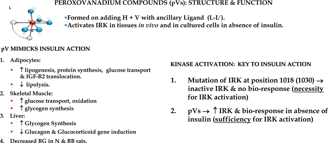
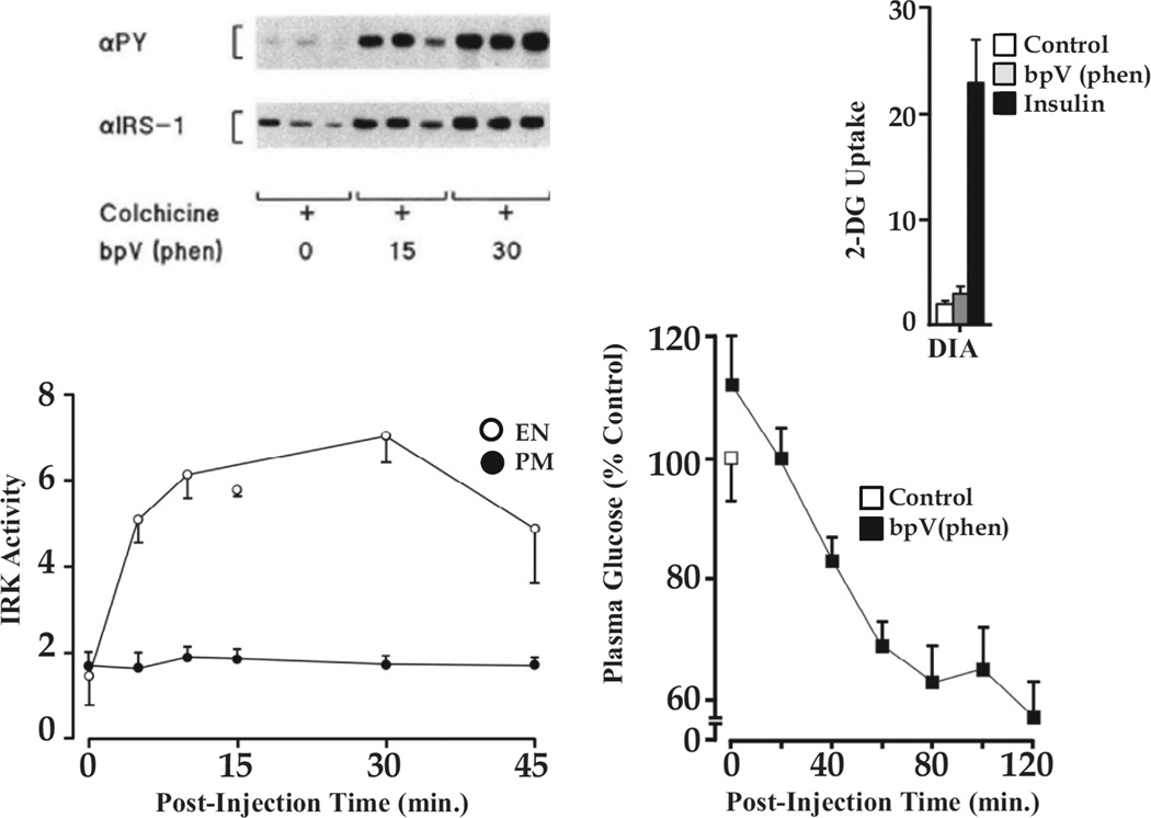
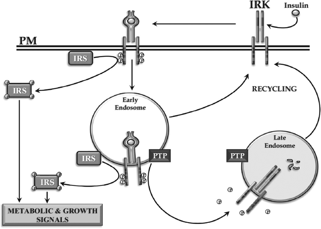
Insulin signalling begins with binding to its cell surface insulin receptor (IR), which is a tyrosine kinase. The insulin receptor kinase (IRK) is subsequently autophosphorylated and activated to tyrosine phosphorylate key cellular substrates that are essential for entraining the insulin response. Although IRK activation begins at the cell surface, it is maintained and augmented following internalization into the endosomal system (ENS). The peroxovanadium compounds (pVs) were discovered to activate the IRK in the absence of insulin and lead to a full insulin response. Thus, IRK activation is both necessary and sufficient for insulin signalling. Furthermore, this could be shown to occur with activation of only the endosomal IRK. The mechanism of pV action was shown to be the inhibition of IRK-associated phosphotyrosine phosphatases (PTPs). Our studies showed that the duration and intensity of insulin signalling are modulated within ENS by the recruitment of cellular substrates to ENS; intra-endosomal acidification, which promotes dissociation of insulin from the IRK; an endosomal acidic insulinase, which degrades intra-endosomal insulin; and IRK-associated PTPs, which dephosphorylate and, hence, deactivate the IRK. Therefore, the internalization of IRKs is central to insulin signalling and its regulation.

La signalisation de l’insuline commence par la liaison de l’insuline à son récepteur (IR) situé à la surface des cellules, soit la tyrosine kinase. La kinase du récepteur de l’insuline (KRI) s’est subséquemment autophosphorylée et activée vers les principaux substrats cellulaires de la tyrosine phosphorylée qui sont essentiels au déclenchement de la réponse à l’insuline. Bien que l’activation de la KRI commence à la surface des cellules, elle est maintenue et augmentée à la suite de l’internalisation du système endosomal (SEN). Il a été découvert que les composés de peroxovanadium (pV) activent la KRI en l’absence d’insuline et mènent à une réponse insulinique complète. Par conséquent, l’activation de la KRI est nécessaire et suffisante à la signalisation de l’insuline. De plus, il pourrait être démontré qu’elle apparaît avec l’activation de la KRI endosomale seulement. Il a été démontré que le mécanisme de l’activité du pV est l’inhibition des phosphotyrosines phosphatases (PTP) associées à la KRI. Nos études ont montré que la durée et l’intensité de la signalisation de l’insuline sont modulées au sein du SEN par le recrutement de substrats cellulaires du SEN; l’acidification intra-endosomale, qui favorise la dissociation de l’insuline de la KRI; une insulinase endosomique acide, qui dégrade l’insuline dans les endosomes; les PTP associées à la KRI, qui déphosphorylent et, donc, désactivent la KRI. Par conséquent, l’internalisation des KRI est essentielle au déclenchement de la signalisation de l’insuline et à sa régulation.

Keywords: endosomal acidic insulinase; endosomes; insulin receptor; insulinase endosomique acide; phosphorylation; phosphotyrosine phosphatase; récepteur de l'insuline; tyrosine kinase.


Figures

Figure 1
Figure 1
Electron microscope radioautographs illustrating the localization of 125I-insulin-specific binding sites (silver grains). Left, insulin binding sites on the endothelium of rat brain microvessels. Right, insulin binding sites over a free nerve terminal (T) and a preterminal axon (A) containing dense core vesicles (arrow) in the external median eminence of the rat hypothalamus.
Figure 2
Figure 2
Insulin binding sites in Golgi fractions (GFs) and PM from rat liver. Top, binding of 125I-insulin to GF and PM before and after freeze-thawing of the cell fractions. Bottom, binding of 125I-insulin vs. 125I-hGH to GF and PM after freeze-thawing. hGH, like all primate growth hormones, binds to both prolactin and GH binding sites. The former receptors predominate in rat liver. These figures were derived from the data in reference (8).
Figure 3
Figure 3
Time course of 125I-insulin uptake into subcellular fractions. Left, concentration over time of injected 125I-insulin in homogenate, microsomes and GF. Results are expressed as a per cent of maximum concentration in GF. Right, EM radioautograph of GF fraction at 2 min post-125I-insulin injection. Silver grains are largely found over the periphery (arrows) of GF vesicles with their characteristic lipoprotein (lp) content. The data are from reference (9).
Figure 4
Figure 4
Time course of IRK concentration and activation following insulin administration. Left, IRK content per mg cell fraction protein was determined by immunoblotting with IRK-specific antibody. Results are expressed as a per cent of the basal levels in ENS and PM (100% at zero time). Right, IRK enzyme activity per mg cell fraction protein (specific activity) was expressed as a per cent of maximum measured in ENS. The data were derived from figures 6 and 9 of reference (13).
Figure 5
Figure 5
Degradation of endocytosed insulin in isolated ENS: requirement for acidic pH. At 5 min after 125I-insulin injection ENS were prepared and incubated in vitro under the noted conditions, and degradation of endosomal insulin was assayed as described in reference . Left, effect on insulin degradation of ATP and agents that alter endosomal proton accumulation. Right, effect of pH of incubation medium on endosomal insulin degradation. The data are from reference (16).
Figure 6
Figure 6
Characteristics of peroxovanadium compounds (pVs). Note that a given compound’s properties are significantly influenced by the nature of the ancillary ligand bound in the inner coordination sphere of vanadium. The major insulin mimetic effects are summarized (left); and the importance of pVs in establishing that IRK activation in the absence of insulin is sufficient for promoting a full insulin response (right).
Figure 7
Figure 7
Activation of the endosomal IRK is sufficient to promote insulin signalling. Left, (bottom) IRK activity in ENS and PM following bpV(phen) administration to rats pretreated with colchicine to block recycling of IRK from ENS to PM. IRK activation is seen exclusively in the ENS; (top) IRS-1 phosphorylation is seen when only endosomal IRK is activated. Right, (bottom) bpV(phen) lowers blood glucose levels; (top) in the absence of any effect to increase glucose uptake as measured by 2-deoxyglucose (2-DG) accumulation in skeletal muscle exemplified by diaphragm (DIA). (bottom) Lowering of blood glucose levels by bpV(phen) in colchicine-treated rats. For details see reference (22).
Figure 8
Figure 8
Internalization of the activated insulin-IRK complex. Signalling from within ENS of the activated IRK is depicted. The role of IRK-associated PTP and endosomal insulin degradation in terminating intracellular insulin signalling is shown.

References

    1. Freychet P, Roth J, Neville DM., Jr Insulin receptors in the liver: Specific binding of (125 I) insulin to the plasma membrane and its relation to insulin bioactivity. Proc Natl Acad Sci U S A. 1971;68:1833–1837. - PMC - PubMed
    1. Posner BI, Kelly PA, Shiu RP, Friesen HG. Studies of insulin, growth hormone and prolactin binding: Tissue distribution, species variation and characterization. Endocrinology. 1974;95:521–531. - PubMed
    1. Bergeron JJ, Levine G, Sikstrom R, et al. Polypeptide hormone binding sites in vivo: Initial localization of 125I-labeled insulin to hepatocyte plasmalemma as visualized by electron microscope radioautography. Proc Natl Acad Sci U S A. 1977;74:5051–5055. - PMC - PubMed
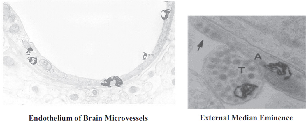
    1. Van Houten M, Posner BI. Insulin binds to brain blood vessels in vivo. Nature. 1979;282:623–625. - PubMed
    1. Van Houten M, Posner BI, Kopriwa BM, Brawer JR. Insulin binding sites localized to nerve terminals in rat median eminence and arcuate nucleus. Science. 1980;207:1081–1083. - PubMed

MeSH terms