Review
. 2016 Oct;32(10):638-659.
doi: 10.1016/j.tig.2016.07.004.

Mechanisms of Photoreceptor Patterning in Vertebrates and Invertebrates

Affiliations
Review

Mechanisms of Photoreceptor Patterning in Vertebrates and Invertebrates

Kayla Viets et al. Trends Genet. 2016 Oct.

Abstract

Across the animal kingdom, visual systems have evolved to be uniquely suited to the environments and behavioral patterns of different species. Visual acuity and color perception depend on the distribution of photoreceptor (PR) subtypes within the retina. Retinal mosaics can be organized into three broad categories: stochastic/regionalized, regionalized, and ordered. We describe here the retinal mosaics of flies, zebrafish, chickens, mice, and humans, and the gene regulatory networks controlling proper PR specification in each. By drawing parallels in eye development between these divergent species, we identify a set of conserved organizing principles and transcriptional networks that govern PR subtype differentiation.

Keywords: Danio rerio; Drosophila melanogaster; Gallus gallus domesticus; Homo sapiens; Mus musculus; chick; color vision; color-detecting; cone; evolution; eye; fruit fly; gene network; human; inner photoreceptor; mosaic; motion-detecting; mouse; outer photoreceptor; photoreceptor; regionalization; regulatory network; retina; rod; stochasticity; zebrafish.


Figures

Figure 1
Figure 1. Retinas are patterned in stochastic/regionalized, regionalized, and ordered mosaics
A1) Schematic of the Drosophila melanogaster (fruit fly) PR mosaic (not to scale). D: Dorsal, V: Ventral, A: Anterior, P: Posterior. A2) Schematic of a Drosophila ommatidium. A3) Schematic of a pale ommatidium. A4) Schematic of a yellow ommatidium. A5) Schematic of a dorsal third yellow ommatidium. A6) Schematic of a dorsal rim ommatidium. A7) Whole-mount immunostain of a Drosophila retina showing the stochastic distribution of ommatidial subtypes. A8) Immunostain showing Rhodopsin 1 (Rh1) expression in the outer PRs. A9) Immunostain showing the stochastic patterning of Rh3 and Rh4 in a section of the Drosophila retina. A10) Immunostain showing the stochastic patterning of Rh5 and Rh6 in a section of the Drosophila retina. A11) Immunostain of the dorsal third of the Drosophila retina, showing coexpression of Rh3 and Rh4 in dorsal third yR7s. A12) Immunostain of the dorsal rim of the Drosophila retina, showing expression of Rh3 in R7s and R8s. B1) Schematic of the Danio rerio (zebrafish) PR mosaic (not to scale). D: Dorsal, V: Ventral, A: Anterior, P: Posterior. B2) Schematic side view of a single unit of the zebrafish retinal pattern. B3) Schematic showing the overlapping, regionalized expression patterns of zebrafish LWS and RH2 opsin subtypes (not to scale). 1: Inner central/dorsal area, 2: Outer central/dorsal area, 3: Inner periphery/ventral area, 4: Outer periphery/ventral area. B4) Immunostain of a section of the zebrafish cone mosaic. Reprinted from Progress in Retinal and Eye Research, Volume 42, M. Hoon, H. Okawa, L. Della Santina, R.O. Wong, Functional architecture of the retina: Development and disease, Pages 44-84, Copyright (2014), with permission from Elsevier. B5) Immunostain of a section of the zebrafish rod mosaic. Reprinted from Developmental Biology, Volume 258, J.M. Fadool, Development of a rod photoreceptor mosaic revealed in transgenic zebrafish, Pages 277-290, Copyright (2003), with permission from Elsevier. C1) Schematic of the Gallus gallus domesticus (chicken) PR mosaic (not to scale). 1: area centralis, 2: dorsal rod free zone, 3: dorsal rod zone, 4: central meridian, 5: ventral rod rich zone. C2) The chicken has five different types of cone cells: red, green, blue, violet, and double cones. Type A double cones contain an auxiliary cone lacking an oil droplet. Type B double cones both have oil droplets. Images adapted from Wai et al., 2006 and Santiago Ramon y Cajal, 2000(46, 254). C3) Light microscope image of oil droplets in the chicken retina. Adapted from Figure 1b from Kram et al., 2010(43). D1) Schematic of the Mus musculus (mouse) PR mosaic (not to scale). D2-D5) Labeled depiction and immunostaining of mouse PRs. Rods shown in yellow (D2), S-cones in blue (D3), M-cones in green (D4), and S/M-cones in blue/green (D5). D6) Immunostain of a whole-mount mouse retina. Green: M-opsin. Blue: S-opsin. D7) Pseudocolored DIC section of whole-mount mouse retina, showing cone and rod distribution. Rods shown in yellow. Blue and green are arbitrarily chosen to represent S- and M-cones, respectively, but each cell could express S-opsin only, M-opsin only, or both S- and M-opsins. Adapted from Jeon et al. 1998(58). Copyright 1998, http://www.jneurosci.org/content/18/21/8936.long, under Creative Commons Attribution 4.0 International Public License and Disclaimer of Warranties (http://creativecommons.org/licenses/by/4.0/legalcode). E1) Schematic of the Homo sapiens (human) PR mosaic (not to scale). 1: foveola, 2: fovea, 3: macula, 4: posterior pole, 5: peripheral rim. E2-E5) Labeled depiction of human PRs. E2: rod, E3: S-cone, E4: L-cone, E5: M-cone. E6) Pseudocolored adaptive optics image of the human fovea. Blue: S-cones, Red: L-cones, Green: M-cones. Adapted from Figure 8B of Williams et al., 2011(77). Copyright 2011, http://www.ncbi.nlm.nih.gov/pmc/articles/PMC3189497/, DOI 10.1016/j.visres.2011.05.002, under Creative Commons Attribution 4.0 International Public License and Disclaimer of Warranties (http://creativecommons.org/licenses/by/4.0/legalcode). E7) Pseudocolored image of human cones in the posterior pole. Yellow: rods, Blue: S-cones. Red and green are arbitrarily chosen to represent L- and M-cones, respectively, but each cell could be either red or green. Adapted from Curcio et al., 1991(67). Copyright 1991, http://onlinelibrary.wiley.com/doi/10.1002/cne.903120411/abstract, DOI 10.1002/cne.903120411, under Creative Commons Attribution 4.0 International Public License and Disclaimer of Warranties (http://creativecommons.org/licenses/by/4.0/legalcode). Note: In Danio rerio and Mus musculus, the optic disc is located temporal to the central retina, and in Gallus gallus domesticus and Homo sapiens retinas, it is located temporal to the foveal center. This area is devoid of photoreceptors and is not represented in the included mosaics.
Figure 2
Figure 2. The gene-regulatory networks controlling PR specification
All gene-regulatory networks have been simplified to emphasize PR factors that are conserved between species. Arrows within gene networks solely represent our current understanding of network relationships and do not imply genetic mechanisms such as direct or indirect transcriptional regulation. A) The basic steps of PR differentiation, which are largely conserved between organisms. B) Drosophila melanogaster. C) Danio rerio. D) Mus musculus. E) Homo sapiens.
Figure 3
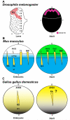
Figure 3. Gradients of signaling molecules determine regionalized retinal development
For A-C, D: dorsal, V: ventral, A: anterior, P: posterior. A) In Drosophila, the diffusible morphogen Wg is expressed in a dorsal patch of the larval eye disc, beginning the signaling cascade leading to expression of Rh3 in the dorsal rim in the adult (See Fig. 2B). B) Gradients of signaling molecules in the mouse retina leading to M (green) and S (blue) opsin expression. Sonic Hedgehog (Shh) is expressed in a ventral to dorsal gradient in both the embryo and the adult. Retinoic acid (RA) is expressed in a ventral to dorsal gradient at embryonic stages, and is produced by the enzymes V1 (ventral, high enzymatic activity) and AHD2 (dorsal, low enzymatic activity). CYP26 degrades RA in a strip through the middle of the retina. In the adult neither V1 nor CYP26 are expressed, so RA is present in a dorsal to ventral gradient. Thyroid hormone (T3) is present throughout the embryonic retina. In the adult, T3 is present in a dorsal to ventral gradient, presumably governed by the presence of the T3 synthesizing enzyme Dio2. BMP is present in a dorsal to ventral gradient in both the embryonic and adult mouse retina. C) In the chicken, RA is expressed in a ventral to dorsal gradient at embryonic stages and is produced by V1 and AHD2, as in mice. This mirrors the ventral to dorsal gradient of rods (black) within the chick retina. In the adult, V1 is not expressed, so RA is present in a dorsal to ventral gradient.
Figure 4
Figure 4. Retinal development proceeds through waves of differentiation
For A-E, A: anterior, P: posterior, D: dorsal, V: ventral, N: nasal, T: temporal. A) In Drosophila, waves of differentiation and mitosis move from posterior to anterior. B) In zebrafish, differentiation proceeds from ventral-nasal to dorsal-temporal in a wave resembling an opening fan. C) In chickens, mice, and humans, differentiation begins in the center of the retina and expands towards the periphery. D) Chicken retinal development also involves a temporal wave of cone maturation. Green and red cones are the earliest to mature, followed by blue and violet cones. E) A ventral-to-dorsal wave of differentiation patterns rods in the chicken retina in a density gradient, excluding the area centralis.
Figure 5
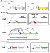
Figure 5. Looping of DNA elements regulates cone subtypes
A) In Drosophila, looping of regulatory elements may cause activation or repression of ss, the key determinant of R7 subtype fate. Sil1: Silencer 1, Enh: Enhancer, Sil2: Silencer 2. B) RA signaling and LCR looping select between opsin subtypes in zebrafish. Numbers in RH2 box indicate the temporal order of RH2 subtype expression. C) LCR looping selects between L-and M-opsin for expression in human L/M-cones.

References

    1. Julius D, Nathans J. Signaling by sensory receptors. Cold Spring Harbor perspectives in biology. 2012;4:a005991. - PMC - PubMed
    1. Osorio D, Vorobyev M. A review of the evolution of animal colour vision and visual communication signals. Vision research. 2008;48:2042–2051. - PubMed
    1. Osorio D, Vorobyev M. Photoreceptor spectral sensitivities in terrestrial animals: adaptations for luminance and colour vision. Proceedings. Biological sciences / The Royal Society. 2005;272:1745–1752. - PMC - PubMed
    1. Mazzoni EO, et al. Iroquois complex genes induce co-expression of rhodopsins in Drosophila. PLoS biology. 2008;6:e97. - PMC - PubMed
    1. Wernet MF, et al. Genetic dissection reveals two separate retinal substrates for polarization vision in Drosophila. Current biology: CB. 2012;22:12–20. - PMC - PubMed

Publication types