Review
. 2016 Sep;18 Suppl 1(Suppl 1):87-96.
doi: 10.1111/dom.12726.

Stress-induced adaptive islet cell identity changes

Affiliations
Review

Stress-induced adaptive islet cell identity changes

V Cigliola et al. Diabetes Obes Metab. 2016 Sep.

Abstract

The different forms of diabetes mellitus differ in their pathogenesis but, ultimately, they are all characterized by progressive islet β-cell loss. Restoring the β-cell mass is therefore a major goal for future therapeutic approaches. The number of β-cells found at birth is determined by proliferation and differentiation of pancreatic progenitor cells, and it has been considered to remain mostly unchanged throughout adult life. Recent studies in mice have revealed an unexpected plasticity in islet endocrine cells in response to stress; under certain conditions, islet non-β-cells have the potential to reprogram into insulin producers, thus contributing to restore the β-cell mass. Here, we discuss the latest findings on pancreas and islet cell plasticity upon physiological, pathological and experimental conditions of stress. Understanding the mechanisms involved in cell reprogramming in these models will allow the development of new strategies for the treatment of diabetes, by exploiting the intrinsic regeneration capacity of the pancreas.

Keywords: adaptive cell plasticity; cell conversion; cell dedifferentiation; cell fate change; cell identity; cell reprogramming; cell transdifferentiation; diabetes; diabetes treatment; islet; pancreas; transgenic.


Figures

Figure 1
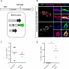
Figure 1. Bihormonal cells in islets of wild-type mice after STZ-induced β-cell ablation
A, Experimental design for STZ-induced diabetes in C57BL/6 nontransgenic mice. B, Bihormonal cells expressing insulin and glucagon are absent in islets of control mice (left panel). After STZ-mediated β-cell loss, insulin+/glucagon+ bihormonal cells appear in the islets (right panel). Scale Bar is 20 μm. C, Percentage of bihormonal cells in C57BL/6 mice. (control group: 0%, STZ-treated group: 0,312±0,043). *p<0.05.
Figure 2
Figure 2. Insulin production in α-cells after STZ- and ALX-induced diabetes
A, Experimental design for tracing α-cells through YFP expression and ablating β-cells with either DT (3 injections of 120 ng), ALX (1 injection of 60 mg/kg), STZ high-dose (1 injection of 200 mg/kg), or STZ low-dose (5 injections of 60 mg/kg). B, Number of β-cells per islet section upon ST, ALX or STZ induced β-cell ablation. *P ≤ 0.05**P ≤ 0.01. C, YFP-traced α-cells expressing insulin, upon STZ-mediated β-cell loss. A fraction of the converted α-cells retain glucagon expression (upper panel). Scale Bar is 20 μm. D, % of YFP-traced α-cells expressing insulin 30 days after DT-, ALX- and STZ-mediated β-cell ablation (control 0.3±0.1%, DT 1.1±0.3%, ALX 0.8±0.09%, STZ high 0.9±0.2%, STZ low 0.6±0.2%) . *P ≤ 0.05**P ≤ 0.01.
Figure 3
Figure 3. Characterization of insulin-expressing α-cells after STZ treatment
A, Experimental design for α-, β-, or δ-cell tracing and STZ-induced β-cell ablation. B, α-, β-, and δ-cells are efficiently traced with YFP in the 3 transgenic lines (black bars). Cells co-expressing glucagon and insulin are YFP-labelled only in the transgenic line allowing α-cell lineage tracing (red bars), confirming their α-cell origin. C, Percentage of YFP+/insulin+ cells retaining glucagon expression 1 month after DT-, ALX- and STZ-mediated β-cell loss. D, Immunofluorescence staining of the β-cell-specific markers Pdx1 (upper panel) and Nkx6.1 (bottom panel) in YFP+ α-cells 30 days after STZ-induced β-cell loss. E, quantification of YFP+ α-cells expressing Pdx1 and Nkx6.1 (Pdx1: control 0.4±0.3% STZ 3.7±0.2%; Nkx6.1: cnt 0.34±0.3 & STZ 6.2±2%). Scale Bar is 20 µm . *P ≤ 0.05
Figure 4
Figure 4. Insulin production in δ-cells after STZ administration
A, Experimental design for lineage-tracing δ-cells and ablating β-cells with STZ (1 injection of 200 mg/kg). B, YFP-traced δ-cells expressing insulin 45 days after STZ treatment. A fraction of the converted δ-cells retains somatostatin expression (upper panel). C, percentage of YFP-traced δ-cells expressing insulin 45 days after STZ-mediated β-cell ablation (control 1.3±0.7%, STZ high 1±0.7%). D, Percentage of YFP+/insulin+ cells retaining somatostatin expression 45 days after STZ-mediated β-cell loss. Scale Bar is 20 μm. *P ≤ 0.05
Figure 5
Figure 5
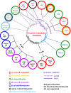
Overview of the adaptive cell identity changes in the pancreas in response to a variety of stress / injury conditions.

References

    1. Guariguata L, Whiting DR, Hambleton I, et al. Global estimates of diabetes prevalence for 2013 and projections for 2035. Diabetes Res Clin Pract. 2014;103:137–149. - PubMed
    1. Campbell-Thompson M, Fu A, Kaddis JS, et al. Insulitis and beta-cell mass in the natural history of type 1 diabetes. Diabetes. 2016;65:719–731. - PMC - PubMed
    1. Teta M, Long SY, Wartschow LM, Rankin MM, Kushner JA. Very slow turnover of beta-cells in aged adult mice. Diabetes. 2005;54:2557–2567. - PubMed
    1. Dor Y, Brown J, Martinez OI, Melton DA. Adult pancreatic beta-cells are formed by self-duplication rather than stem-cell differentiation. Nature. 2004;429:41–46. - PubMed
    1. Bonner-Weir S, Guo L, Li WC, et al. Islet neogenesis: a possible pathway for beta-cell replenishment. Rev Diabet Stud. 2012;9:407–416. - PMC - PubMed

LinkOut - more resources