. 2016 Sep 12:6:33099.
doi: 10.1038/srep33099.

Molecular evolutionary dynamics of cytochrome P450 monooxygenases across kingdoms: Special focus on mycobacterial P450s

Affiliations

Molecular evolutionary dynamics of cytochrome P450 monooxygenases across kingdoms: Special focus on mycobacterial P450s

Mohammad Parvez et al. Sci Rep. .

Abstract

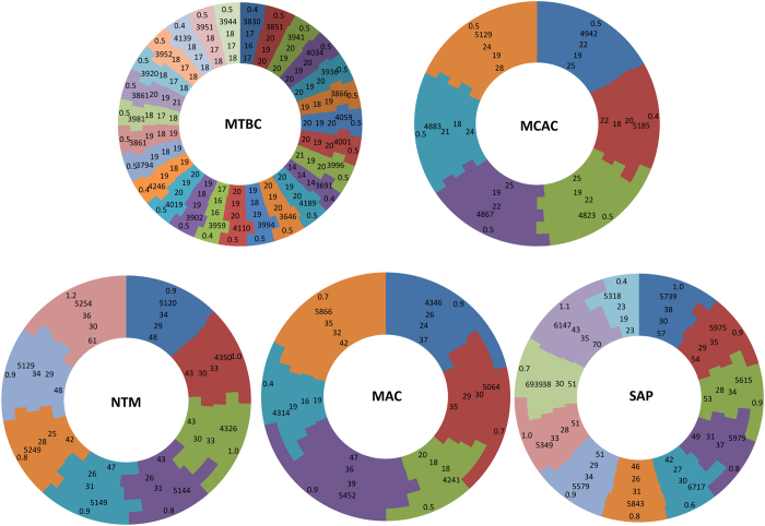
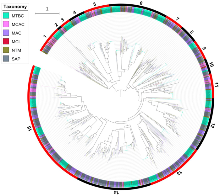
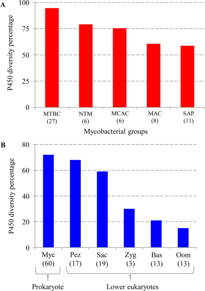
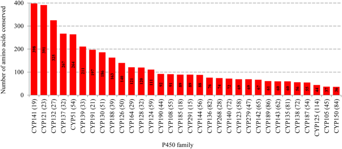
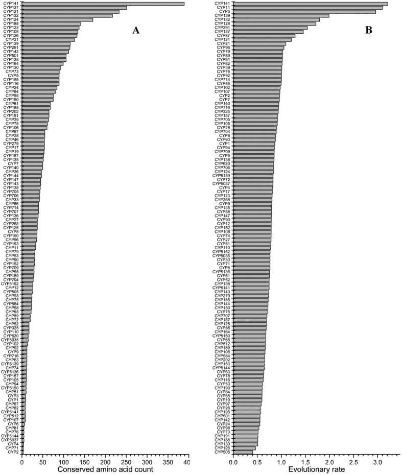
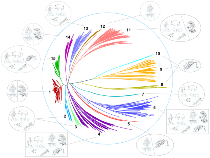
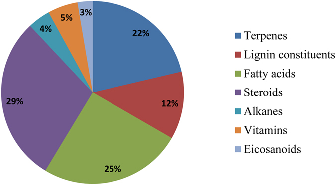
Since the initial identification of cytochrome P450 monooxygenases (CYPs/P450s), great progress has been made in understanding their structure-function relationship, diversity and application in producing compounds beneficial to humans. However, the molecular evolution of P450s in terms of their dynamics both at protein and DNA levels and functional conservation across kingdoms still needs investigation. In this study, we analyzed 17 598 P450s belonging to 113 P450 families (bacteria -42; fungi -19; plant -28; animal -22; plant and animal -1 and common P450 family -1) and found highly conserved and rapidly evolving P450 families. Results suggested that bacterial P450s, particularly P450s belonging to mycobacteria, are highly conserved both at protein and DNA levels. Mycobacteria possess the highest P450 diversity percentage compared to other microbes and have a high coverage of P450s (≥1%) in their genomes, as found in fungi and plants. Phylogenetic and functional analyses revealed the functional conservation of P450s despite belonging to different biological kingdoms, suggesting the adherence of P450s to their innate function such as their involvement in either generation or oxidation of steroids and structurally related molecules, fatty acids and terpenoids. This study's results offer new understanding of the dynamic structural nature of P450s.


Figures

Figure 1
Figure 1. Comparative analysis of P450s in mycobacteria.
Mycobacterial species were grouped into different groups such as MTBC (Mycobacterium tuberculosis complex), MCAC (Mycobacterium chelonae-abscessus complex), MAC (Mycobacterium avium complex), NTM (Nontuberculous mycobacteria) and SAP (Saprophytes). Each color in the circle represents a mycobacterial species in that group. The numerical order from the inside to the outside of the circle is as follows: number of P450s, number of P450 families, number of P450 subfamilies, number of ORFs in an organism and percentage of P450s compared to ORFs of an organism. Considering the presence of a single P450 in MCL (Mycobacteria causing leprosy) species, this group is omitted from comparative analysis. Details on species and their P450s were listed in Supplementary Table S3.
Figure 2
Figure 2. Phylogenetic analysis of P450s in mycobacteria.
A phylogenetic tree was constructed with 1772 mycobacterial P450s. The inner circle is the phylogenetic tree based on the consensus sequences of the mycobacterial P450s against the Pfam seed PF00067. The branches with different colors show their taxonomic groups: MTBC (Mycobacterium tuberculosis complex), MCAC (Mycobacterium chelonae-abscessus complex), MAC (Mycobacterium avium complex), MCL (Mycobacteria causing leprosy), NTM (Nontuberculous mycobacteria) and SAP (Saprophytes). Ancestral branches with children that had identical colors were assigned the same color as the children. The middle circle shows the corresponding CYPs, which are covered by different colors to show their taxonomic groups. Each taxon links the branch with a dotted line. The outermost numbers indicate the 15 clades based on this study, and their ranges are marked by alternating red and black. Distribution of P450s families into different clans was listed in Supplementary Table S5. A high-resolution phylogenetic tree is provided in the supplementary Fig. S1.
Figure 3
Figure 3. P450 diversity percentage analysis.
(A) Comparative analysis of P450 diversity percentage between different mycobacterial categories. (B) Comparative analysis of P450 diversity percentage between microbes such as prokaryote mycobacterial species and lower eukaryote fungi and oomycetes. The number of species in each of the groups (A) or microbe (B) used for this analysis is shown in parenthesis. Detailed analysis of the P450 diversity percentage values were presented in Supplementary Table S6. Abbreviations: MTBC, Mycobacterium tuberculosis complex; MCAC, Mycobacterium chelonae-abscessus complex; MAC, Mycobacterium avium complex; MCL, Mycobacteria causing leprosy; NTM, nontuberculous mycobacteria; SAP, Saprophytes; Myc, Mycobacterial species; Sac, Saccharomycetes; Pez, Pezizomycetes; Bas, Basidiomycetes; Zyg, Zygomycetes and Oom, Oomycetes.
Figure 4
Figure 4. Conserved amino acid analysis in mycobacterial P450 families.
Numbers of amino acids that are conserved in member P450s were determined using PROMALS3D and presented in the figure with conservation index where the number “9” indicates conserved amino acid in P450 family members. Number of member P450s analyzed for each P450 family is presented in parenthesis next to P450 family. A PROMALS3D analysis of member P450s and the conservation index scores for each mycobacterial P450 family were presented in Supplementary Table S7.
Figure 5
Figure 5. Protein-level (A) and DNA-level (B) P450s structural dynamic analysis.
Structural dynamics for 17 598 P450s belonging to 113 P450 families from different biological kingdoms were analyzed. (A) Protein-level structure dynamics were assessed based on the number of conserved amino acids present in each P450 family. The P450 families in the graph are presented such that the P450 family CYP141 that has the highest number of conserved amino acids is on top of the graph and the lowest number of conserved amino acids observed for the CYP2 family is on the bottom of the graph. A detailed analysis of the number of conserved amino acids and number of member P450s used and their hosts (biological kingdoms) and conservation ranking for each member of the family is presented in Supplementary Table S8. (B) Evolutionary rate analysis of P450s. Evolutionary rates were estimated based on their cDNA sequences under the Tamura-Nei model. A discrete Gamma distribution was used to model the evolutionary rate differences, as X axis of Fig. 5B presents, and more details are provided in Methods section. A discrete Gamma distribution was used to model the evolutionary rate differences. The P450 families in the graph are presented such that the P450 family CYP141 showing the lowest evolutionary rate is on top of the graph and the highest evolutionary rate observed for the CYP505 family is on the bottom of the graph. A detailed analysis of evolutionary rates for each of the P450 family and their ranking is presented in Supplementary Table S9.
Figure 6
Figure 6. Phylogenetic analysis of 17 598 P450s belonging to 113 families from different biological kingdoms such as bacteria, fungi, animals and plants.
All 113 P450 families were grouped under 15 clans based on their phylogenetic relationships and following the methods described elsewhere. Clans were presented in different colors and the host containing the respective P450 families that grouped under each clan is shown in schematic diagrams. Schematic diagrams are representative only and the P450 family is not necessarily confined to the depicted animals. Cartoon figures for plants, bacteria and fungi are shown as representative of their kingdoms. The tree was viewed by Hypertree in fisheye pattern. Detailed information on clan-level grouping of 113 P450 families is presented in Supplementary Table S10 and functional data on P450s at family level is presented in Supplementary Table S11.
Figure 7
Figure 7. Classification of P450s based on their main substrate class.
Percentage of P450s involved in oxidation of particular substrate class is shown in the figure. The functional classification is presented in a broader terms of substrates as described elsewhere. Detailed information on classification of P450 family members into different substrate class is presented in Supplementary Table S12.

References

    1. Nelson D. R. A world of cytochrome P450s. Phil. Trans. R. Soc. B. Biol. Sci. 368, 161220120430 (2013). - PMC - PubMed
    1. Nelson D. R. The cytochrome P450 homepage. Human Genomics 4, 59–65 (2009). - PMC - PubMed
    1. Guengerich F. P. Human cytochrome P450 enzymes In Cytochrome P450: Structure, mechanism, and biochemistry 4th edn. (eds Oritz de Montellano P. R.) Ch. 9, 523–786. (Springer International Publishing, 2015).
    1. Sezutsu H., Le Goff G. & Feyereisen R. Origins of P450 diversity. Phil. Trans. R. Soc. B. Biol. Sci. 368, 20120428 (2013). - PMC - PubMed
    1. Kelly S. L. & Kelly D. E. Microbial cytochrome P450: Biodiversity and biotechnology, where do cytochrome P450 come from, what do they do and what can they do for us? Phil. Trans. R. Soc. B. Biol. Sci. 368, 161220120430 (2013). - PMC - PubMed

Publication types

Substances

LinkOut - more resources