. 2016 Sep 12:6:33239.
doi: 10.1038/srep33239.

Soil bacteria as sources of virulence signal providers promoting plant infection by Phytophthora pathogens

Affiliations

Soil bacteria as sources of virulence signal providers promoting plant infection by Phytophthora pathogens

Ping Kong et al. Sci Rep. .

Abstract

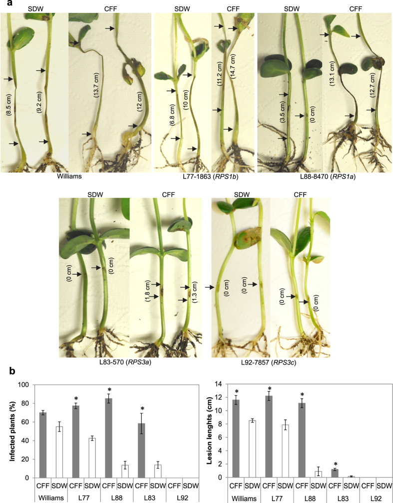
Phytophthora species are known as "plant destroyers" capable of initiating single zoospore infection in the presence of a quorum of chemical signals from the same or closely related species of oomycetes. Since the natural oomycete population is too low to reach a quorum necessary to initiate a disease epidemic, creation of the quorum is reliant on alternate sources. Here, we show that a soil bacterial isolate, Bacillus megaterium Sb5, promotes plant infection by Phytophthora species. In the presence of Sb5 exudates, colonization of rhododendron leaf discs by 12 Phytophthora species/isolates was significantly enhanced, single zoospores of P. nicotianae infected annual vinca and P. sojae race 25 successfully attacked a non-host plant, Nicotiana benthamiana as well as resistant soybean cultivars with RPS1a or RPS3a. Sb5 exudates, most notably the fractions larger than 3 kDa, promoted plant infection by improving zoospore swimming, germination and plant attachment. Sb5 exudates also stimulated infection hypha growth and upregulated effector gene expression. These results suggest that environmental bacteria are important sources of virulence signal providers that promote plant infection by Phytophthora species, advancing our understanding of biotic factors in the environmental component of the Phytophthora disease triangle and of communal infection of plant pathogens.


Figures

Figure 1
Figure 1. Soil bacterial products enhance colonization of rhododendron leaf discs by Phytophthora nicotianae.
Leaf discs were submerged in a suspension at 10 zoospores/ml in an autoclaved filtrate of bacterial cell suspension of individual isolates (Sb1, Sb3, Sb4, Sb5 or Sb6) prepared from potato dextrose agar (PDA), autoclaved soil water extract (ASWE), sterile distilled water (SDW) or rinsate of PDA plate surface with SDW (SDWms). The discs were examined for colonization 48 h after being plated on PARP-V8 agar. The graph depicts colonization rates (%) of the treatments. Each column is a mean of three repeating experiments. Bars depict standard error. Columns with the same letter are not different according to LSD test at P = 0.05.
Figure 2
Figure 2. Sb5 cell-free filtrate (CFF) promotes infection of annual vinca (Catharanthus roseus) leaves by single zoospores of Phytophthora nicotianae.
Individual leaves were inoculated at ten different sites each with a 10-μl drop of zoospore suspension (100/ml) prepared with CFF, sterile distilled water (SDW) or autoclaved soil water extract (ASWE). (a) Symptoms and (b) Infection rates at 72 h after inoculation. Each column is a mean of sites infected from three repeating experiments. Bars depict standard error. Columns with the same letter are not different according to LSD test at P = 0.05.
Figure 3
Figure 3. Sb5 cell-free filtrate (CFF) affects the responses of soybean differentials to Phytophthora sojae race 25.
(a) Hypersensitive response of hypocotyls 4 days after being injected with the pathogen mycelia suspended in CFF or sterile distilled water (SDW). Length of lesion at the inoculated site is marked between arrows. CFF treatment resulted in expanded lesions on two susceptible varieties (Williams and L77-1863) and one resistant variety (L88-8470) and necrotic lesions on the resistant variety L83-570 but not L92-7857. (b) Infection rate and lesion size are means of three repeating experiments. Bars in columns depict standard errors. *depicts significant difference between the CFF treatment and the SDW control according to t-test assuming unequal variances (P = 0.05).
Figure 4
Figure 4. Sb5 cell-free filtrate (CFF) promotes infection of a nonhost plant, Nicotiana benthamiana by Phytophthora sojae.
(a) Symptoms on detached leaves 72 h after inoculation on four sites of a leaf with each site having a 20-μl drop containing 250 zoospores in CFF or SDW. (b) Disease symptoms (arrows) developed on seedlings after their roots were submerged in a mixture suspension of P. sojae at 3,200 zoospores/ml and Sb5 at 106 cells/ml for 6 days but not on any other seedlings whose roots were submerged in suspension of Sb5 or P. sojae alone at the same concentration. (c) Percent seedlings diseased after their roots were submerged in mixture suspensions of zoospores (Zsp) at 3,200/ml and different concentrations of Sb5 from 106 to zero cell/ml. Each point is a mean of three replicates. Means with the same letter are not different according to LSD test at P = 0.05.
Figure 5
Figure 5. Effect of Sb5 cell-free filtrate (CFF) on zoospore attachment to Arabidopsis thaliana (Col-0) roots.
(a) Photomicrographs taken after roots were incubated 40 min in suspension of P. nicotianae or P. sojae at 1,000 zoospores/ml prepared with CFF or SDW. Attached cysts were indicated with arrows. Bars = 50 μm. (b) Number of attached cysts per microscopic field. Each column is a mean of six fields. *depicts significant difference between the CFF treatment and SDW controls according to t-test assuming equal variances (P = 0.05).
Figure 6
Figure 6. Effect of Sb5 cell-free filtrate (CFF) fractions on zoospore morphogenesis and infection of annual vinca (Catharanthus roseus) by Phytophthora nicotianae.
Top: Photomicrographs of cyst morphogenesis taken after 16-h incubation of 500 zoospores in 100 μl SDW, CFF or one of the four CFF fractions on a depression slide. Finger-like projections and vesicles formed in CFF and its fractions with molecular weight >3 kDa. c, g, p and v indicate cyst, germling, finger-like projections and vesicles, respectively. Bar = 50 μm. Bottom: Percent leaf sites infected following inoculation with ten 10 μl-drops of SDW, CFF or one of its fractions with each containing approximately 1 zoospore and 72-h incubation at 23 °C. Each column represents a mean of three repeating experiments. Bars depict standard errors. Columns with the same letter are not statistically different according to LSD at P = 0.05.
Figure 7
Figure 7. Effector gene expression in plants after inoculation with and without presence of Sb5 cell-free filtrate (CFF).
Real-time PCRs were conducted with RNA from hypocotyls of Phytophthora sojae race 25 soybean differentials (L77-1863, L83-570, L88-8470, L92-7857 and Williams) inoculated with mycelia of P. sojae mixed with CFF or sterile distilled water (SDW), and from Nicotiana benthaminana roots (Nb) that were submerged in a mixture suspension of P. sojae at 3,200 zoospores/ml and Sb5 at 106 cells/ml or SDW. Average Ct (threshold cycles) of PCR runs with P. sojae effector genes Avr1a, Avr3a, Avr1b1 or Avr3c in both CFF treatment and control was normalized with mean value of the average Ct with three reference genes, actin A, β-tubulin and ubiquitin in the same sample (dCt = Cteffector − Ctreference). Transcription levels of effector genes in the CFF treatment were measured with 2−ddCt after the calibration of the dCt with that of the control (ddCt = dCtSb5 − dCtcontrol). Each point is a mean of three PCR runs. Vertical lines depict the range of transcript level of individual genes by plant and cultivar.

References

    1. Deacon J. W. & Donaldson S. P. Molecular recognition in the homing responses of zoosporic fungi, with special reference to Pythium and Phytophthora. Mycol. Res. 97, 1153–1171 (1993).
    1. Tyler B. M. Molecular basis of recognition between Phytophthora pathogens and their hosts. Annu. Rev. Phytopathol. 40, 137–167 (2002). - PubMed
    1. Latijnhouwers M., de Wit P. & Govers F. Oomycetes and fungi: similar weaponry to attack plants. Trends Microbiol. 11, 462–469 (2003). - PubMed
    1. Judelson H. S. & Blanco F. A. The spores of Phytophthora: Weapons of the plant destroyer. Nature Reviews Microbiology 3, 47–58 (2005). - PubMed
    1. Kamoun S. A catalogue of the effector secretome of plant pathogenic oomycetes. Annu. Rev. Phytopathol. 44, 41–60 (2006). - PubMed

Publication types

LinkOut - more resources