. 2016 Sep 12:6:32514.
doi: 10.1038/srep32514.

Local and Circulating Endothelial Cells Undergo Endothelial to Mesenchymal Transition (EndMT) in Response to Musculoskeletal Injury

Affiliations

Local and Circulating Endothelial Cells Undergo Endothelial to Mesenchymal Transition (EndMT) in Response to Musculoskeletal Injury

Shailesh Agarwal et al. Sci Rep. .

Abstract

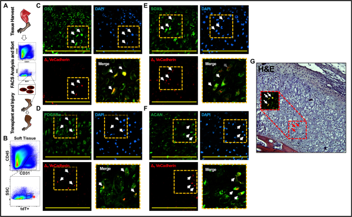
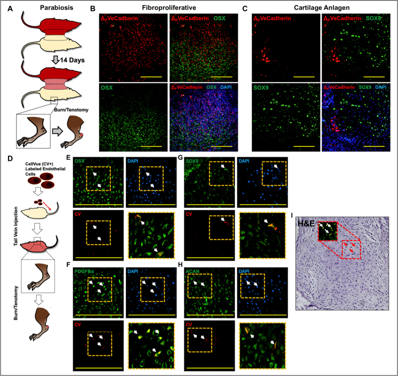
Endothelial-to-mesenchymal transition (EndMT) has been implicated in a variety of aberrant wound healing conditions. However, unambiguous evidence of EndMT has been elusive due to limitations of in vitro experimental designs and animal models. In vitro experiments cannot account for the myriad ligands and cells which regulate differentiation, and in vivo tissue injury models may induce lineage-independent endothelial marker expression in mesenchymal cells. By using an inducible Cre model to mark mesenchymal cells (Scx-creERT/tdTomato + ) prior to injury, we demonstrate that musculoskeletal injury induces expression of CD31, VeCadherin, or Tie2 in mesenchymal cells. VeCadherin and Tie2 were expressed in non-endothelial cells (CD31-) present in marrow from uninjured adult mice, thereby limiting the specificity of these markers in inducible models (e.g. VeCadherin- or Tie2-creERT). However, cell transplantation assays confirmed that endothelial cells (ΔVeCadherin/CD31+/CD45-) isolated from uninjured hindlimb muscle tissue undergo in vivo EndMT when transplanted directly into the wound without intervening cell culture using PDGFRα, Osterix (OSX), SOX9, and Aggrecan (ACAN) as mesenchymal markers. These in vivo findings support EndMT in the presence of myriad ligands and cell types, using cell transplantation assays which can be applied for other pathologies implicated in EndMT including tissue fibrosis and atherosclerosis. Additionally, endothelial cell recruitment and trafficking are potential therapeutic targets to prevent EndMT.


Figures

Figure 1
Figure 1. VeCadherin-cre-labeled mice suggest EndMT in a model of trauma-induced HO.
(A) Burn/tenotomy results in trauma-induced HO (tHO) at the tendon transection site; (B) RNA Seq shows up-regulated transcript levels for Tgfβ, fibroblast growth factor (FGF), Snai1, and Twist1; (C) VeCadherin-cre/tdTomato lineage-tracing mice show presence of tdTomato + cells in the fibroproliferative region expressing PDGFRα, Osterix (OSX) but not SOX9 or Aggrecan (ACAN); D) VeCadherin-cre/tdTomato lineage-tracing mice show presence of tdTomato+ cells in the cartilage region expressing PDGFRα, Osterix (OSX), SOX9, and Aggrecan (ACAN).
Figure 2
Figure 2. Trauma induces endothelial marker expression in non-endothelial cells.
(A) Flow cytometry confirms relative absence of Scleraxis-cre cells in marrow (0.015% +/−0.007%); (B) Site of tissue harvest from Scx-cre/ROSA26mTmG mice; (C) Immunostaining of vessel from Scx-cre/ROSA26mTmG mice with VeCadherin, CD31, and Tie2 confirms that endothelial cells are not marked by the Scleraxis-lineage; (D) Immunostaining of tendon from Scx-cre/ROSA26mTmG mice with VeCadherin, CD31, and Tie2 confirms that cells of the Scleraxis-lineage within the hindlimb soft tissue are not endothelial cells; (E) Experimental design to mark non-endothelial, mesenchymal cells using Scx-creERT2/tdTomato + mice; (F) tdTomato + cells (ΔSCX) from Scx-creERT2/tdTomato + mice express VeCadherin after injury; (G) tdTomato + cells (ΔSCX) from Scx-creERT2/tdTomato + mice express CD31 after injury; (H) tdTomato + cells (ΔSCX) from Scx-creERT2/tdTomato + mice express Tie2 after injury. Yellow scale bars = 200 um.
Figure 3
Figure 3
(A) Schematic showing cell transplantation experiments; (B) FACS sorting of endothelial cells (tdTomato+/CD31+/CD45−) from the hindlimb musculature of uninjured VeCadherin-cre/tdTomato + mice; (C) tdTomato + cells are present within the cartilage anlagen and express Osterix (OSX); (D) tdTomato + cells are present within the cartilage anlagen and express PDGFRα; (E) tdTomato + cells are present within the cartilage anlagen and express SOX9; (F) tdTomato + cells are present within the cartilage anlagen and express Aggrecan (ACAN); (G) H&E showing histologic character of tdTomato + cells undergoing EndMT with chondrocyte appearance and OSX expression as in (C).
Figure 4
Figure 4
(A) Schematic showing parabiosis of VeCadherin-cre/tdTomato mouse with unlabeled wild type mouse and injury on unlabeled wild type mouse; (B) tdTomato + (ΔVeCadherin) cells in parabiosis model express Osterix (OSX); (C) tdTomato + (ΔVeCadherin) cells in parabiosis model express SOX9; (D) Schematic showing tail vein injection of cultured, Cell-Vue-labeled (CV+) endothelial cells after trauma; (E) CV + endothelial cells are present in the injured site and express Osterix (OSX) with merge image in lower right quadrant; (F) CV + endothelial cells are present within the cartilage anlagen and express PDGFRα with merge image in lower right quadrant; (G) CV + endothelial cells are present within the cartilage anlagen and express SOX9 with merge image in lower right quadrant; (H) CV + endothelial cells are present within the cartilage anlagen and express Aggrecan (ACAN) with merge image in lower right quadrant; (I) H&E showing histologic character of CV + cells undergoing EndMT with chondrocyte appearance and PDGFRα expression as in (F).

References

    1. Cooley B. C. et al.. TGF-beta signaling mediates endothelial-to-mesenchymal transition (EndMT) during vein graft remodeling. Science translational medicine 6, 227ra234 (2014). - PMC - PubMed
    1. Kovacic J. C., Mercader N., Torres M., Boehm M. & Fuster V. Epithelial-to-mesenchymal and endothelial-to-mesenchymal transition: from cardiovascular development to disease. Circulation 125, 1795–1808 (2012). - PMC - PubMed
    1. Zeisberg E. M. et al.. Endothelial-to-mesenchymal transition contributes to cardiac fibrosis. Nature medicine 13, 952–961 (2007). - PubMed
    1. Aisagbonhi O. et al.. Experimental myocardial infarction triggers canonical Wnt signaling and endothelial-to-mesenchymal transition. Disease models & mechanisms 4, 469–483 (2011). - PMC - PubMed
    1. Chen P. Y. et al.. Endothelial-to-mesenchymal transition drives atherosclerosis progression. The Journal of clinical investigation 2015 (2015). - PMC - PubMed

Publication types

MeSH terms