Case Reports
. 2016 Oct 6;99(4):894-902.
doi: 10.1016/j.ajhg.2016.07.018. Epub 2016 Sep 8.

NAXE Mutations Disrupt the Cellular NAD(P)HX Repair System and Cause a Lethal Neurometabolic Disorder of Early Childhood

Affiliations
Case Reports

NAXE Mutations Disrupt the Cellular NAD(P)HX Repair System and Cause a Lethal Neurometabolic Disorder of Early Childhood

Laura S Kremer et al. Am J Hum Genet. .

Abstract

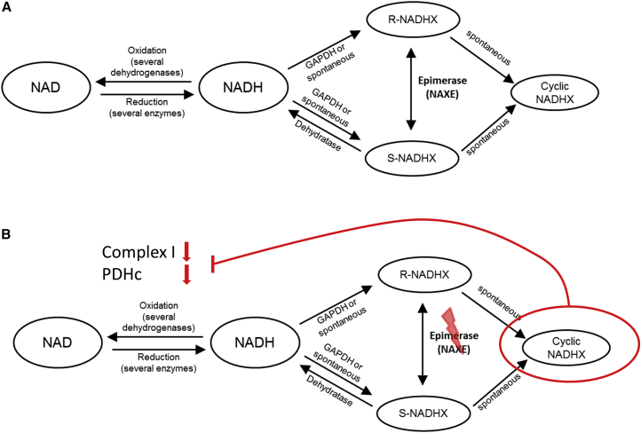
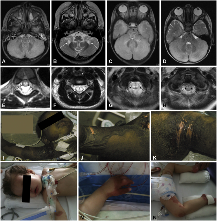
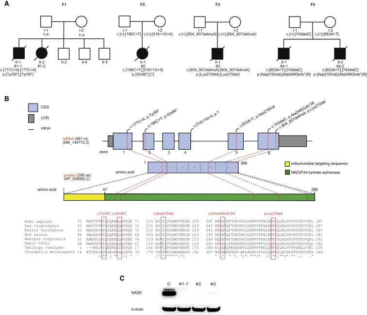
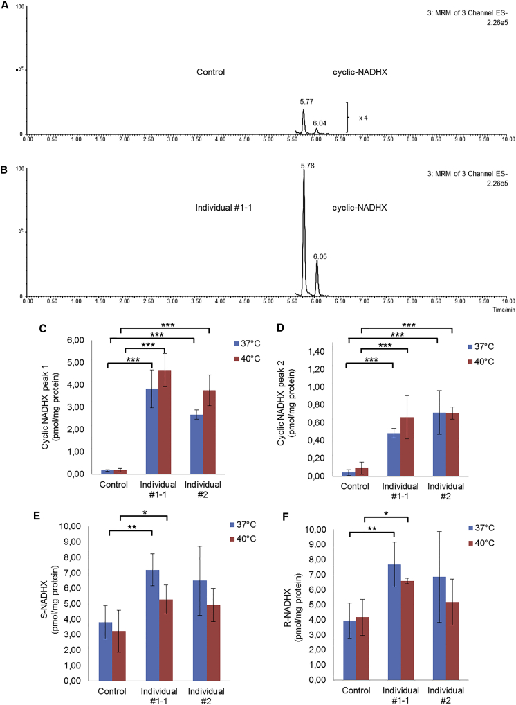
To safeguard the cell from the accumulation of potentially harmful metabolic intermediates, specific repair mechanisms have evolved. APOA1BP, now renamed NAXE, encodes an epimerase essential in the cellular metabolite repair for NADHX and NADPHX. The enzyme catalyzes the epimerization of NAD(P)HX, thereby avoiding the accumulation of toxic metabolites. The clinical importance of the NAD(P)HX repair system has been unknown. Exome sequencing revealed pathogenic biallelic mutations in NAXE in children from four families with (sub-) acute-onset ataxia, cerebellar edema, spinal myelopathy, and skin lesions. Lactate was elevated in cerebrospinal fluid of all affected individuals. Disease onset was during the second year of life and clinical signs as well as episodes of deterioration were triggered by febrile infections. Disease course was rapidly progressive, leading to coma, global brain atrophy, and finally to death in all affected individuals. NAXE levels were undetectable in fibroblasts from affected individuals of two families. In these fibroblasts we measured highly elevated concentrations of the toxic metabolite cyclic-NADHX, confirming a deficiency of the mitochondrial NAD(P)HX repair system. Finally, NAD or nicotinic acid (vitamin B3) supplementation might have therapeutic implications for this fatal disorder.

Keywords: NAD(P)HX; energy metabolism; metabolite repair; mitochondrial.


Figures

Figure 1
Figure 1
Schematic Overview of NAD/NADH Metabolism and the NAD(P)HX Repair System (A) NAD(P)HX repair system at metabolic equilibrium under normal/healthy conditions. (B) Putative consequences of NAXE deficiency, leading to increased formation of cyclic NADHX, a toxic inhibitor of cellular NADH dehydrogenases.
Figure 2
Figure 2
Unifying Neuroimaging Findings and Skin Manifestations in Children with NAXE Mutations (A) Brain MRI (axial view, T2-weighted) of individual #1-1, showing diffuse cerebellar edema. (B) Brain MRI of individual #1-2 (axial view, T2-weighted), demonstrating symmetrical signal alterations in cerebellar white matter and cerebellar peduncles. (C) Brain MRI (axial view, T2-weighted) in individual #2 showing diffuse cerebellar edema comparable to the findings in (A), leading to nearly fatal brain herniation. (D) Brain MRI (axial view, T2-weighted) of individual #4-1, also demonstrating diffuse cerebellar edema. (E–H) Spinal MRI (axial view, T2-weighted) in individuals #1-1 (cervical spine) (E), #1-2 (thoracic spine) (F), #2 (cervical spine) (G), and #4-1 (cervical spine) (H), showing symmetrical signal abnormalities of the spinal cord, indicating various degrees of myelopathy. Please note that additional imaging findings can be found in Figure S1. (I–K) Extensive skin lesions in individual #1-2. Onset of skin manifestation was sub-acute within 2–3 weeks after the start of neurological symptoms. Large bullous skin lesions developed (J shows popliteal fossa), partially leading to erosion and eruption of the skin (K shows axilla). (L–N) Skin lesions in individual #2. Please also note the critical clinical condition of both affected individuals as indicated in (I) and (L).
Figure 3
Figure 3
Genetic Findings in Four NAXE-Deficient Families with Consequences on the Protein Level (A) Pedigrees of four families identified with recessive inherited mutations in NAXE. (B) Genomic organization of NAXE with known conserved protein domains in the gene product, location of mutations within NAXE, and phylogenetic conservation of amino acid residues affected by mutations; positions of mutations are highlighted in red. (C) Western blot analysis of fibroblast from index case subjects with NAXE mutations and a control cell line. Fibroblasts were cultured in Dulbecco’s Modified Eagle’s medium (Life Technologies) supplemented with 10% fetal bovine serum and 1% penicillin/streptomycin (both: Life Technologies) at 37°C in an atmosphere of 5% CO2 in air. Whole cell lysates were prepared with cell lysis reagent CelLytic M (Sigma Aldrich). A polyclonal antibody against NAXE detecting amino acid sequence 218 to 286 (HPA048164, Sigma Aldrich) was used. A monoclonal antibody against beta-actin (Sigma Aldrich) served as loading control. Secondary antibodies were anti-mouse and anti-rabbit (GE Healthcare). Signal was detected using BM chemiluminescence blotting substrate (Roche).
Figure 4
Figure 4
Cellular Consequences of NAXE Deficiency (A and B) Typical examples of chromatograms of cyclic-NADHX analyzed by HPLC-ESI-MS/MS in a control cell line (A) and in the affected individual #1-1 (B). Chromatograms clearly show that the levels of cyclic-NADHX are increased in NAXE-deficient cells compared to controls. The two peaks that we measured for cyclic-NADHX most likely depict the R- and the S-form of the metabolite. In the control cell line (B), the peaks were additionally 4-fold magnified for better visualization. (C and D) Quantitative results of the cyclic-NADHX measurements under normal (37°C) and heat-stressed (40°C) conditions for 24 hr. (E and F) Quantitative results of S- and R-NADHX measurements. Control bar graphs depict pooled data obtained from two independent healthy cell lines. All experimental data were obtained in at least three independent experiments. Student’s t test was used to determine the statistical significance. Statistics: ∗∗∗p < 0.001, ∗∗p < 0.01, and p < 0.05 relative to controls. Error bars indicate standard deviation.

References

    1. Linster C.L., Van Schaftingen E., Hanson A.D. Metabolite damage and its repair or pre-emption. Nat. Chem. Biol. 2013;9:72–80. - PubMed
    1. Yoshida A., Dave V. Inhibition of NADP-dependent dehydrogenases by modified products of NADPH. Arch. Biochem. Biophys. 1975;169:298–303. - PubMed
    1. Marbaix A.Y., Tyteca D., Niehaus T.D., Hanson A.D., Linster C.L., Van Schaftingen E. Occurrence and subcellular distribution of the NADPHX repair system in mammals. Biochem. J. 2014;460:49–58. - PubMed
    1. Marbaix A.Y., Noël G., Detroux A.M., Vertommen D., Van Schaftingen E., Linster C.L. Extremely conserved ATP- or ADP-dependent enzymatic system for nicotinamide nucleotide repair. J. Biol. Chem. 2011;286:41246–41252. - PMC - PubMed
    1. Giaever G., Chu A.M., Ni L., Connelly C., Riles L., Véronneau S., Dow S., Lucau-Danila A., Anderson K., André B. Functional profiling of the Saccharomyces cerevisiae genome. Nature. 2002;418:387–391. - PubMed

Publication types

MeSH terms

Substances