. 2016 Oct 6;99(4):802-816.
doi: 10.1016/j.ajhg.2016.07.013. Epub 2016 Sep 8.

GRIN2D Recurrent De Novo Dominant Mutation Causes a Severe Epileptic Encephalopathy Treatable with NMDA Receptor Channel Blockers

Affiliations

GRIN2D Recurrent De Novo Dominant Mutation Causes a Severe Epileptic Encephalopathy Treatable with NMDA Receptor Channel Blockers

Dong Li et al. Am J Hum Genet. .

Abstract

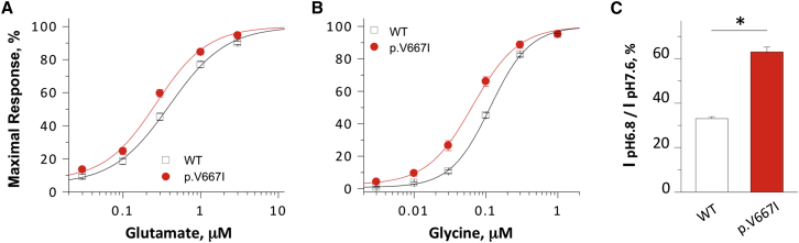
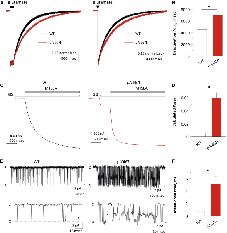
N-methyl-D-aspartate receptors (NMDARs) are ligand-gated cation channels that mediate excitatory synaptic transmission. Genetic mutations in multiple NMDAR subunits cause various childhood epilepsy syndromes. Here, we report a de novo recurrent heterozygous missense mutation-c.1999G>A (p.Val667Ile)-in a NMDAR gene previously unrecognized to harbor disease-causing mutations, GRIN2D, identified by exome and candidate panel sequencing in two unrelated children with epileptic encephalopathy. The resulting GluN2D p.Val667Ile exchange occurs in the M3 transmembrane domain involved in channel gating. This gain-of-function mutation increases glutamate and glycine potency by 2-fold, increases channel open probability by 6-fold, and reduces receptor sensitivity to endogenous negative modulators such as extracellular protons. Moreover, this mutation prolongs the deactivation time course after glutamate removal, which controls the synaptic time course. Transfection of cultured neurons with human GRIN2D cDNA harboring c.1999G>A leads to dendritic swelling and neuronal cell death, suggestive of excitotoxicity mediated by NMDAR over-activation. Because both individuals' seizures had proven refractory to conventional antiepileptic medications, the sensitivity of mutant NMDARs to FDA-approved NMDAR antagonists was evaluated. Based on these results, oral memantine was administered to both children, with resulting mild to moderate improvement in seizure burden and development. The older proband subsequently developed refractory status epilepticus, with dramatic electroclinical improvement upon treatment with ketamine and magnesium. Overall, these results suggest that NMDAR antagonists can be useful as adjuvant epilepsy therapy in individuals with GRIN2D gain-of-function mutations. This work further demonstrates the value of functionally evaluating a mutation, enabling mechanistic understanding and therapeutic modeling to realize precision medicine for epilepsy.


Figures

Figure 1
Figure 1
Genetic and Protein Alteration of GRIN2D and GluN2D (A) Pedigrees and genotypes of a de novo mutation c.1999G>A (p.Val667Ile) identified in both unrelated kindreds. (B) Schematic topology represents a GluN2D subunit, where asterisk indicates the position of the p.Val667Ile variant. The Val667 residue is highly conserved across vertebrate species and all GluN2 subunits. (C) The red asterisk in the cartoon indicating the domain arrangement of a GluN2D subunit shows the position of Val667 in the transmembrane domain (M3) (left). A homology model of GluN2D subunit, shown as space fill, was built from the GluN1/GluN2B crystallographic data, using Muscle for sequence alignment, Modeler to build the homology model, and the Schrödinger Modeling for model refinement (middle). The location on the M3 transmembrane helix is show in an expanded panel (right).
Figure 2
Figure 2
The Mutant GluN2D-p.Val667Ile Changes NMDAR Pharmacological Properties (A and B) The composite glutamate (in the presence of 100 μM glycine) (A) and glycine (in the presence of 100 μM glutamate) (B) concentration-response curves show an increased agonist potency in the mutant receptors compared to the WT receptors. (C) Summary of proton sensitivity of WT GluN2D and mutant receptors, evaluated by current ratio at pH 6.8 to pH 7.6. p < 0.05 compared to WT GluN2D, unpaired t test. Error bars show SEM, which were calculated from 20 and 28 independent recordings for WT and mutant receptors, respectively.
Figure 3
Figure 3
The Mutant GluN2D-p.Val667Ile Changes Response Time Course and Channel Open Probability (A) The p.Val667Ile mutation prolongs deactivation time course of GluN2D receptors. Normalized representative current response of di-heteromeric NMDARs (WT GluN2D and GluN2D-p.Val667Ile) to 1 mM glutamate applied for 1.5 s (left) or 5 ms (right) (VHOLD −60 mV); 50 μM glycine was present in all solutions. (B) Summary of weighted deactivation tau of WT GluN2D and mutant receptors. Error bars show SEM, which were calculated from six independent recordings. (C) Representative current traces evoked by agonists (100 μM glutamate and glycine) followed by 200 μM MTSEA were determined by TEVC recordings from Xenopus oocytes expressing GluN1-p.Ala652Cys/GluN2D (left) and GluN1-p.Ala652Cys/GluN2D-p.Val667Ile receptors (right). (D) Summary of estimated POPEN of WT GluN2D and mutant receptors determined in oocytes. Error bars show SEM, which were calculated from 22 and 32 independent recordings of WT and mutant receptors, respectively. (E) Representative current traces of steady-state recordings from an outside-out patch containing WT (left) and the mutant (right) NMDARs. Unitary currents were activated by 1 mM glutamate and 50 μM glycine. Abbreviations are as follows: C, close; O, open. (F) Summary of channel mean open time of WT GluN2D and mutant receptors. p < 0.05 compared to WT GluN2D, unpaired t test. Error bars show SEM, which were calculated from three and four independent recordings for WT and mutant receptors, respectively.
Figure 4
Figure 4
Transfection of GluN2D-p.Val667Ile into Cultured Cortical Neurons Induces Excitotoxicity, which Can Be Prevented by Memantine (A) Confocal images display morphological features of rat cortical neurons transfected with a GFP-expressing plasmid (1.5/2.5 μg/mL of total transfected DNA), along with either WT GluN2D or GluN2D-p.Val667Ile (1/2.5 μg/mL of total transfected DNA); the latter was supplemented with either vehicle or memantine (50 μM). Note the prominent dendritic swelling induced by expression of GluN2D-p.Val667Ile, but not by WT GluN2D, which was mitigated by memantine treatment. Left column images were acquired 24 hr after transfection at 20× total magnification; areas boxed in red are magnified to 100 × in the adjacent panels. Scale bars represent 10 μM. (B) Histograms representing mean cell viability values as a percent of control groups. Main graph: neuronal cultures were transfected with luciferase cDNA (0.625/2.5 μg/mL of total transfected DNA) to assay cell viability, along with either PCIneo-vector, WT GluN2D, or GluN2D-p.Val667Ile cDNA (1/2.5 μg/mL of total transfected DNA). Each transfection condition was performed in the presence or either vehicle (–) or memantine (50 μM; +). Luciferase assays were performed 48 hr after transfection. Each experiment was carried out in quadruplicate, and five independent experiments were performed. Each condition was normalized to its corresponding vector-transfected group to obtain relative viability values (e.g., memantine-treated, vector-expressing group was used to normalize memantine-treated GluN2D-expressing group). Histograms are displayed as mean ± SEM of viability (percent control) values for each condition: WT GluN2D (–), 83.3 ± 9.5; WT GluN2D (+), 118.6 ± 6.3; GluN2D-p.Val667Ile (–), 46.7 ± 7.0; GluN2D-p.Val667Ile (+), 152.2 ± 16.6; ∗∗p < 0.01, ∗∗∗p < 0.001; ANOVA/Bonferroni. Inset: cell viability assays were confirmed by cell counting. Neuronal cultures were transfected with a GFP-expressing plasmid (1.5/2.5 μg/mL of total transfected DNA), along with either WT GluN2D or GluN2D-p.Val667Ile cDNA (1/2.5 μg/mL of total transfected DNA). Each transfection condition was performed in the presence or either vehicle (–) or memantine (50 μM; +). Cells were fixed with 4% paraformaldehyde 72 hr after transfection and counted by a person blinded to the experimental groups. 30 image fields from 3 coverslips per condition were obtained at 20× magnification in four independent experiments. The number of neurons per coverslip was averaged for each condition. GluN2D-p.Val667Ile groups (with and without memantine) were standardized to WT GluN2D-transfected control to obtain relative viability values expressed as percent control for each experiment. Data are displayed as mean ± SEM of viability (percent control) for each condition: GluN2D-p.Val667Ile (–), 75.3 ± 9.6; GluN2D-p.Val667Ile (+), 108.1 ± 13.7; p < 0.05; paired t test.
Figure 5
Figure 5
Screening of FDA-Approved NMDAR Antagonists (A–C) Composite concentration-response curves for memantine (A), ketamine (B), and Mg2+ (C) inhibition on the mutant and WT current responses to maximal effective concentration (100 μM) of glutamate and glycine determined by TEVC recordings from oocytes. (D) Current-voltage (I-V) curves in the presence of 0, 0.3 mM, 1.0 mM, and 3.0 mM of Mg2+.
Figure 6
Figure 6
EEG before and after Treatments Are Presented (A) The top row is the pre-treatment EEG (baseline at 6 years old). The best awake background is shown (A) with a 7–8 Hz posterior theta-alpha rhythm present for intermittent periods. When she went to sleep (A′, A″), there was an absence of normal sleep architecture (A′) and at time the recording became hypsarrythmic with high amplitude, multifocal sharps, and a lack of organization (A″). (B) The second row presents the post memantine treatment EEG, 3 months after starting treatment, on a stable dose for 6 weeks. Again, the best awake background (B) is presented, but this had fewer periods with a posterior theta alpha rhythm and more frequent interruption by bifrontal or lateralized (arrows in B) epileptiform sharps. After treatment, when asleep, the EEG was continuously high voltage, multifocal sharps, and lacked any discernable organization (B′). (C) The third row presents the EEG after being off memantine and admitted for status epilepticus. The EEG shows continuous generalized spike and slow wave discharges at ∼3 Hz. After failing first and second line i.v. medications, she was placed on a continuous midazolam infusion (C′) with slowing of the spike and wave but status persisted. Changing to pentobarbital resulted in burst suppression, but when pentobarbital was weaned, the continous spike and slow wave returned. She was put under pentobarbital and ketamine and burst suppression returned (C″). (D) Wean of pentobarbital again resulted in return of spike wave. Addition of i.v. magnesium to ketamine resulted in acute sessation of spike wave without EEG suppression (D′). Finally, lowering the IV Mg2+ and ketamine resulted in persistence of a slow and better organized EEG (D″). This EEG background continued when Mg2+ and ketamine were changed to oral administration (not shown). (E) Weeks after transitioning to oral ketamine and Mg2+, she remains clinically seizure free.

Comment in

References

    1. Srivastava S., Cohen J.S., Vernon H., Barañano K., McClellan R., Jamal L., Naidu S., Fatemi A. Clinical whole exome sequencing in child neurology practice. Ann. Neurol. 2014;76:473–483. - PubMed
    1. Yang Y., Muzny D.M., Reid J.G., Bainbridge M.N., Willis A., Ward P.A., Braxton A., Beuten J., Xia F., Niu Z. Clinical whole-exome sequencing for the diagnosis of mendelian disorders. N. Engl. J. Med. 2013;369:1502–1511. - PMC - PubMed
    1. Lee H., Deignan J.L., Dorrani N., Strom S.P., Kantarci S., Quintero-Rivera F., Das K., Toy T., Harry B., Yourshaw M. Clinical exome sequencing for genetic identification of rare Mendelian disorders. JAMA. 2014;312:1880–1887. - PMC - PubMed
    1. Yang Y., Muzny D.M., Xia F., Niu Z., Person R., Ding Y., Ward P., Braxton A., Wang M., Buhay C. Molecular findings among patients referred for clinical whole-exome sequencing. JAMA. 2014;312:1870–1879. - PMC - PubMed
    1. Allen A.S., Berkovic S.F., Cossette P., Delanty N., Dlugos D., Eichler E.E., Epstein M.P., Glauser T., Goldstein D.B., Han Y., Epi4K Consortium. Epilepsy Phenome/Genome Project De novo mutations in epileptic encephalopathies. Nature. 2013;501:217–221. - PMC - PubMed

MeSH terms