. 2016 Jun:36:156-246.
doi: 10.3767/003158516X691582. Epub 2016 Apr 29.

Generic hyper-diversity in Stachybotriaceae

Affiliations

Generic hyper-diversity in Stachybotriaceae

L Lombard et al. Persoonia. 2016 Jun.

Abstract

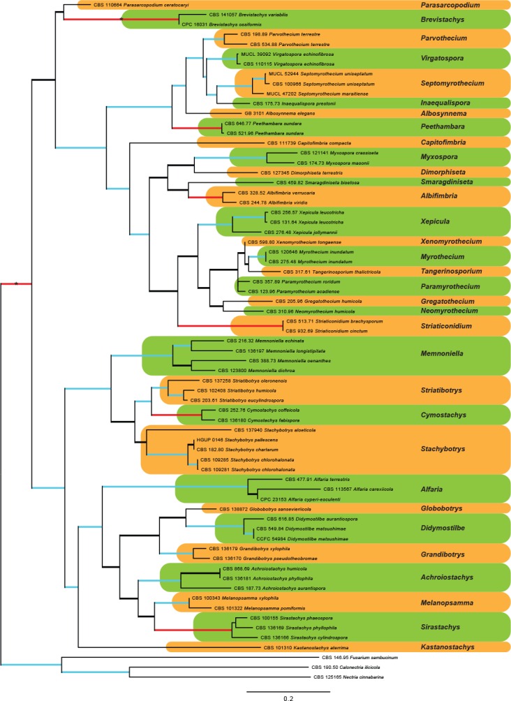
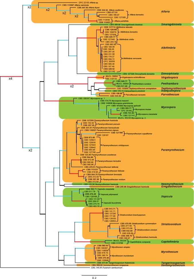
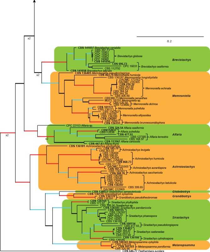
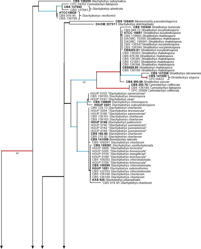
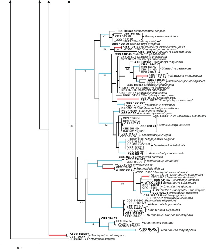
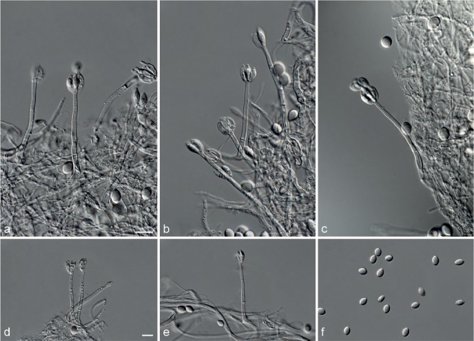
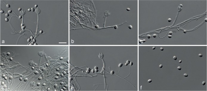
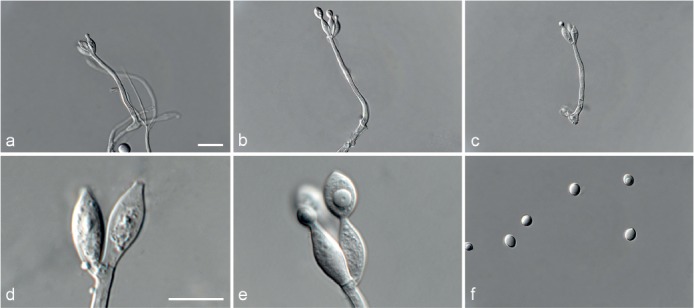
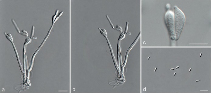
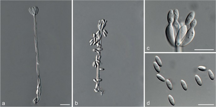
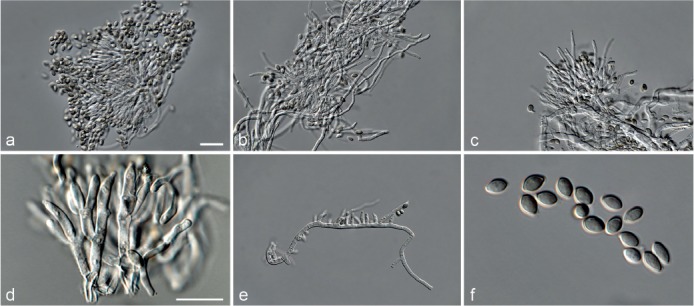
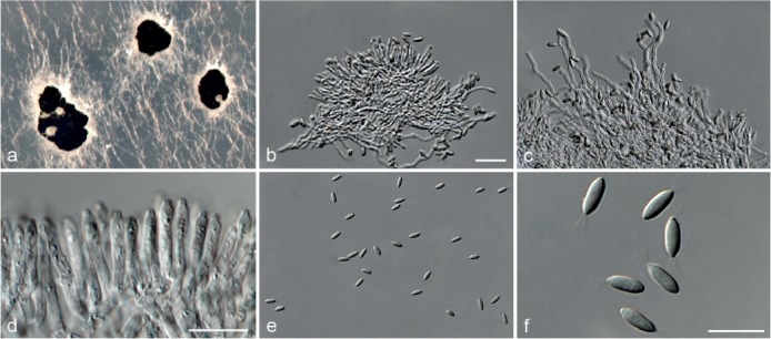
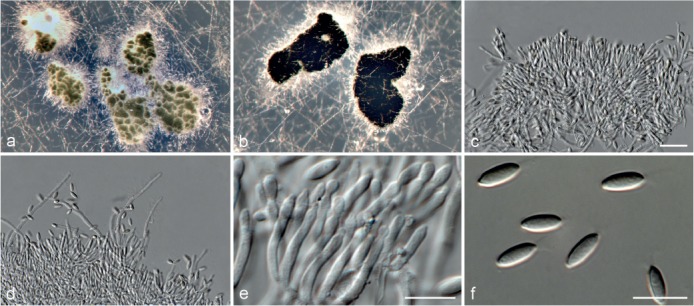
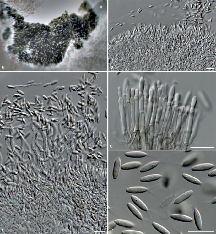
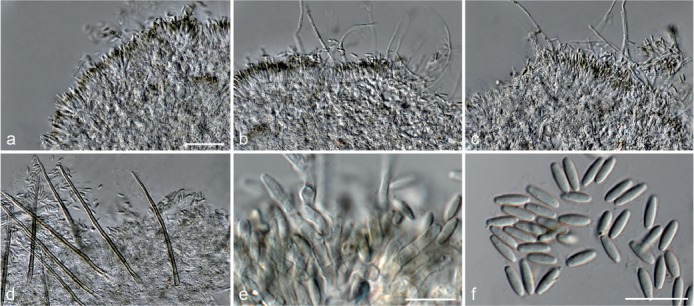
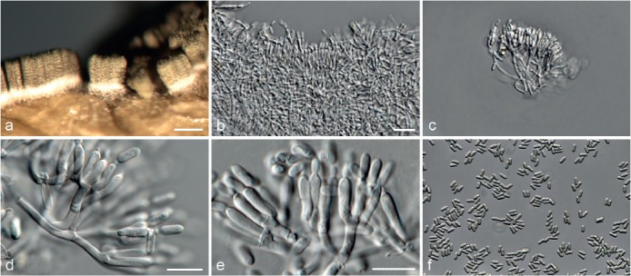
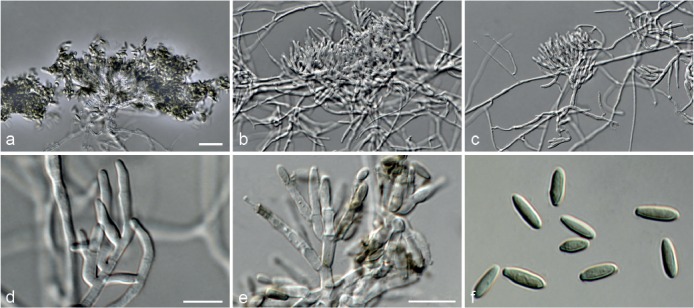
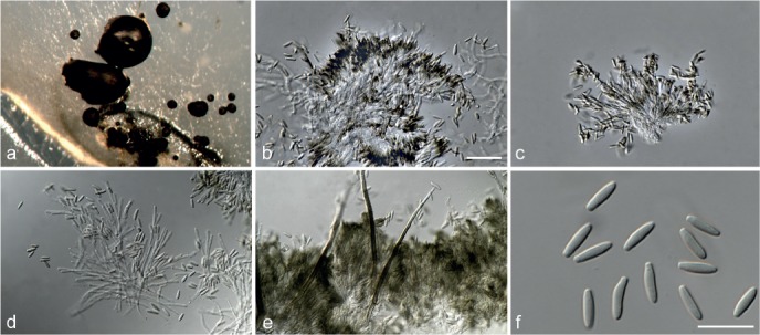
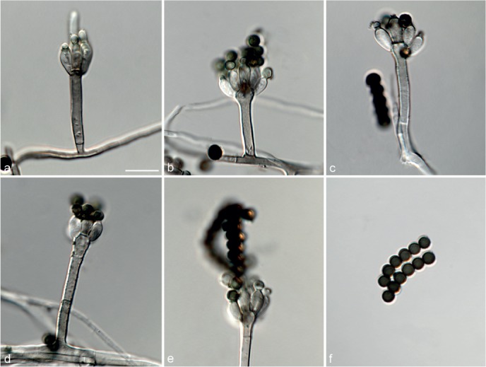
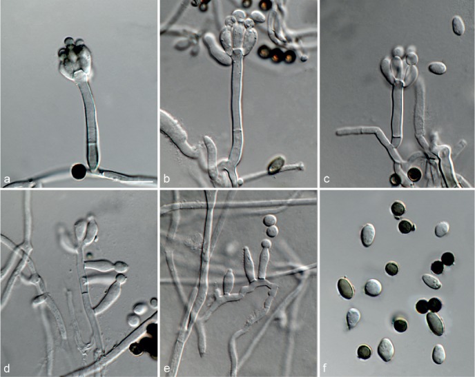
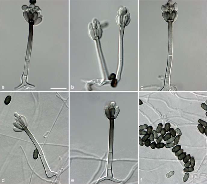
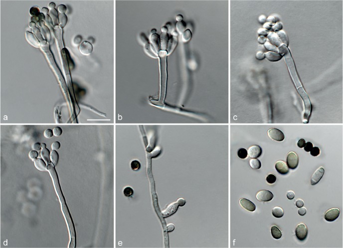
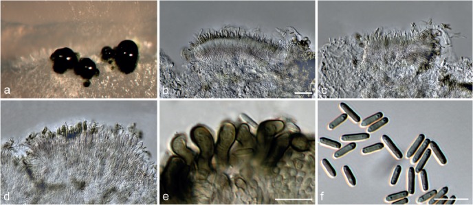
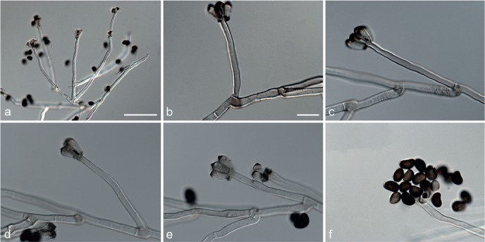
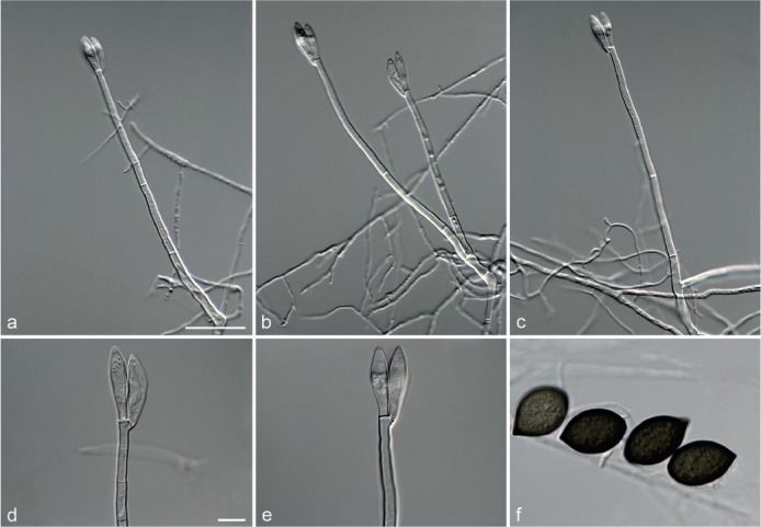
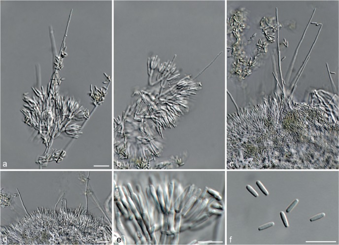
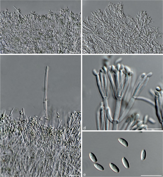
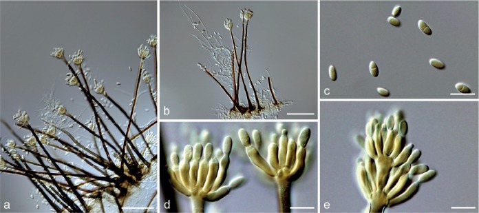
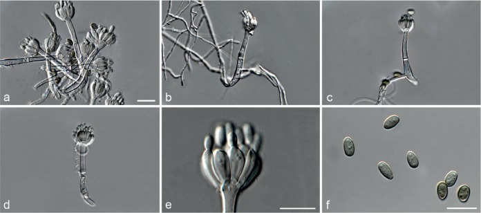
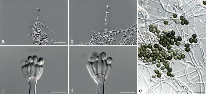
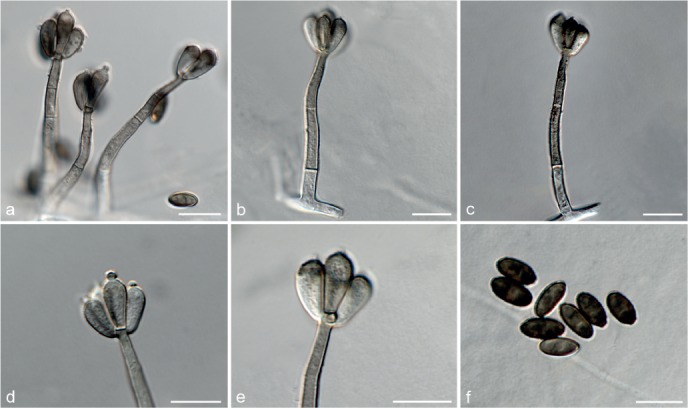
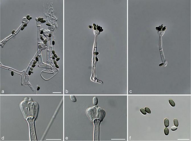
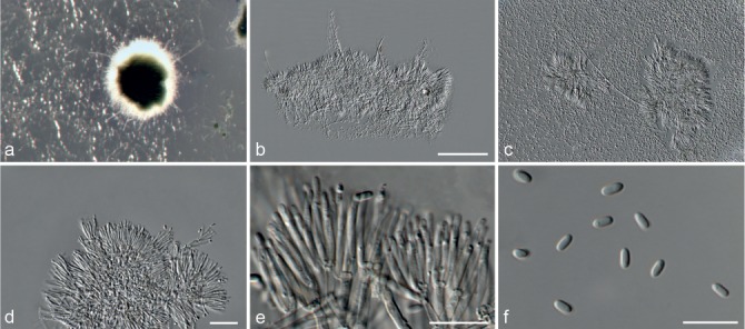
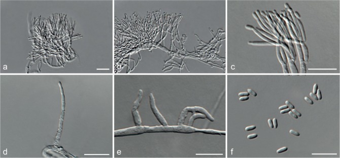
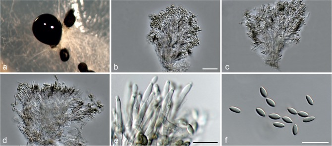
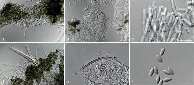
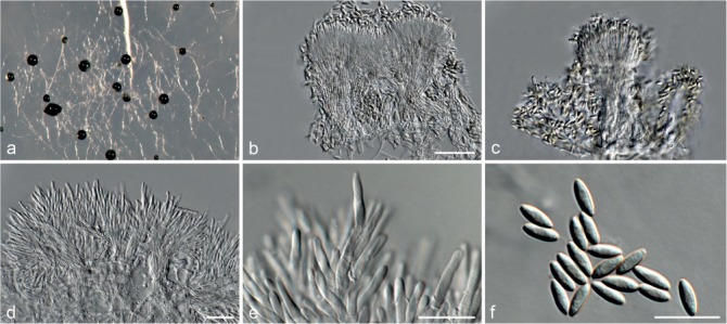
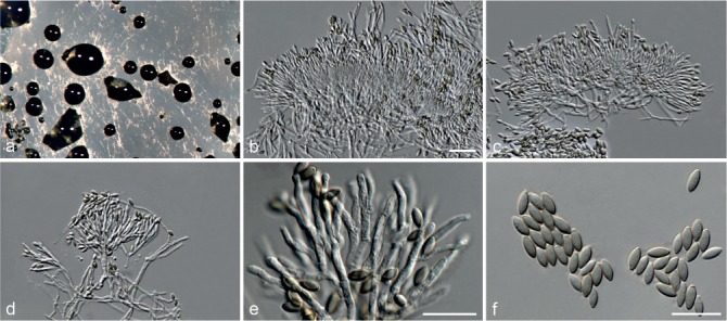
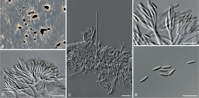
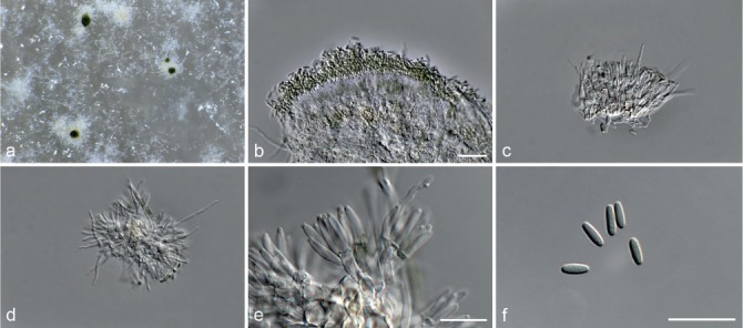
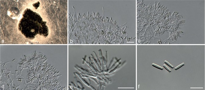
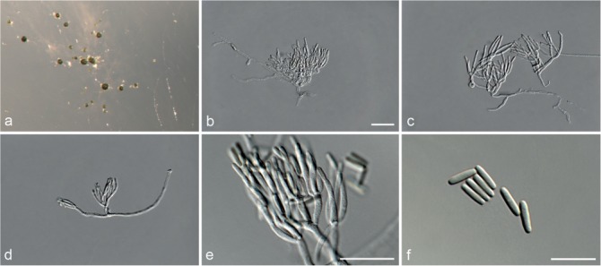
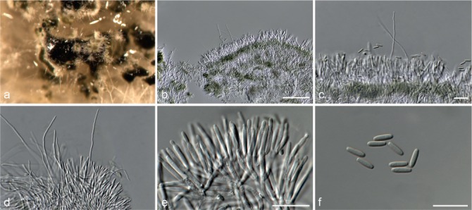
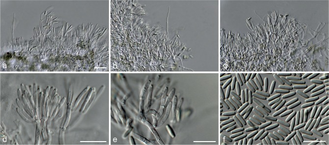
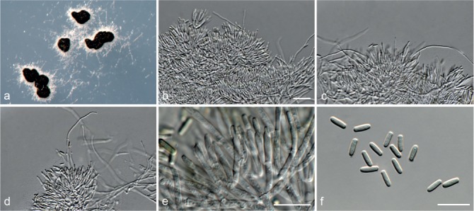
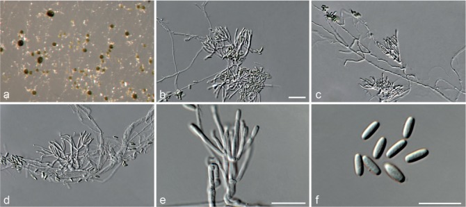
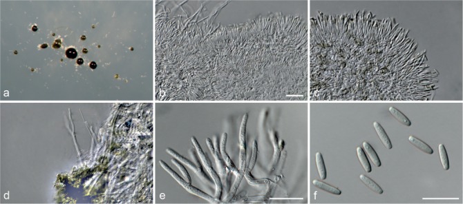
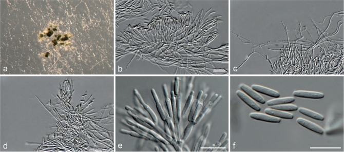
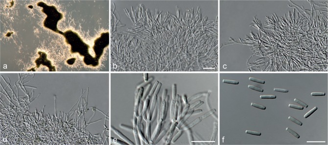
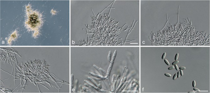
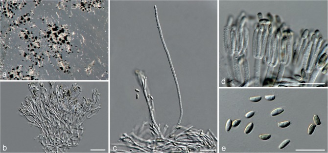
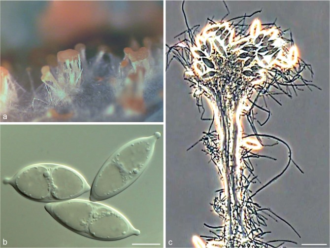
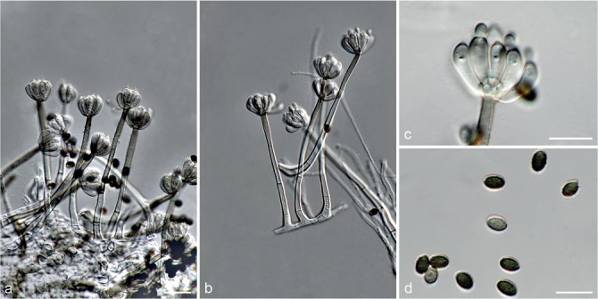
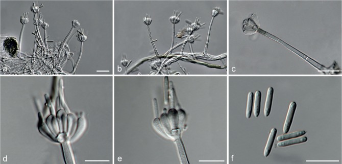
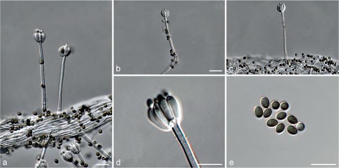
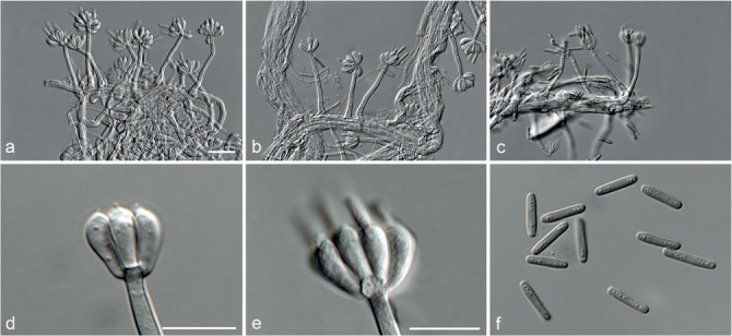
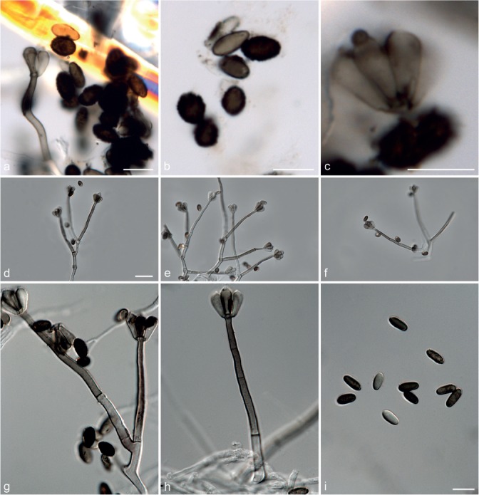
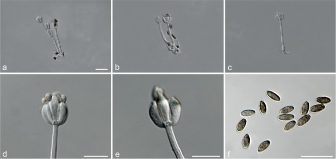
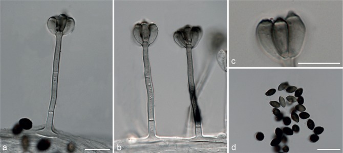
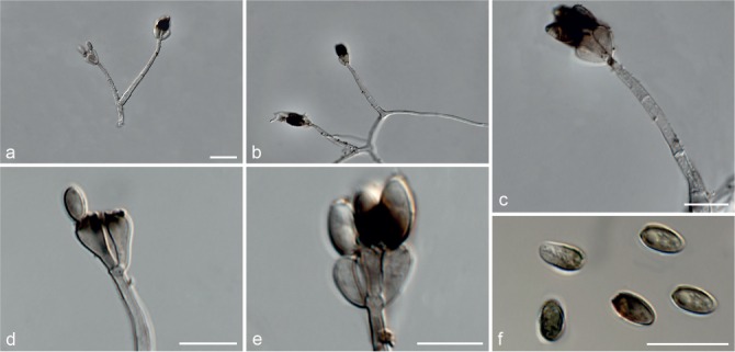
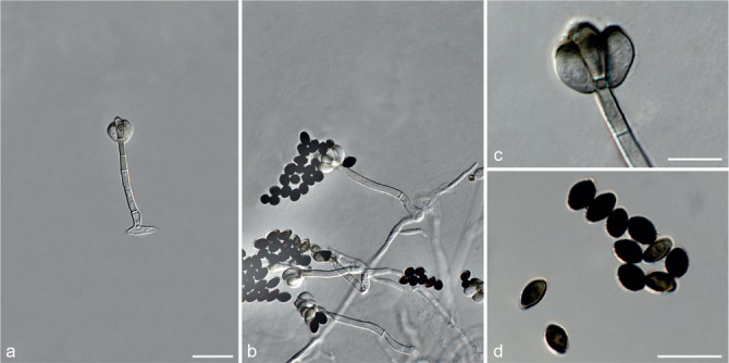
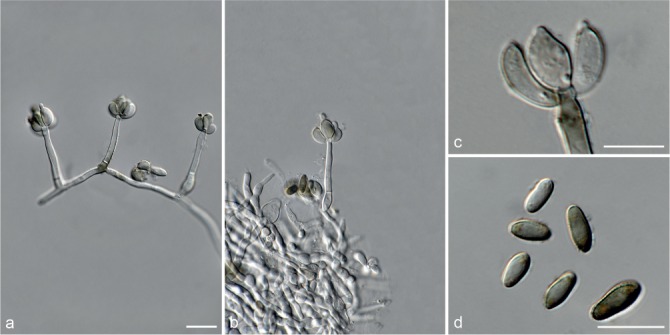
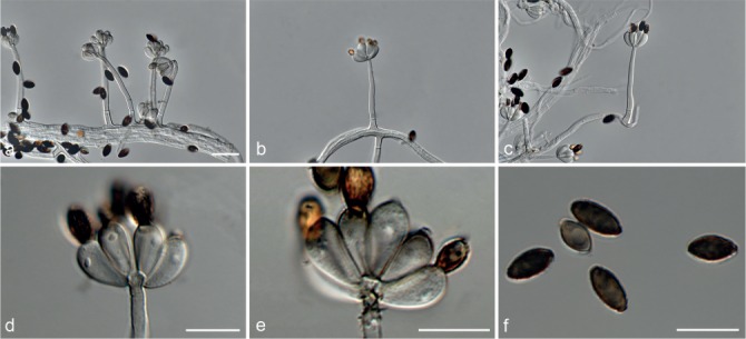
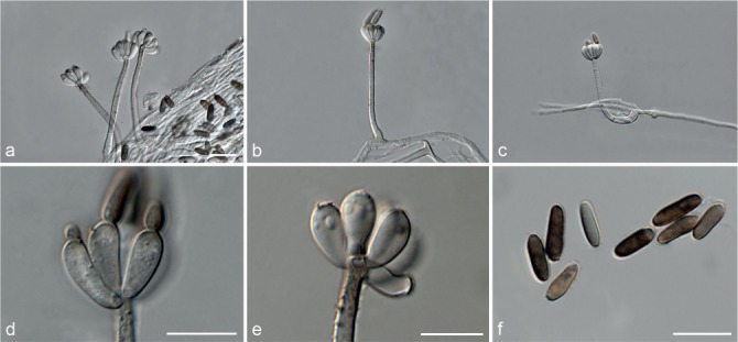
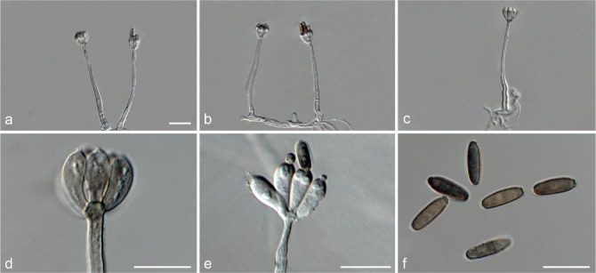
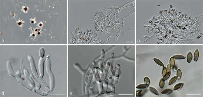
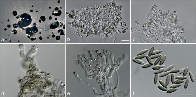
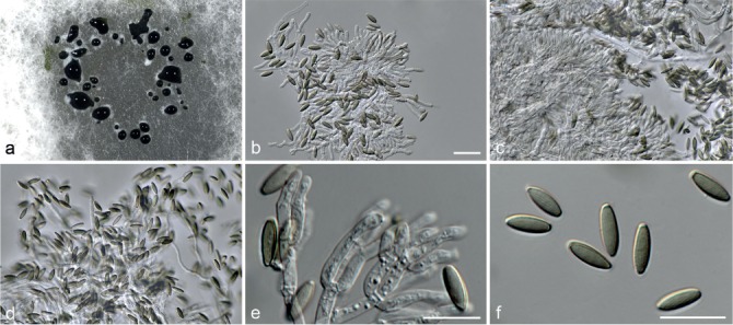
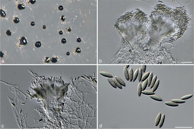
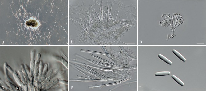
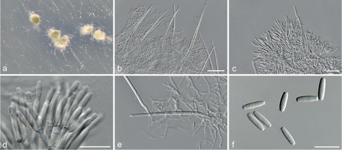
The family Stachybotriaceae was recently introduced to include the genera Myrothecium, Peethambara and Stachybotrys. Members of this family include important plant and human pathogens, as well as several species used in industrial and commercial applications as biodegraders and biocontrol agents. However, the generic boundaries in Stachybotriaceae are still poorly defined, as type material and sequence data are not readily available for taxonomic studies. To address this issue, we performed multi-locus phylogenetic analyses using partial gene sequences of the 28S large subunit (LSU), the internal transcribed spacer regions and intervening 5.8S nrRNA (ITS), the RNA polymerase II second largest subunit (rpb2), calmodulin (cmdA), translation elongation factor 1-alpha (tef1) and β-tubulin (tub2) for all available type and authentic strains. Supported by morphological characters these data resolved 33 genera in the Stachybotriaceae. These included the nine already established genera Albosynnema, Alfaria, Didymostilbe, Myrothecium, Parasarcopodium, Peethambara, Septomyrothecium, Stachybotrys and Xepicula. At the same time the generic names Melanopsamma, Memnoniella and Virgatospora were resurrected. Phylogenetic inference further showed that both the genera Myrothecium and Stachybotrys are polyphyletic resulting in the introduction of 13 new genera with myrothecium-like morphology and eight new genera with stachybotrys-like morphology.

Keywords: biodegraders; generic concept; human and plant pathogens; indoor mycobiota; multi-gene phylogeny; species concept; taxonomy.


Figures

Fig. 1
Fig. 1
The ML consensus tree inferred from the combined LSU and rpb2 sequence alignments. Thickened branches indicate branches present in the ML, MP and Bayesian consensus trees. Branches with ML-BS & MP-BS = 100 % and PP = 1.0 are in red. Branches with ML-BS & MP-BS ≥ 75 % and PP ≥ 0.95 are in blue. The scale bar indicates 0.2 expected changes per site. Coloured blocks represent the accepted genera. The tree is rooted to Calonectria ilicicola (CBS 190.50), Fusarium sambucinum (CBS 146.95) and Nectria cinnabarina (CBS 125165).
Fig. 2
Fig. 2
The ML consensus tree inferred from the combined cmdA, ITS, rpb2 and tub2 sequence alignments of the Myrothecium s.l. dataset. Thickened branches indicate branches present in the ML, MP and Bayesian consensus trees. Branches with ML-BS & MP-BS = 100 % and PP = 1.0 are in red. Branches with ML-BS & MP-BS ≥ 75 % and PP ≥ 0.95 are in blue. The scale bar indicates 0.2 expected changes per site. The tree is rooted to Fusarium sambucinum (CBS 146.95). Epi- and ex-type strains are indicated in bold.
Fig. 3
Fig. 3
The ML consensus tree inferred from the combined cmdA, ITS, rpb2, tef1 and tub2 sequence alignments of the Stachybotrys s.l. dataset. Thickened branches indicate branches present in the ML, MP and Bayesian consensus trees. Branches with ML-BS & MP-BS = 100 % and PP = 1.0 are in red. Branches with ML-BS & MP-BS ≥ 75 % and PP ≥ 0.95 are in blue. The scale bar indicates 0.2 expected changes per site. The tree is rooted to Peethambara sundara (CBS 646.77). Epi- and ex-type strains are indicated in bold.
Fig. 3
Fig. 3
The ML consensus tree inferred from the combined cmdA, ITS, rpb2, tef1 and tub2 sequence alignments of the Stachybotrys s.l. dataset. Thickened branches indicate branches present in the ML, MP and Bayesian consensus trees. Branches with ML-BS & MP-BS = 100 % and PP = 1.0 are in red. Branches with ML-BS & MP-BS ≥ 75 % and PP ≥ 0.95 are in blue. The scale bar indicates 0.2 expected changes per site. The tree is rooted to Peethambara sundara (CBS 646.77). Epi- and ex-type strains are indicated in bold.
Fig. 4
Fig. 4
The ML consensus tree inferred from the ITS sequence alignment of the Stachybotrys s.l. dataset. Thickened branches indicate branches present in the ML, MP and Bayesian consensus trees. Branches with ML-BS & MP-BS = 100 % and PP = 1.0 are in red. Branches with ML-BS & MP-BS ≥ 75 % and PP ≥ 0.95 are in blue. The scale bar indicates 0.1 expected changes per site. The tree is rooted to Peethambara sundara (CBS 646.77). Epi- and ex-type strains are indicated in bold.
Fig. 4
Fig. 4
The ML consensus tree inferred from the ITS sequence alignment of the Stachybotrys s.l. dataset. Thickened branches indicate branches present in the ML, MP and Bayesian consensus trees. Branches with ML-BS & MP-BS = 100 % and PP = 1.0 are in red. Branches with ML-BS & MP-BS ≥ 75 % and PP ≥ 0.95 are in blue. The scale bar indicates 0.1 expected changes per site. The tree is rooted to Peethambara sundara (CBS 646.77). Epi- and ex-type strains are indicated in bold.
Fig. 5
Fig. 5
Achroiostachys aurantispora (DAOMC 225565). a–c. Conidiophores; d. conidiophores on substrate; e. conidia. — Scale bar = 10 μm (apply to b–d).
Fig. 6
Fig. 6
Achroiostachys betulicola (CBS 136397). a–e. Conidiophores; f. conidia. — Scale bars = 10 μm.
Fig. 7
Fig. 7
Achroiostachys humicola (CBS 868.73). a–e. Conidiophores; f. conidia. — Scale bar = 10 μm.
Fig. 8
Fig. 8
Achroiostachys levigata (CBS 185.79). a–c. Conidiophores; d–e. conidiogenous cells; f. conidia. — Scale bars: a = 10 μm (apply to b–c, f); d = 10 μm (apply to e).
Fig. 9
Fig. 9
Achroiostachys phyllophila (CBS 136181). a–b. Conidiophores; c. conidiogenous cells; d. conidia. — Scale bars: a = 10 μm (apply to b); c–d = 10 μm.
Fig. 10
Fig. 10
Achroiostachys saccharicola (CBS 268.76). a–b. Conidiophores; c. conidiogenous cells; d. conidia. — Scale bars: a = 10 μm (apply to b); c–d = 10 μm.
Fig. 11
Fig. 11
Albifimbria lateralis (CBS 117712). a–c. Sporodochial conidiomata; d. conidiogenous cells; e. simple conidiomata; f. conidia. — Scale bars: a = 20 μm (apply to b–c, e–f); d = 10 μm.
Fig. 12
Fig. 12
Albifimbria terrestris (CBS 126186). a. Sporodochial conidiomata on SNA; b. sporodochial conidiomata; c. setae; d. conidiogenous cells; e–f. conidia. — Scale bars: b = 20 μm (apply to c, e); d, f = 10 μm.
Fig. 13
Fig. 13
Albifimbria verrucaria (CBS 328.52). a–b. Sporodochial conidiomata on SNA; c–d. sporodochial conidiomata; e. conidiogenous cells; f. conidia. — Scale bars: c = 20 μm (apply to d); e–f = 10 μm.
Fig. 14
Fig. 14
Albifimbria viridis (CBS 449.71). a. Sporodochial conidiomata on SNA; b. sporodochial conidiomata; c. setae; d. conidiogenous cells; e. conidia. — Scale bars = 10 μm.
Fig. 15
Fig. 15
Alfaria caricicola (CBS 113567). a. Sporodochial conidiomata; b–c. sporodochia with Type I setae; d. Type II setae; e. conidiogenous cells; f. conidia; Scale bars: a = 20 μm (apply to b–d); e–f = 10 μm.
Fig. 16
Fig. 16
Alfaria ossiformis (CBS 324.54). a. Sporodochia with dry conidial pillars; b–c. sporodochial conidiomata; d–e. conidiogenous cells; f. conidia. — Scale bars: a = 100 μm; b = 20 μm (apply to c, f); d–e = 10 μm.
Fig. 17
Fig. 17
Alfaria terrestris (CBS 477.91). a–c. Sporodochial conidiomata; d–e. conidiogenous cells; f. conidia. — Scale bars: a = 20 μm (apply to b–c, f); d–e = 10 μm.
Fig. 18
Fig. 18
Alfaria thymi (CBS 447.83). a. Sporodochial conidiomata on SNA; b–c. sporodochia; d. conidiogenous cells; e. setae; f. conidia. — Scale bars: b = 20 μm (apply to c–e); f = 10 μm.
Fig. 19
Fig. 19
Brevistachys globosa (CBS 141056). a–e. Conidiophores; f. conidia in chains. — Scale bar = 10 μm (apply to all).
Fig. 20
Fig. 20
Brevistachys lateralis (CBS 141058). a–c. Conidiophores; d. conidiogenous cells carried laterally on the stipe of a conidiophore; e. conidiogenous cells carried laterally on a hyphae; f. conidia. — Scale bar = 10 μm (apply to all).
Fig. 21
Fig. 21
Brevistachys ossiformis (CBS 696.73). a–e. Conidiophores; f. conidia in chains. — Scale bar = 10 μm.
Fig. 22
Fig. 22
Brevistachys variabilis (CBS 141057). a–d. Conidiophores; e. conidiogenous cells carried laterally on a hyphae; f. conidia. — Scale bar = 10 μm.
Fig. 23
Fig. 23
Capitofimbria compacta (CBS 111739). a. Sporodochial conidiomata on SNA; b–c. sporodochia; d–e. marginal hyphae of the sporodochia; f. conidia. — Scale bars: b = 20 μm (apply to c–d); e–f = 10 μm.
Fig. 24
Fig. 24
Cymostachys coffeicola (CBS 252.76). a–e. Conidiophores; f. conidia. — Scale bars: a = 50 μm; b = 10 μm (apply to c–f).
Fig. 25
Fig. 25
Cymostachys fabispora (CBS 136180). a–c. Conidiophores; d–e. conidiogenous cells; f. conidia. — Scale bars: a = 20 μm (apply to b–c); d–f = 10 μm.
Fig. 26
Fig. 26
Dimorphiseta terrestris (CBS 127345). a. Sporodochial conidiomata; b. sporodochia with Type I setae; c. sporodochia with Type II setae; d. conidiogenous cells; e. conidia. — Scale bars: a = 20 μm (apply to b); c–e = 10 μm.
Fig. 27
Fig. 27
Grandibotrys pseudotheobromae (CBS 136170). a–c. Conidiophores; d–e. conidiogenous cells; f. conidia. — Scale bars: a = 20 μm (apply to b–c); d = 10 μm (apply to e–f).
Fig. 28
Fig. 28
Grandibotrys xylophila (CBS 136179). a–c. Conidiophores; d–e. conidiogenous cells; f. conidia. — Scale bars: a = 20 μm (apply to b–c); d = 10 μm (apply to e–f).
Fig. 29
Fig. 29
Gregatothecium humicola (CBS 205.96). a–b. Penicillate conidiophores; c–d. sporodochial conidiophores; e. conidiogenous cells; f. conidia. — Scale bars: a = 20 μm (apply to b–c); d = 20 μm; e–f = 10 μm.
Fig. 30
Fig. 30
Inaequalispora prestonii (CBS 175.73). a–b. Sporodochial conidiomata; c. seta; d. conidiogenous cells; e. conidia. — Scale bars: a = 20 μm (apply to b); c = 20 μm; d, e = 10 μm.
Fig. 31
Fig. 31
Kastanostachys aterrima (CBS 101310). a–b. Conidiophores; c. conidia; d–e. conidiogenous cells. — Scale bars: a–b = 50 μm; c–e = 10 μm.
Fig. 32
Fig. 32
Melanopsamma pomiformis (CBS 101322). a–d. Conidiophores; e. conidiogenous cells; f. conidia. — Scale bars: a = 10 μm (apply to b); e–f = 10 μm.
Fig. 33
Fig. 33
Melanopsamma xylophila (CBS 100343). a–b. Conidiophores; c–d. conidiogenous cells; e. conidia. — Scale bars: a = 50 μm (apply to b); c–e = 10 μm.
Fig. 34
Fig. 34
Memnoniella brunneoconidiophora (CBS 136191). a–c. Conidiophores; d–e. conidiogenous cells; f. conidia. — Scale bars = 10 μm.
Fig. 35
Fig. 35
Memnoniella dichroa (CBS 526.50). a–c. Conidiophores; d–e. conidiogenous cells; f. conidia. — Scale bars: a = 20 μm (apply to b–c); d–f = 10 μm.
Fig. 36
Fig. 36
Memnoniella echinata (CBS 216.32). a–c. Conidiophores; d–e. conidiogenous cells; f. conidia in chains. — Scale bars = 10 μm.
Fig. 37
Fig. 37
Memnoniella ellipsoidea (CBS 136201). a–c. Conidiophores; d–e. conidiogenous cells; f. conidia. — Scale bars: a = 20 μm (apply to b–c); d–f = 10 μm.
Fig. 38
Fig. 38
Memnoniella humicola (CBS 463.74). a–c. Conidiophores; d–e. conidiogenous cells; f. conidia. — Scale bar: a = 20 μm (apply to b–c); d–f = 10 μm.
Fig. 39
Fig. 39
Memnoniella pseudonilagirica (CBS 136405). a–d. Conidiophores; e. conidiogenous cells; f. conidia. — Scale bars: a = 50 μm (apply to b); c = 20 μm (apply to d, f); e = 10 μm.
Fig. 40
Fig. 40
Memnoniella putrefolia (CBS 101177). a–c. Conidiophores; d–e. conidiogenous cells; f. conidia. — Scale bars: a = 20 μm; b = 10 μm (apply to c, f); d–e = 10 μm.
Fig. 41
Fig. 41
Myrothecium inundatum (CBS 275.48). a. Sporodochial conidiomata on SNA; b–d. sporodochia; e. conidiogenous cells; f. conidia. — Scale bars: b = 100 μm (apply to c); d = 20 μm; e–f = 10 μm.
Fig. 42
Fig. 42
Myrothecium simplex (CBS 582.93). a–b. Sporodochial conidiomata; c. conidiogenous cells; d–e. simple conidiomata; f. conidia. — Scale bar: a = 20 μm (apply to b); c–f = 10 μm.
Fig. 43
Fig. 43
Myxospora aptrootii (CBS 101263). a. Conidiomata on SNA; b–d. synnematous conidiomata; e. conidiogenous cells; f. conidia. — Scale bars: b = 50 μm (apply to c–d); e–f = 10 μm.
Fig. 44
Fig. 44
Myxospora crassiseta (CBS 731.83). a–b. Sporodochial conidiomata; c. conidiogenous cells; d–e. setae; f. conidia. — Scale bars: a = 50 μm; b = 20 μm (apply to d–e); c, f = 10 μm.
Fig. 45
Fig. 45
Myxospora graminicola (CBS 116538). a. Conidiomata on SNA; b–d. synnematous conidiomata; e. conidiogenous cells; f. conidia. — Scale bars: b = 50 μm (apply to c); d = 20 μm; e–f = 10 μm.
Fig. 46
Fig. 46
Myxospora musae (CBS 265.71). a. Sporodochial conidiomata on SNA; b–d. sporodochia; e. conidiogenous cells; f. conidia. — Scale bars: b = 20 μm (apply to c–d); e–f = 10 μm.
Fig. 47
Fig. 47
Neomyrothecium humicola (CBS 310.96). a. Sporodochial conidiomata on SNA; b. sporodochia; c. setae; d. conidiogenous cells; e. conidia. — Scale bars: b, d–e = 10 μm; c = 20 μm.
Fig. 48
Fig. 48
Paramyrothecium breviseta (CBS 544.75). a. Sporodochial conidiomata on SNA; b–d. sporodochia; e. conidiogenous cells; f. conidia. — Scale bars: b = 20 μm (apply to c–d); e–f = 10 μm.
Fig. 49
Fig. 49
Paramyrothecium cupuliforme (CBS 127789). a. Sporodochial conidiomata on SNA; b–d. sporodochia; e. conidiogenous cells; f. conidia. — Scale bars: b = 20 μm (apply to c–d); e–f = 10 μm.
Fig. 50
Fig. 50
Paramyrothecium foeniculicola (CBS 331.51). a. Sporodochial conidiomata on SNA; b–d. sporodochia; e. conidiogenous cells; f. conidia. — Scale bars: b = 20 μm (apply to c–d); e–f = 10 μm.
Fig. 51
Fig. 51
Paramyrothecium foliicola (CBS 113121). a. Sporodochial conidiomata on SNA; b–d. sporodochia; e. conidiogenous cells; f. conidia. — Scale bars: b = 20 μm (apply to c–d); e–f = 10 μm.
Fig. 52
Fig. 52
Paramyrothecium humicola (CBS 127295). a–c. Sporodochial conidiomata; d–e. conidiogenous cells; f. conidia. — Scale bars: a = 20 μm (apply to b–c); d, f = 10 μm.
Fig. 53
Fig. 53
Paramyrothecium nigrum (CBS 116537). a. Sporodochial conidiomata on SNA; b–d. sporodochia; e. conidiogenous cells; f. conidia. — Scale bars: b = 20 μm (apply to c–d); e–f = 10 μm.
Fig. 54
Fig. 54
Paramyrothecium parvum (CBS 257.35). a. Sporodochial conidiomata on SNA; b–d. sporodochia; e. conidiogenous cells; f. conidia. — Scale bars: b = 20 μm (apply to c–d); e–f = 10 μm.
Fig. 55
Fig. 55
Paramyrothecium roridum (CBS 357.89). a. Sporodochial conidiomata on SNA; b–c. sporodochia; d. setae; e. conidiogenous cells; f. conidia. — Scale bars: b = 20 μm (apply to c–d); e–f = 10 μm.
Fig. 56
Fig. 56
Paramyrothecium tellicola (CBS 478.91). a. Sporodochial conidiomata on SNA; b–c. sporodochia; e. conidiogenous cells; f. conidia. — Scale bars: b = 20 μm (apply to c–d); e–f = 10 μm.
Fig. 57
Fig. 57
Paramyrothecium terrestris (CBS 564.86). a. Sporodochial conidiomata on SNA; b–d. sporodochia; e. conidiogenous cells; f. conidia. — Scale bars: b = 20 μm (apply to c–d); e–f = 10 μm.
Fig. 58
Fig. 58
Paramyrothecium viridisporum (CBS 873.85). a. Sporodochial conidiomata on SNA; b–d. sporodochia; e. conidiogenous cells; f. conidia. — Scale bars: b = 20 μm (apply to c–d); e–f = 10 μm.
Fig. 59
Fig. 59
Parvothecium terrestre (CBS 198.89). a. Sporodochial conidiomata on SNA; b. sporodochia; c. setae; d. conidiogenous cells; e. conidia. — Scale bars: b = 20 μm; c–e = 10 μm.
Fig. 60
Fig. 60
Peethambara sundara (CBS 521.96). a. Synnemata on substrate; b. conidia; c. synnemata. — Scale bars: b = 10 μm; c = 50 μm.
Fig. 61
Fig. 61
Sirastachys castanedae (CBS 136403). a–b. Conidiophores; c. conidiogenous cells; d. conidia. — Scale bars: a = 20 μm (apply to b); c–d = 10 μm.
Fig. 62
Fig. 62
Sirastachys cylindrospora (CBS 136166). a–c. Conidiophores; d–e. conidiogenous cells; f. conidia. — Scale bars: a = 10 μm (apply to b); c–f = 10 μm.
Fig. 63
Fig. 63
Sirastachys pandanicola (CBS 136545). a–c. Conidiophores; d. conidiogenous cells; e. conidia. — Scale bar: a, d, e = 10 μm; b = 20 μm (apply to c).
Fig. 64
Fig. 64
Sirastachys phaeospora (CBS 100155). a–c. Conidiophores; d. conidiogenous cells; e. conidia. — Scale bars: a, d–e = 10 μm; b = 20 μm.
Fig. 65
Fig. 65
Sirastachys phyllophila (CBS 136169). a–b. Conidiophores; c. conidiogenous cells; d. conidia. — Scale bars = 10 μm.
Fig. 66
Fig. 66
Sirastachys pseudolongispora (CBS 100154). a–c. Conidiophores; d–e. conidiogenous cells; f. conidia. — Scale bars: a = 10 μm (apply to b–c); d–f = 10 μm.
Fig. 67
Fig. 67
Stachybotrys chartarum. a–c. DAOM 51026. a. Conidiophore; b. conidia; c. conidiogenous cells. — d–i. CBS 182.80; d–h. Conidiophores; i. conidia. — Scale bars: a–c = 10 μm (apply to g–h); d = 20 μm (apply to e–f); i = 10 μm.
Fig. 68
Fig. 68
Stachybotrys dolichophialis (DAOM 227011). a–c. Conidiophores; d–e. conidiogenous cells; f. conidia. — Scale bars: a = 20 μm (apply to b–c); d–f = 10 μm.
Fig. 69
Fig. 69
Stachybotrys limonispora (CBS 128809). a–b. Conidiophores; c. conidiogenous cells; d. conidia. — Scale bars: a = 10 μm (apply to b); c–d = 10 μm.
Fig. 70
Fig. 70
Stachybotrys phaeophialis (KAS 525). a–c. Conidiophores; d–e. conidiogenous cells; f. conidia. — Scale bars: a = 10 μm (apply to b); c–f = 10 μm.
Fig. 71
Fig. 71
Stachybotrys subsylvatica (CBS 126205). a–b. Conidiophores; c. conidiogenous cells; d. conidia. — Scale bars: a = 10 μm (apply to b); c–d = 10 μm.
Fig. 72
Fig. 72
Striatibotrys atypica (CBS 141059). a–b. Conidiophores; c. conidiogenous cells; d. conidia. — Scale bars: a = 20 μm (apply to b); c–d = 10 μm.
Fig. 73
Fig. 73
Striatibotrys humicola (CBS 102408). a–c. Conidiophores; d–e. conidiogenous cells; f. conidia. — Scale bars: a = 10 μm (apply to b–c); d–f = 10 μm.
Fig. 74
Fig. 74
Striatibotrys rhabdospora (CBS 528.80). a–c. Conidiophores; d–e. conidiogenous cells; f. conidia. — Scale bars: a = 20 μm (apply to b–c); d–f = 10 μm.
Fig. 75
Fig. 75
Striatibotrys yuccae (CBS 390.68). a–c. Conidiophores; d–e. conidiogenous cells; f. conidia. — Scale bars: a = 10 μm (apply to b–c); d–f = 10 μm.
Fig. 76
Fig. 76
Striaticonidium brachysporum (CBS 513.71). a. Sporodochial conidiomata on SNA; b, c. sporodochia; d–e. conidiogenous cells; f. conidia. — Scale bars: b = 20 μm (apply to c); d–f = 10 μm.
Fig. 77
Fig. 77
Striaticonidium cinctum (CBS 932.69). a. Sporodochial conidiomata on SNA; b–c. sporodochia; d. setae; e. conidiogenous cells; f. conidia. — Scale bars: b = 20 μm (apply to c); d–f = 10 μm.
Fig. 78
Fig. 78
Striaticonidium humicola (CBS 258.76). a. Sporodochial conidiomata on SNA; b–c. sporodochia; d. setae; e. conidiogenous cells; f. conidia. — Scale bars: b = 20 μm (apply to c–d); e–f = 10 μm.
Fig. 79
Fig. 79
Striaticonidium synnematum (CBS 479.85). a. Conidiomata on SNA; b–c. synnematous conidiomata; d. conidia. — Scale bars: b = 10 μm (apply to c); d = 10 μm.
Fig. 80
Fig. 80
Tangerinosporium thalitricola (CBS 317.61). a. Sporodochia with orange conidial masses on colony surface; b–c. sporodochial conidiomata; d. conidiogenous cells; e. seta; f. conidia. — Scale bars: b = 20 μm (apply to c, e); d, f = 10 μm.
Fig. 81
Fig. 81
Xepicula crassiseta (CBS 392.71). a. Sporodochial conidiomata on SNA; b–c. sporodochia; d. conidiogenous cells; e. setae; f. conidia. — Scale bars: b = 100 μm; c = 20 μm (apply to e); d, f = 10 μm.
Fig. 82
Fig. 82
Xepicula jollymannii (CBS 276.48). a. Sporodochial conidiomata on SNA; b–c. sporodochia; d. conidiogenous cells; e. setae; f. conidia. — Scale bars: b = 100 μm; c = 20 μm (apply to e); d, f = 10 μm.

References

    1. Abbas HK, Johnson BB, Shier WT, et al. 2002. Phytotoxicity and mammalian cytotoxicity of macrocyclic trichothecene mycotoxins from Myrothecium verrucaria. Phytochemistry 59: 309–313. - PubMed
    1. Agarwal SC. 1980. A new species of Myrothecium from Indian alkaline soils. Current Science 49: 281–282.
    1. Alves JL, Barreto RW, Pereira OL. 2010. Additions to the mycobiota of the invasive weed Miconia calvescens (Melastomataceae). Mycologia 102: 69–82. - PubMed
    1. Amagata T, Rath C, Rigot JF, et al. 2003. Structures and cytotoxic properties of trichoverroids and their macrolide analogues produced by saltwater culture of Myrothecium verrucaria. Journal of Medical Chemistry 46: 4342–4350. - PubMed
    1. Andersen B, Nielsen KF, Thrane U, et al. 2003. Molecular and phenotypic descriptions of Stachybotrys chlorohalonata sp. nov. and two chemotypes of Stachybotrys found in water-damaged buildings. Mycologia 95: 1227–1238. - PubMed

LinkOut - more resources