

An unusual pain in the hip
- PMID: 27617105
- PMCID: PMC5015422
- DOI: 10.1093/omcr/omw072
An unusual pain in the hip
Abstract
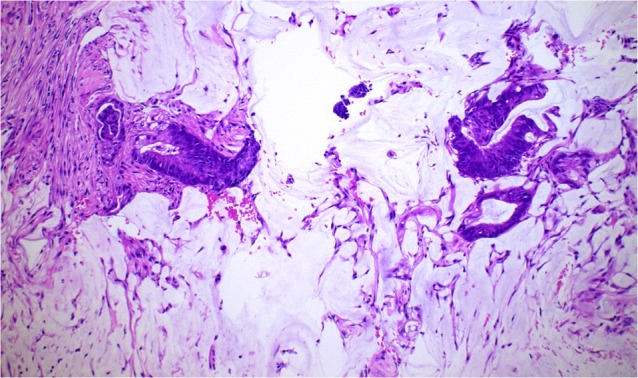
A 68-year-old previously healthy man presented with increasing right hip pain of 6 months duration. On examination he was found to have a hard mass in the right hip arising from the pelvic bone. Imaging studies were in keeping with a sarcoma arising from the right iliac bone. However, biopsy of this bony lesion confirmed this to be a metastatic adenocarcinoma rather than a primary bone malignancy. Further imaging and a subsequent colonoscopy revealed the primary to be a colonic adenocarcinoma. The unique and unusual nature of this case was the presentation as a solitary bony metastasis from a colonic primary. There is no previously documented report in the literature of such a rare presentation of a colonic adenocarcinoma as a solitary bony lesion mimicking a primary sarcoma in the absence of other signs or symptoms.
Keywords: adenocarcinoma; colon; hip; metastasis; pelvic; solitary.
Figures


References
-
- Cancer Research UK. Bowel cancer statistics, Cancer Research UK. 2013.
-
- Disibio G, French SW. Metastatic patterns of cancers: results from a large autopsy study. Arch Pathol Lab Med 2008;132:931–9. - PubMed
-
- Santini D, Tampellini M, Vincenzi B, Ibrahim T, Ortega C, Virzi V, et al. . Natural history of bone metastasis in colorectal cancer: final results of a large Italian bone metastases study. Ann Oncol 2012;23:2072–7. - PubMed
Publication types
LinkOut - more resources
Full Text Sources
Other Literature Sources
