. 2016 Oct;23(10):899-905.
doi: 10.1038/nsmb.3293. Epub 2016 Sep 12.

Glycan shield and epitope masking of a coronavirus spike protein observed by cryo-electron microscopy

Affiliations

Glycan shield and epitope masking of a coronavirus spike protein observed by cryo-electron microscopy

Alexandra C Walls et al. Nat Struct Mol Biol. 2016 Oct.

Abstract

The threat of a major coronavirus pandemic urges the development of strategies to combat these pathogens. Human coronavirus NL63 (HCoV-NL63) is an α-coronavirus that can cause severe lower-respiratory-tract infections requiring hospitalization. We report here the 3.4-Å-resolution cryo-EM reconstruction of the HCoV-NL63 coronavirus spike glycoprotein trimer, which mediates entry into host cells and is the main target of neutralizing antibodies during infection. The map resolves the extensive glycan shield obstructing the protein surface and, in combination with mass spectrometry, provides a structural framework to understand the accessibility to antibodies. The structure reveals the complete architecture of the fusion machinery including the triggering loop and the C-terminal domains, which contribute to anchoring the trimer to the viral membrane. Our data further suggest that HCoV-NL63 and other coronaviruses use molecular trickery, based on epitope masking with glycans and activating conformational changes, to evade the immune system of infected hosts.


Conflict of interest statement

The authors declare no competing financial interests.

Figures

Figure 1
Figure 1. Cryo-EM structure of the HCoV-NL63 S trimer.
(a) Representative micrograph of frozen-hydrated HCoV-NL63 S particles (defocus 3.4 μm). Scale bar, 355 Å. (b) Five selected class averages showing the particles along different orientations. Scale bar, 60 Å. (c,d) 3D map filtered at 3.4-Å resolution and colored by protomer. Two orthogonal views of the S trimer from the side (c) and from the top, facing toward the viral membrane, (d) are shown. (e,f) Ribbon diagrams showing the HCoV-NL63 S atomic model, oriented as in c and d, respectively.
Figure 2
Figure 2. Organization of the HCoV-NL63 S-protein glycan shield, revealed by cryo-EM and MS.
(a,b) Ribbon diagrams showing two orthogonal views of the S trimer, from the side (a) and from the top (b), facing toward the viral membrane. Glycans are shown as dark-blue spheres. (c) Residue-level schematic of N-linked glycans. The most extensive glycan structure detected by MS at each site is represented except for glycans observed only by cryo-EM, for which the resolved sugar moieties are shown. FP, fusion peptide, HR1, heptad-repeat 1 region; HR2, heptad-repeat 2 region (shown with a dashed line because it is not resolved in the map); TM, transmembrane domain (the striated texture indicates regions that are not part of the construct); GlcNac, N-acetylglucosamine; Man, mannose; Fuc, fucose.
Figure 3
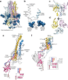
Figure 3. Architecture of the complete coronavirus fusion machinery.
(a) Ribbon diagram of the S2 trimer, colored by protomer with glycans rendered as dark-blue spheres. (b) Zoomed-in view of the S2′ trigger-loop region comprising the central helix and the fusion peptide (light blue). N-linked glycans are shown as dark-blue spheres. The polypeptide segment corresponding to the putative cleavage site is poorly resolved in the density, and this part of the model should be considered to be hypothetical. (c,d) Ribbon diagrams showing two orthogonal views of the S2′ C-terminal region, which is assembled from the connector domains and stem helices. (e,f) Ribbon diagrams of the HCoV-NL63 S2 subunit (e) and of the RSV F protein (f). Conserved structural elements are colored identically to highlight the similar 3D organization of these two fusion machineries, whereas nonconserved regions are colored gray. The topology diagrams underscore the similar topology of the HCoV-NL63 S connector domain and the equivalent RSV F domain, although the tertiary structures of these domains are different, and several structural motifs have been added to the latter domain throughout evolution. The RSV F secondary-structural elements are annotated according to ref. . The N- and C-terminal extremities of the polypeptide segments are indicated.
Figure 4
Figure 4. Evolution of the α-coronavirus S-glycoprotein fold appears to correlate with tissue tropism.
(a) Schematic representation of several α-coronavirus S-glycoprotein S1 subunits, highlighting the presence of one or several domains 0 (blue), as compared with β-coronaviruses. HCoV-NL63 (GenBank YP_003767.1), 229-rel. CoV 1 (GenBank ALK28775.1), 229-rel. CoV 2 (GenBank ALK28765.1), HCoV-229E (GenBank NP_073551.1), porcine epidemic diarrhea virus (PEDV; GenBank AAK38656.1), transmissible gastroenteritis virus strain Purdue P115 (TGEV; GenBank ABG89325.1), porcine respiratory coronavirus strain ISU-1 (PRCV; GenBank ABG89317.1), feline enteric coronavirus strain UU23 (FECV-UU23; GenBank ADC35472.1) and feline infectious peritonitis coronavirus strain UU21 (FIPV-UU21; GenBank ADL71466.1). The β-coronavirus MHV S1 subunit is shown for comparison. Domains A–D are indicated for MHV and HCoV-NL63. (b) Ribbon diagram of the HCoV-NL63 S1 subunit. (c) Ribbon diagram of the MHV S1 subunit. (dg) Ribbon diagrams of HCoV-NL63 domain 0 (d), domain A (e), MHV domain A (f) and rotavirus VP8* (g), showing their structural similarity, which suggests common ancestry. HCoV-NL63 domain 0 and A probably arose from a duplication event.
Figure 5
Figure 5. Potential immune-evasion strategy used by HCoV-NL63.
(a) Ribbon diagram of the HCoV-NL63 S trimer, highlighting the conformation of the S1 subunit. Domains 0, A, B, C and D are colored for one protomer. (b) The HCoV-NL63 receptor-binding loops are buried via interactions with domain A of the same protomer (including the glycan moiety at Asn358) and are not available to engage host-cell receptors. Superimposition of the HCoV-NL63 (purple) and MHV (light gray) S1 subunits via their C domains highlights that their B domains feature opposite orientations related by an ∼180° rotation, thus suggesting a putative trajectory for the conformational changes that must occur to engage the host-cell receptor. Only domain B is shown for MHV S. (c) Comparison of the HCoV-NL63 domain-B structure in our cryo-EM-derived model (purple) with the crystal structure of the same domain in complex with ACE2 (green and dark gray), showing that the receptor-binding loop containing residues 531–539 substantially changes its conformation after binding. (d) ACE2 binding ELISA showing that isolated HCoV-NL63 domain B (HCoV-NL63 S1-B-mFc) binds ACE2 with higher affinity than does the full-length S1 domain (HCoV-NL63 S1-mFc). SARS-CoV S1 (HCoV-NL63 S1-mFc) is a positive control. HCoV-NL63 S1 domain 0 (HCoV-NL63 S1-0-mFc) and PEDV S1 (PEDV S1-mFc), which do not bind ACE2, are negative controls. Mean values and s.d. of three independent experiments are shown.

References

    1. Zumla A, Chan JF, Azhar EI, Hui DS, Yuen KY. Coronaviruses: drug discovery and therapeutic options. Nat. Rev. Drug Discov. 2016;15:327–347. doi: 10.1038/nrd.2015.37. - DOI - PMC - PubMed
    1. Vijay R, Perlman S. Middle East respiratory syndrome and severe acute respiratory syndrome. Curr. Opin. Virol. 2016;16:70–76. doi: 10.1016/j.coviro.2016.01.011. - DOI - PMC - PubMed
    1. Ge XY, et al. Isolation and characterization of a bat SARS-like coronavirus that uses the ACE2 receptor. Nature. 2013;503:535–538. doi: 10.1038/nature12711. - DOI - PMC - PubMed
    1. Sabir JS, et al. Co-circulation of three camel coronavirus species and recombination of MERS-CoVs in Saudi Arabia. Science. 2016;351:81–84. doi: 10.1126/science.aac8608. - DOI - PubMed
    1. Yang Y, et al. Receptor usage and cell entry of bat coronavirus HKU4 provide insight into bat-to-human transmission of MERS coronavirus. Proc. Natl. Acad. Sci. USA. 2014;111:12516–12521. doi: 10.1073/pnas.1405889111. - DOI - PMC - PubMed

MeSH terms