. 2016 Nov;125(5):929-942.
doi: 10.1097/ALN.0000000000001342.

Neural Correlates of Wakefulness, Sleep, and General Anesthesia: An Experimental Study in Rat

Affiliations

Neural Correlates of Wakefulness, Sleep, and General Anesthesia: An Experimental Study in Rat

Dinesh Pal et al. Anesthesiology. 2016 Nov.

Abstract

Background: Significant advances have been made in our understanding of subcortical processes related to anesthetic- and sleep-induced unconsciousness, but the associated changes in cortical connectivity and cortical neurochemistry have yet to be fully clarified.

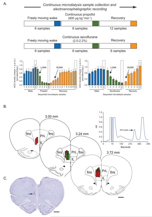
Methods: Male Sprague-Dawley rats were instrumented for simultaneous measurement of cortical acetylcholine and electroencephalographic indices of corticocortical connectivity-coherence and symbolic transfer entropy-before, during, and after general anesthesia (propofol, n = 11; sevoflurane, n = 13). In another group of rats (n = 7), these electroencephalographic indices were analyzed during wakefulness, slow wave sleep (SWS), and rapid eye movement (REM) sleep.

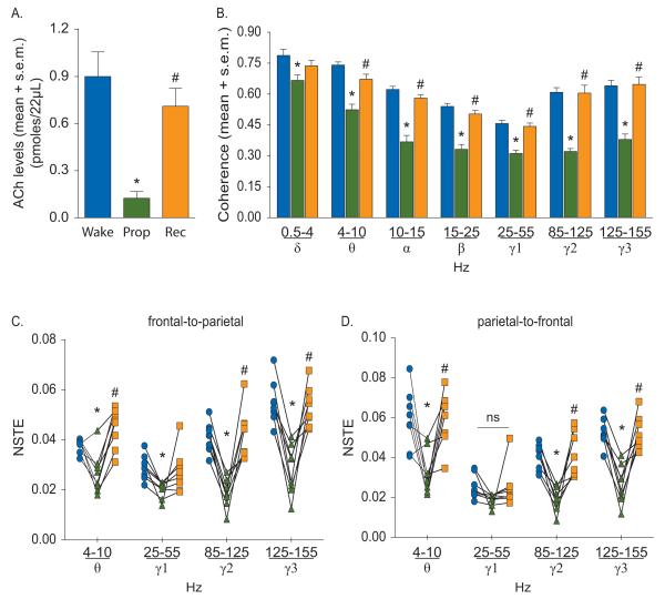
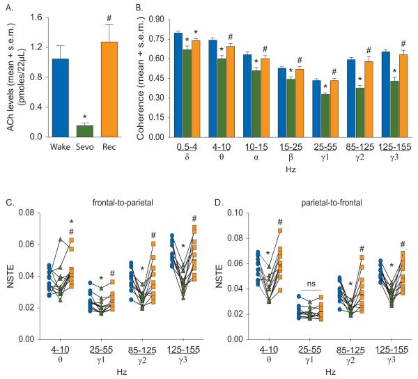
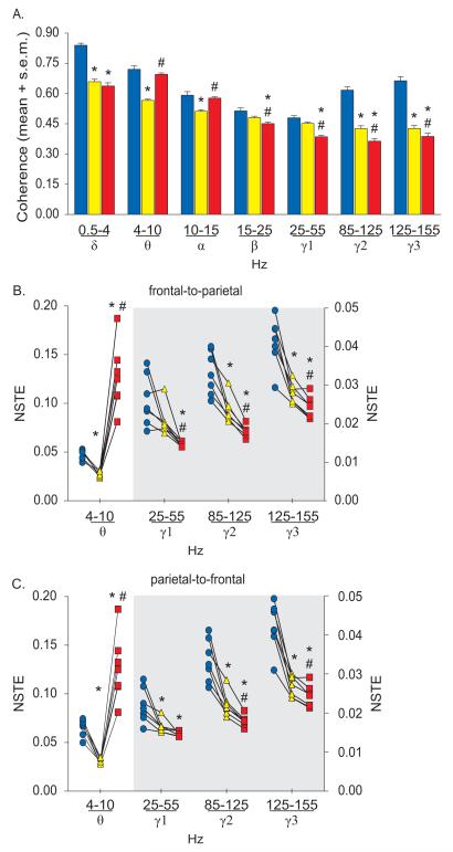
Results: Compared to wakefulness, anesthetic-induced unconsciousness was characterized by a significant decrease in cortical acetylcholine that recovered to preanesthesia levels during recovery wakefulness. Corticocortical coherence and frontal-parietal symbolic transfer entropy in high γ band (85 to 155 Hz) were decreased during anesthetic-induced unconsciousness and returned to preanesthesia levels during recovery wakefulness. Sleep-wake states showed a state-dependent change in coherence and transfer entropy in high γ bandwidth, which correlated with behavioral arousal: high during wakefulness, low during SWS, and lowest during REM sleep. By contrast, frontal-parietal θ connectivity during sleep-wake states was not correlated with behavioral arousal but showed an association with well-established changes in cortical acetylcholine: high during wakefulness and REM sleep and low during SWS.

Conclusions: Corticocortical coherence and frontal-parietal connectivity in high γ bandwidth correlates with behavioral arousal and is not mediated by cholinergic mechanisms, while θ connectivity correlates with cortical acetylcholine levels.


Figures

Figure 1
Figure 1
A) Schematic illustrating the design for microdialysis experiments and temporal course of electroencephalogram and acetylcholine (ACh) data collection. Each ACh sample epoch represents microdialysate collected over a period of 12.5 min. The electroencephalographic data were analyzed in the corresponding 12.5 min epochs. Blue, green and orange bars represent wake, anesthetic-induced unconsciousness (propofol/sevoflurane), and post-anesthetic recovery wake epochs, respectively. The epochs highlighted with broken lines were selected for statistical comparisons. Arrows indicate the approximate time points at which rats showed loss of righting reflex (LORR) and recovery of righting reflex (RORR). B) Reconstruction diagram shows a cascade of coronal brain section drawings from the rat brain atlas to illustrate the location (vertical cylinders) of microdialysis probes (1 mm) within the prefrontal cortex. Green cylinders represent the microdialysis probes from the propofol experiments while the red cylinders represent those from the sevoflurane experiments. Numbers above the coronal brain section drawings are the anterior-posterior stereotaxic coordinates relative to Bregma. The chromatogram above the reconstruction diagram shows signal to noise ratio and the retention time for ACh. C) Cresyl violet stained representative coronal brain section through prefrontal cortex shows the dialysis probe track and the site of microdialysis. Arrow indicates the location of ventral tip of the microdialysis membrane. fmi - forceps minor corpus callosum, IL - infralimbic area, PrL - prelimbic area. Scale bar - 1 mm.
Figure 2
Figure 2
Effect of propofol on cortical acetylcholine (ACh) (A), corticocortical coherence (B), and frontal-parietal directed connectivity (C-D). The symbol-line plots (C-D) show the individual rat data while the significance symbols represent the outcome of group level statistical testing using repeated measures analysis of variance with Tukey’s multiple comparisons test. Significance symbols denote a statistical difference at an alpha of p<0.05. The actual p values are reported in the text in the results section. *significant as compared to wake, #significant as compared to propofol-induced unconsciousness, blue bars/circles: wake, green bars/triangles: propofol-induced unconsciousness, orange bars/squares: post-propofol recovery wake. ns: not significant, NSTE: normalized symbolic transfer entropy, Prop: propofol, Rec: recovery wake, s.e.m.: standard error of the mean.
Figure 3
Figure 3
Effect of sevoflurane on cortical acetylcholine (ACh) (A), corticocortical coherence (B), and frontal-parietal directed connectivity (C-D). The symbol-line plots (C-D) show the individual rat data while the significance symbols represent the outcome of group level statistical testing using repeated measures analysis of variance with Tukey’s multiple comparisons test. Significance symbols denote a statistical difference at an alpha of p<0.05. The actual p values are reported in the text in the results section. *significant as compared to wake, #significant as compared to sevoflurane-induced unconsciousness, blue bars/circles: wake, green bars/triangles: sevoflurane-induced unconsciousness, orange bars/squares: post-sevoflurane recovery wake. ns: not significant, NSTE: normalized symbolic transfer entropy, Rec: recovery wake, s.e.m.: standard error of the mean, Sevo: sevoflurane.
Figure 4
Figure 4
Corticocortical coherence (A) and frontal-parietal directed connectivity (B-C) during wakefulness, slow wave sleep, and rapid eye movement sleep. The symbol-line plots (B-C) show the individual rat data while the significance symbols represent the outcome of group level statistical testing using repeated measures analysis of variance with Tukey’s multiple comparisons test. The right axis in (B) and (C) applies only to the grey shaded area. Significance symbols denote a statistical difference at an alpha of p<0.05. The actual p values are reported in the text in the results section. *significant as compared to wake, #significant as compared to slow wave sleep, blue bars/circles: wake, yellow bars/triangles: slow wave sleep, red bars/squares: rapid eye movement sleep. NSTE: normalized symbolic transfer entropy, s.e.m.: standard error of the mean.

Comment in

References

    1. Boveroux P, Vanhaudenhuyse A, Bruno MA, Noirhomme Q, Lauwick S, Luxen A, Degueldre C, Plenevaux A, Schnakers C, Phillips C, Brichant JF, Bonhomme V, Maquet P, Greicius MD, Laureys S, Boly M. Breakdown of within- and between-network resting state functional magnetic resonance imaging connectivity during propofol-induced loss of consciousness. Anesthesiology. 2010;113:1038–53. - PubMed
    1. Ferrarelli F, Massimini M, Sarasso S, Casali A, Riedner BA, Angelini G, Tononi G, Pearce RA. Breakdown in cortical effective connectivity during midazolam-induced loss of consciousness. Proc Natl Acad Sci U S A. 2010;107:2681–86. - PMC - PubMed
    1. Lewis LD, Weiner VS, Mukamel EA, Donoghue JA, Eskandar EN, Madsen JR, Anderson WS, Hochberg LR, Cash SS, Brown EN, Purdon PL. Rapid fragmentation of neuronal networks at the onset of propofol-induced unconsciousness. Proc Natl Acad Sci U S A. 2012;109:E3377–86. - PMC - PubMed
    1. Casali AG, Gosseries O, Rosanova M, Boly M, Sarasso S, Casali KR, Casarotto S, Bruno MA, Laureys S, Tononi G, Massimini M. A theoretically based index of consciousness independent of sensory processing and behavior. Sci Transl Med. 2013;5:198ra05. - PubMed
    1. Schroeder KE, Irwin ZT, Gaidica M, Bentley JN, Patil PG, Mashour GA, Chestek CA. Disruption of corticocortical information transfer during ketamine anesthesia in the primate brain. Neuroimage. 2016;134:459–65. - PMC - PubMed