. 2016 Sep 8;8(9):265.
doi: 10.3390/toxins8090265.

Effective Botulinum Toxin Injection Guide for Treatment of Temporal Headache

Affiliations

Effective Botulinum Toxin Injection Guide for Treatment of Temporal Headache

You-Jin Choi et al. Toxins (Basel). .

Abstract

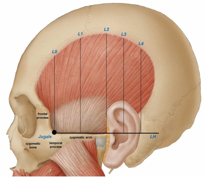
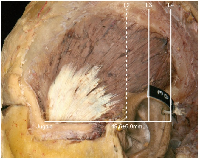
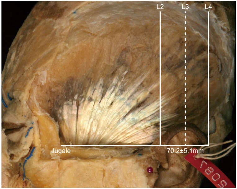
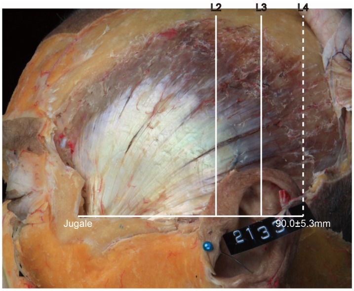
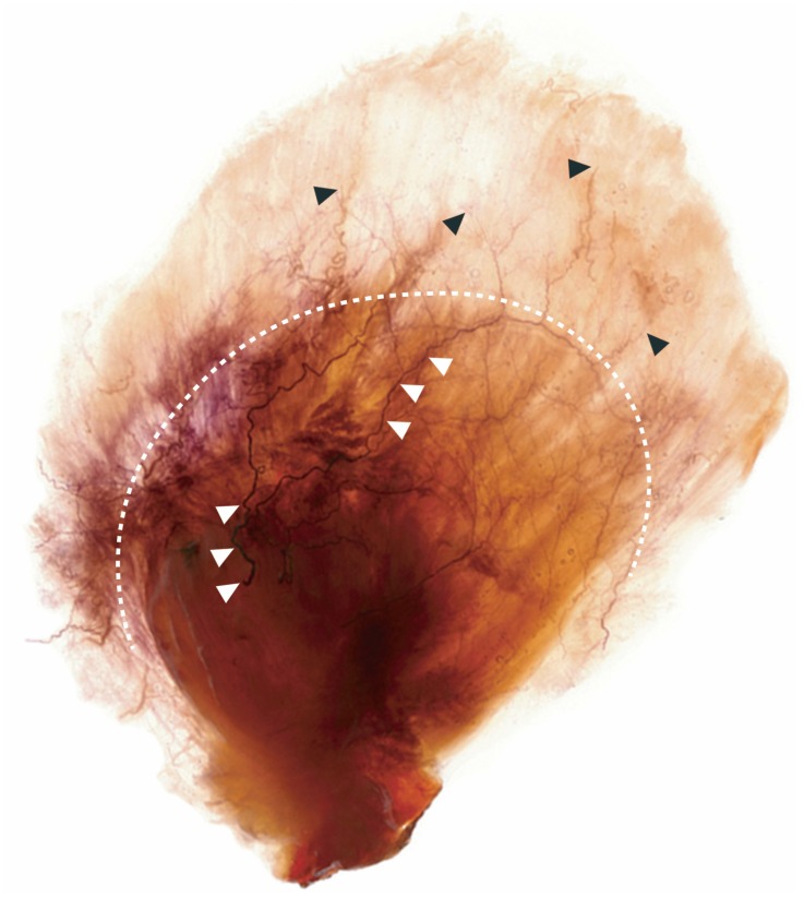
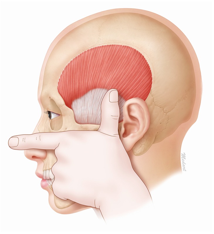
This study involved an extensive analysis of published research on the morphology of the temporalis muscle in order to provide an anatomical guideline on how to distinguish the temporalis muscle and temporalis tendon by observing the surface of the patient's face. Twenty-one hemifaces of cadavers were used in this study. The temporalis muscles were dissected clearly for morphological analysis between the temporalis muscle and tendon. The posterior border of the temporalis tendon was classified into three types: in Type I the posterior border of the temporalis tendon is located in front of reference line L2 (4.8%, 1/21), in Type II it is located between reference lines L2 and L3 (85.7%, 18/21), and in Type III it is located between reference lines L3 and L4 (9.5%, 2/21). The vertical distances between the horizontal line passing through the jugale (LH) and the temporalis tendon along each of reference lines L0, L1, L2, L3, and L4 were 29.7 ± 6.8 mm, 45.0 ± 8.8 mm, 37.7 ± 11.1 mm, 42.5 ± 7.5 mm, and 32.1 ± 0.4 mm, respectively. BoNT-A should be injected into the temporalis muscle at least 45 mm vertically above the zygomatic arch. This will ensure that the muscle region is targeted and so produce the greatest clinical effect with the minimum concentration of BoNT-A.

Keywords: Sihler staining; botulinum toxin type A; injection site; migraine; temporalis muscle.


Conflict of interest statement

The authors declare no conflict of interest.

Figures

Figure 1
Figure 1
Reference points and lines based on surface anatomical structures. Jugale: A landmark on the skull at which the temporal and frontal processes of the zygomatic bone meet. LH: The horizontal line passing through the jugale. L0: The vertical line passing through the jugale. L1: The line evenly dividing L0 and L2. L2: The vertical line passing through the anterior outer margin of the ear. L3: The line evenly dividing L2 and L4. L4: The vertical line passing through the posterior outer margin of the ear.
Figure 2
Figure 2
Type I, in which the posterior border of the temporalis tendon is located in front of L2 (4.8%, 1/21). The distance between the jugale and L2 was 49.6 ± 6.0 mm.
Figure 3
Figure 3
Type II, in which the posterior border of the temporalis tendon is located between L2 and L3 (85.7%, 18/21). The distance between the jugale and L3 was 70.2 ± 5.1 mm.
Figure 4
Figure 4
Type III, in which the posterior border of the temporalis tendon is located between L3 and L4 (9.5%, 2/21). The distance between the jugale and L4 was 90.0 ± 5.3 mm.
Figure 5
Figure 5
The temporalis muscle and tendon could be clearly distinguished after Sihler’s staining (white dotted line). The temporalis tendon appeared as a fan beginning at the point where it is inserted into the temporalis muscle. The temporalis muscle occupied the remaining area. The nerve trunk of the deep temporal nerve traversed the temporalis tendon (white arrows). The nerve endings of the deep temporal nerve mainly dispersed in the temporalis muscle (black arrows).
Figure 6
Figure 6
The position of the temporalis tendon can be easily determined first aligning the thumb and first finger in a flat stretched-out state and then placing the second finger on the inferior margin of the zygomatic arch. The tip of the thumb is then located approximately 45 mm from the superior margin of the zygomatic arch. However, since every clinician varies in thumb length, we recommend that a clinician use the above-mentioned method after taking into consideration the difference when comparing the point 45 mm from the patient’s zygomatic arch with that of the location of his/her own thumb.

References

    1. Carruthers J., Carruthers A. Botulinum toxin (Botox) chemodenervation for facial rejuvenation. Facial Plast Surg. Clin. North Am. 2001;9:197–204. - PubMed
    1. Göbel H., Heinze A., Heinze-Kuhn K., Jost W.H. Evidence-based medicine: Botulinum toxin A in migraine and tension type headache. J. Neurol. 2001;248:34–38. doi: 10.1007/PL00007818. - DOI - PubMed
    1. Heckmann M., Ceballos-Baumann A.O., Plewig G. Botulinum toxin A for axillary hyperhidrosis (excessive sweating) N. Engl. J. Med. 2001;344:488–493. doi: 10.1056/NEJM200102153440704. - DOI - PubMed
    1. Dodick V.W., Blumenfeld A., Silberstein S.D. Botulinum neurotoxin for the treatment of migraine and other primary headache disorders. Clin. Dermatol. 2004;22:76–81. doi: 10.1016/j.clindermatol.2003.12.031. - DOI - PubMed
    1. Chan V.W., McCabe E.J., MacGregor D.L. Botox treatment for migraine and chronic daily headache in adolescents. J. Neurosci. Nurs. 2009;41:235–243. doi: 10.1097/JNN.0b013e3181aaa98f. - DOI - PubMed

Publication types

Substances