

doi: 10.1038/nm.4193.
Epub 2016 Sep 12.
Fetal brain lesions after subcutaneous inoculation of Zika virus in a pregnant nonhuman primate
Affiliations
- PMID: 27618651
- PMCID: PMC5365281
- DOI: 10.1038/nm.4193
Item in Clipboard
Fetal brain lesions after subcutaneous inoculation of Zika virus in a pregnant nonhuman primate
Nat Med.
2016 Nov.
Abstract
We describe the development of fetal brain lesions after Zika virus (ZIKV) inoculation in a pregnant pigtail macaque. Periventricular lesions developed within 10 d and evolved asymmetrically in the occipital-parietal lobes. Fetal autopsy revealed ZIKV in the brain and significant cerebral white matter hypoplasia, periventricular white matter gliosis, and axonal and ependymal injury. Our observation of ZIKV-associated fetal brain lesions in a nonhuman primate provides a model for therapeutic evaluation.
Figures
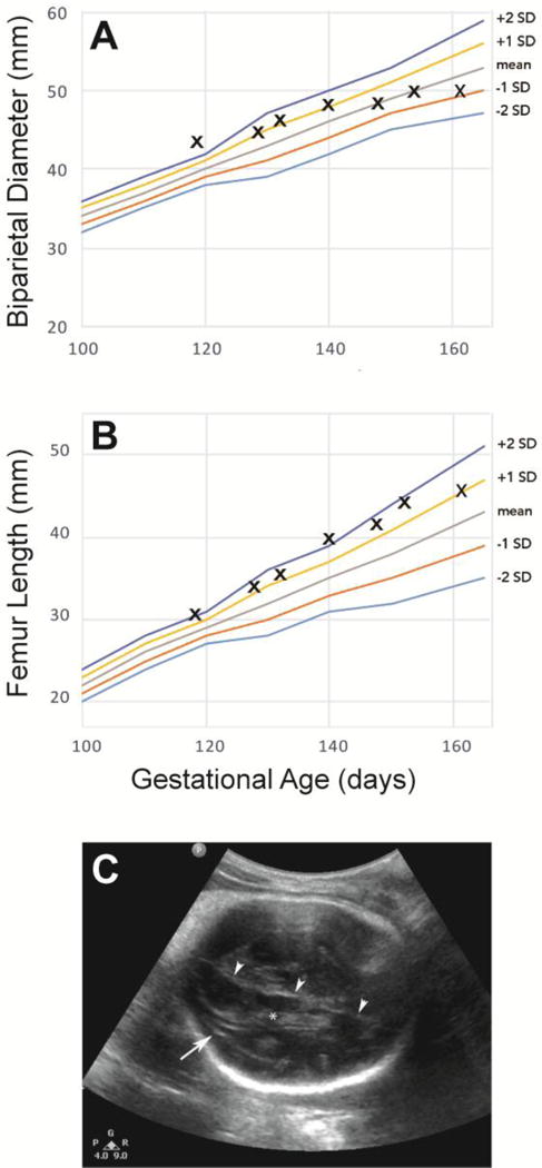
Fetal biometry demonstrates a lag in BPD growth (A) in contrast to continued growth of the femur length (B). An area of linear heterogeneity (arrow) developed in the fetal brain adjacent to the lateral ventricle in the posterior right brain (C). Note the choroid plexus in the lateral ventricle (*) and falx (arrowheads).
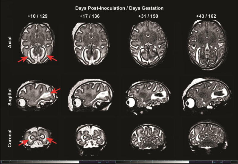
Serial MRI images of the fetal brain over time from 10 to 43 days post-inoculation (129 to 162 days gestation) demonstrate a bilateral periventricular T2 hyperintense foci that evolves over time with concomitant loss of volume in the posterior brain.
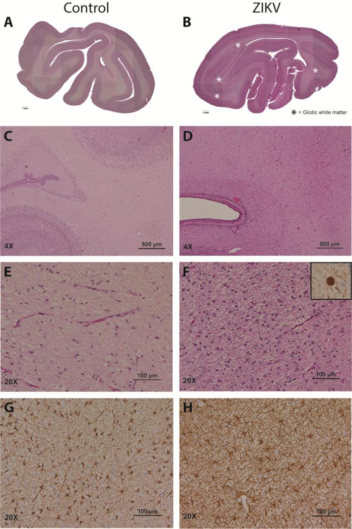
In contrast with sections from similar regions of a two-week younger control brain (A, C, E, G), the occipital cortex of the ZIKV-infected fetus (B, D, F, H) showed marked white matter hypoplasia and gliosis (pink bands indicated by asterisks). Gliosis in the deep white matter corresponded to abundant reactive astrocytes (D, F versus C, E) with increased GFAP immunoreactivity (H versus G). Axonal spheroids, a marker of severe axon injury, were seen only in the ZIKV fetus; the spheroids were confirmed to express neurofilament protein (F, inset).
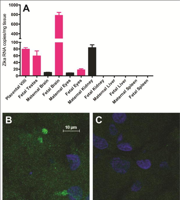
ZIKV RNA levels were measured by a Taqman qRT-PCR assay and calculated as mean viral copy number per mg of tissue using a standard curve (analyzed in triplicate; A). Error bars reflect standard deviation in technical replicates. Confocal fluorescent microscopy was performed on fetal brain sections stained with a primary antibody against the Zika virus E protein (B) and with secondary antibody alone (C). Nuclei were counterstained with DAPI (blue). All images are at the same scale.
References
-
- Gulland A. Zika virus is a global public health emergency, declares WHO. BMJ. 2016;352:i657. - PubMed
-
- Petersen LR, Jamieson DJ, Powers AM, Honein MA. Zika Virus. N Engl J Med. 2016;374:1552–1563. - PubMed
-
- Rasmussen SA, Jamieson DJ, Honein MA, Petersen LR. Zika Virus and Birth Defects - Reviewing the Evidence for Causality. N Engl J Med. 2016 - PubMed
Publication types
MeSH terms
Substances
Grants and funding
LinkOut - more resources
Full Text Sources
Other Literature Sources
Medical
Molecular Biology Databases
