. 2016 Sep 13;12(1):202.
doi: 10.1186/s12917-016-0809-2.

Development and evaluation of real-time loop-mediated isothermal amplification assay for rapid detection of cystic echinococcosis

Affiliations

Development and evaluation of real-time loop-mediated isothermal amplification assay for rapid detection of cystic echinococcosis

Mohamed E Ahmed et al. BMC Vet Res. .

Abstract

Background: Cystic echinococcosis (CE) or hydatidosis, caused by the larval stage of Echinococcus granulosus (EG)-complex, is a neglected parasitic disease of public health importance. The disease is endemic in many African and Mediterranean countries including the Sudan. The objective of the present study was to develop and evaluate a real-time loop-mediated isothermal amplification (LAMP) assay for simple and rapid detection of CE in humans and domestic live stock in Sudan.

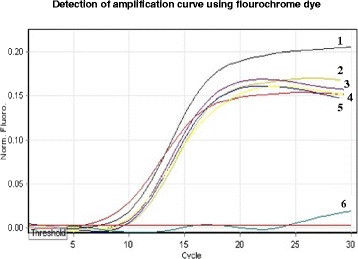
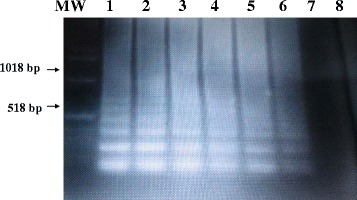
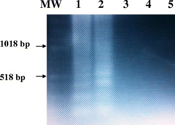
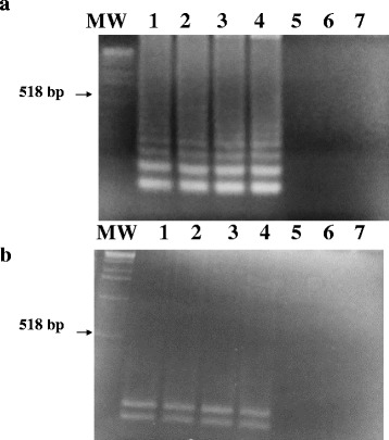
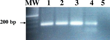
Methods: A set of six LAMP primers, designed from the mitochondrial NADH-1 gene of EG cattle strain of genotype 5 (G5), was used as a target for LAMP assay. The assay was performed at a constant temperature (63 °C), with a real-time follow-up using a LightCycler and fluorochrome dye. Following amplification cycles in a simple water bath, LAMP products were observed for color change by naked eye and were visualized under UV light source using agarose gel electrophoresis.

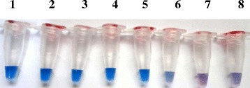
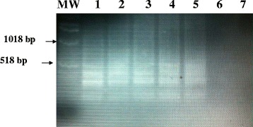
Results: The real-time LAMP assay identified a variety of hydatid cysts strains recovered in the Sudan, including Echinococcus canadenses (G6) and Echinococcus ortleppi (G5). Real-time LAMP positive results were detected by the presence of an amplification curve, whereas negative results were indicated by absence of fluorescence detection. Positive LAMP results appeared as a bluish-colored reaction as observed by naked eye, whereas negative LAMP results were observed as purple-colored reaction. The sensitivity studies indicated that the LAMP assay detected as little as a 10 fg of parasite DNA. There was 100 % agreement between results of the LAMP assay and our previously described nested PCR when testing 10-fold serial dilution of DNA extracted from EG-complex hydatid cyst. However, there was no cross-reactivity with other parasites including cysticercus bovis, Fasciola gigantica, and Schistosoma bovis and nucleic acid free samples.

Conclusion: The developed LAMP assay would be expected to prove highly significant in epidemiological surveys of CE in developing countries or areas of resource-poor settings for both ease of use and cost.

Keywords: Cystic echinococcosis; Echinococcus granulosus-complex; Hydatid cysts; LAMP; Sudan.


Figures

Fig. 1
Fig. 1
Detection by the naked eye of color change using serial dilutions of known concentration of E. ortleppi DNA recovered from a dromedary camel in Sudan. Blue color indicates positive LAMP result whereas purple color indicates negative LAMP result. Tube 1–8: 10-fold serial dilutions of 1.0 ng,100 pg, 10 pg, 1 pg, 100 fg, 10 fg, 1.0 fg, and DNA-free sample (negative control), respectively
Fig. 2
Fig. 2
Real-time monitoring of LAMP assay using lightCycler and a fluorochrome dye. The detection of amplification curves using 1.0 pg DNA from hydatid cysts strains recovered from different animal species. Curve 1: hydatid cyst of cattle origin; curve 2–4: hydatid cyst of camel origin; curve 5: Hydatid cyst of human origin; curve 6: negative control
Fig. 3
Fig. 3
Sensitivities of the LAMP assay for detection of EG-complex hydatid cyst using ethidium bromide-stained agarose gel electrophoresis. The LAMP assay was performed with serial dilutions of known concentration of E. ortleppi DNA recovered from a dromedary camel in Sudan.. Lane MW: molecular weight marker; Lane 1–7: 10-fold serial dilutions of 100 pg, 10 pg, 1 pg, 100 fg, 10 fg, 1.0 fg, of parasite DNA, respectively. Lane 8: nucleic acid-free sample (negative control)
Fig. 4
Fig. 4
Visualization of Lamp products from fresh and archived hydatid cyst samples onto 2 % agarose gel using simple water bath. Lanes MW: Molecular marker; Lane 1: fresh sample of hydatid cyst of cattle origin; Lane 2: fresh sample of hydatid cyst camel origin; Lane 3–4: archived sample of hydatid cyst of camel origin; Lane 5–6: archived sample of hydatid cyst of human origin; Lane 7: nucleic acid-free water
Fig. 5
Fig. 5
Specificity of the LAMP primers for the detection of EG-complex using E. ortleppi DNA recovered from hydatid cyst of a dromedary camel in the Sudan and analyzed in a 2 % agarose gel. Lanes MW: Molecular marker; Lane 1: 1.0 pg E. ortleppi (G5) DNA (positive control); Lane 2: 1.0 pg E. canadensis (G6) DNA (positive control); Lane 3: cysticercus bovis: Lane 4: Fasciola gigantic; Lane 5: Schistosoma bovis
Fig. 6
Fig. 6
Restriction enzyme digestion of the LAMP products from hydatid cyst strains. a Visualization of the LAMP products from hydatid cyst strains. Lane MW: molecular weight marker; lanes 1and 2: 1.0 pg DNA from E. ortleppi (G5) DNA; Lane3 and 4: 1.0 pg DNA from E. canadensis (G6) DNA: Lane 5: cysticercus bovis: Lane 6: Fasciola gigantica; Lane 7: Schistosoma bovis. b Visualization of the restriction patterns of the digested LAMP products using Eco R1 restriction enzyme for the above gel
Fig. 7
Fig. 7
Specificity of the LAMP outer primers (F3 and B3) for amplification of the Sudanese strains of EG-complex using conventional PCR. Visualization of the 200-bp specific DNA PCR products on ethidium bromide-stained agarose gels. Lane MW: molecular weight marker; lanes 1–2: 1.0 pg E.ortleppi (G5) DNA (positive control); Lane 3–4: 1.0 pg E.canadensis (G6) DNA; Lane 5: nucleic acid-free water

References

    1. Ahmed ME, Eltom KH, Musa NO, Ali IA, Elamin FM, Grobusch MP, Aradaib IE. First report on circulation of Echinococcus ortleppi in the one humped camel (Camelus dromedaries), Sudan. BMC Vet Res. 2013;9:127. doi: 10.1186/1746-6148-9-127. - DOI - PMC - PubMed
    1. Wahlers K, Menezes CN, Wong ML, Zeyhle E, Ahmed ME, Ocaido M, Stijnis C, Romig T, Kern P, Grobusch MP. Cystic echinococcosis in sub-Saharan Africa. Lancet Infect Dis. 2012;12:871–80. doi: 10.1016/S1473-3099(12)70155-X. - DOI - PubMed
    1. Abdel Malek E. Check-list of helminth parasites of domestic animals in the Sudan. Indian Vet J. 1959;36:28–286.
    1. Eisa AM, Mustfa AA, Soliman KN. Preliminary report on cysticercosis and hydatidosis in the Southern Sudan. Sud J Vet Sc Anim Husb. 1962;3:97–102.
    1. Eisa AM, El Khawat SE, Slepnev NK. Proceedings of the 8th Veterinary Conference. Khartoum: Livestock and animal production in the Sudan; 1977. A survey of parasites of dogs in Khartoum province.