. 2016 Sep 13;14(1):21.
doi: 10.1186/s12964-016-0144-z.

Vasodilator-Stimulated Phosphoprotein (VASP)-dependent and -independent pathways regulate thrombin-induced activation of Rap1b in platelets

Affiliations

Vasodilator-Stimulated Phosphoprotein (VASP)-dependent and -independent pathways regulate thrombin-induced activation of Rap1b in platelets

Peter M Benz et al. Cell Commun Signal. .

Abstract

Background: Vasodilator-Stimulated Phosphoprotein (VASP) is involved in the inhibition of agonist-induced platelet aggregation by cyclic nucleotides and the adhesion of platelets to the vascular wall. αIIbβ3 is the main integrin responsible for platelet activation and Rap1b plays a key role in integrin signalling. We investigated whether VASP is involved in the regulation of Rap1b in platelets since VASP-null platelets exhibit augmented adhesion to endothelial cells in vivo.

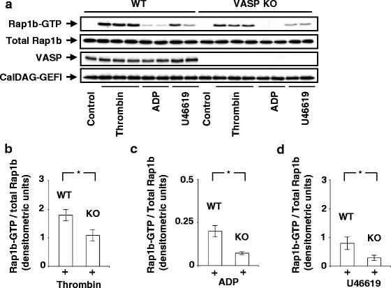
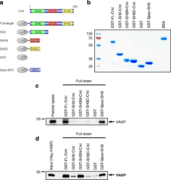
Methods: Washed platelets from wild type and VASP-deficient mice were stimulated with thrombin, the purinergic receptors agonist ADP, or the thromboxane A2 receptor agonist U46619 and Rap1b activation was measured using the GST-RalGDS-RBD binding assay. Interaction of VASP and Crkl was investigated by co-immunoprecipitation, confocal microscopy, and pull-down assays using Crkl domains expressed as GST-fusion proteins.

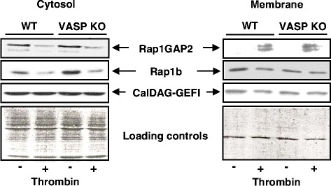
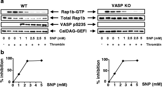
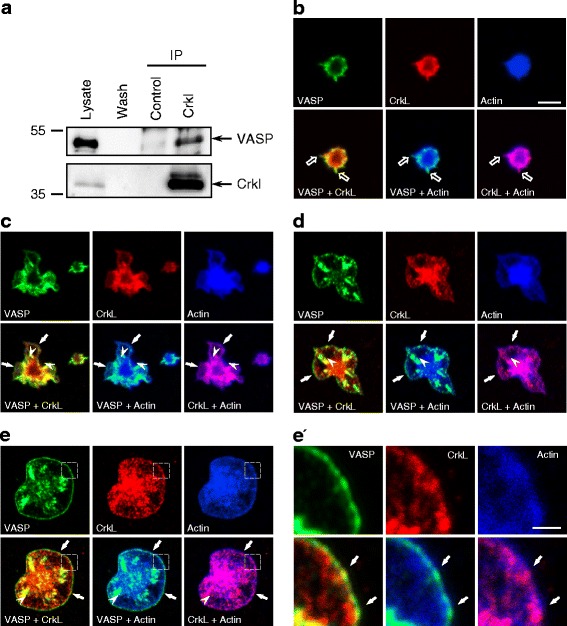
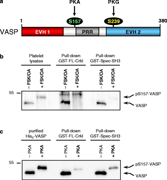
Results: Surprisingly, we found that activation of Rap1b in response to thrombin, ADP, or U46619 was significantly reduced in platelets from VASP-null mice compared to platelets from wild type mice. However, inhibition of thrombin-induced activation of Rap1b by nitric oxide (NO) was similar in platelets from wild type and VASP-null mice indicating that the NO/cGMP/PKG pathway controls inhibition of Rap1b independently from VASP. To understand how VASP regulated Rap1b, we investigated association between VASP and the Crk-like protein (Crkl), an adapter protein which activates the Rap1b guanine nucleotide exchange factor C3G. We demonstrated the formation of a Crkl/VASP complex by showing that: 1) Crkl co-immunoprecipitated VASP from platelet lysates; 2) Crkl and VASP dynamically co-localized at actin-rich protrusions reminiscent of focal adhesions, filopodia, and lamellipodia upon platelet spreading on fibronectin; 3) recombinant VASP bound directly to the N-terminal SH3 domain of Crkl; 4) Protein Kinase A (PKA) -mediated VASP phosphorylation on Ser157 abrogated the binding of Crkl.

Conclusions: We identified Crkl as a novel protein interacting with VASP in platelets. We propose that the C3G/Crkl/VASP complex plays a role in the regulation of Rap1b and this explains, at least in part, the reduced agonist-induced activation of Rap1b in VASP-null platelets. In addition, the fact that PKA-dependent VASP phosphorylation abrogated its interaction with Crkl may provide, at least in part, a rationale for the PKA-dependent inhibition of Rap1b and platelet aggregation.

Keywords: Crkl; Platelets; Rap1b; VASP; cAMP; cGMP.


Figures

Fig. 1
Fig. 1
Agonist-induced Rap1b activation is reduced in VASP-null platelets. a Platelets (2 x10⁸) from wild type (WT) or VASP-null platelets (VASP KO) were unstimulated (control) or stimulated with thrombin (0.01 U/ml, 30s), ADP (10 μM, 1 min), or U46619 (1 μM, 1 min) (non-aggregating conditions). Thereafter, platelets were lysed and GST-RalGDS-RBD pull-down assays were performed as described in Methods. Proteins bound to GST-RalGDS-RBD were separated by 12 % SDS-PAGE, transferred to PVDF membranes, which were subjected to immunoblotting with anti-Rap1b Abs. The insets show representative Western Blots. Levels of Rap1b, VASP, or CalDAG-GEFI in the whole lysates used for the Rap1b pull-down assays were measured by Western blot analysis using appropriate Abs. The diagrams (b-d) illustrate densitometric analysis of the relative activities of Rap1b. Levels of Rap1b-GTP/total Rap1b in WT and VASP KO platelets stimulated with thrombin (b) (n = 5), ADP (c) (n = 3), or U46619 (d) (n = 3) were quantified by densitometry analysis using ImageJ software. *P < 0.05
Fig. 2
Fig. 2
Translocation of CalDAG-GEFI and Rap1GAP2 is independent of VASP. Platelets from wild-type (WT) or VASP-deficient mice (VASP KO) stimulated or not with thrombin (0.01 U/ml, 30s) were broken by sonication in detergent-free buffer. Lysates were spun at 100,000 g for 1 h. Cytosolic (supernatant) and crude membrane fractions (pellet) were collected and re-suspended in Laemmli buffer. Proteins were separated by 12 % SDS-PAGE, transferred to PVDF membranes, which were subjected to immunoblotting with one of the following Abs: anti-Rap1GAP2, anti-Rap1b or anti-CalDAG-GEFI. The Western blots show the results of one representative experiment (out of three). Protein loading controls (Ponceau S red staining of the blots) are shown in the bottom panels
Fig. 3
Fig. 3
NO-dependent inhibition of Rap1b activation is not impaired in VASP-null platelets. a Platelets (2 × 10⁸) from WT mice (left panel) or VASP KO mice (right panel) were pretreated for 2 min with SNP (1–5 μM) and then stimulated with thrombin (0.01 U/ml, 30s). Cells were lysed and the levels of Rap1b-GTP/total Rap1b were measured as described in the legend to Fig. 1. PKG-mediated phosphorylation of VASP at serine 235 (pS235-VASP) and levels of CalDAG-GEFI in whole lysates were measured using VASP phospho-specific or anti-CalDAG-GEFI Abs, respectively. The Western blots show the results of one representative experiment (out of three). b Quantification of the percent inhibition by SNP (1–5 μM) of thrombin-induced activation of Rap1b in both WT and VASP KO platelets. Please note that the blot in panel A (right) was more exposed than the blot in panel A (left)
Fig. 4
Fig. 4
Crkl and VASP dynamically interact in human platelets. (a) Lysates of human platelets were immunoprecipitated (IP) with Abs against Crkl or isotype control antibodys (control) as described in Methods. Lysate, wash fraction (Wash), and precipitated material were analyzed by Western blotting with anti-VASP (upper panel) or anti-Crkl Abs (lower panel), respectively. The arrows on the right hand side indicate the position of VASP and Crkl. One representative experiment (out of three) is shown. B-E, Washed human platelets were seeded onto fibronectin-coated glass slides for 2 min (b), 5 min (c), 12 min (d), or 30 min (e) before fixation and staining with anti-VASP-specific Abs (green), anti-Crkl-specific Abs (red) or with fluorescent conjugated phalloidin to visualize actin fibers (blue). The top panels in each section show individual stainings; lower panels show co-localization staining of VASP and Crkl, VASP and actin, and Crkl and actin. Black arrows, white arrowheads, and white arrows indicate filopodia-, focal adhesion-, and lamellipodia-like membrane protrusions, respectively. All images have a dimension of 20 × 20 μm to better visualize platelet spreading, scale bar in B is 5 μm. In , magnified views of the indicated areas in e are shown; scale bar 1 μm
Fig. 5
Fig. 5
The N-terminal SH3 domain of Crkl interacts directly with VASP. a schematic diagramm of the Crkl domain organization and the different GST-Crkl and GST-Spec-SH3 fusion proteins used in this study. b Coomassie blue-stained gel of the purified GST-fusion proteins depicted in (a). BSA (4 μg) was loaded onto the same gel to allow relative quantification of the purified proteins. c, d GST pull-down assay with lysates of human platelets (c) or recombinant, purified His6-VASP (d). Equal amounts of platelet lysate (c) or recombinant VASP (d) were incubated with equimolar amounts of the depicted, immobilized GST fusion proteins or GST alone. After extensive washing, precipitated material was analyzed by Western blotting with anti-VASP specific Abs. The position of VASP is indicated on the right hand side by an arrow. The Western blots show the results of one representative experiment (out of five)
Fig. 6
Fig. 6
PKA-mediated VASP phosphorylation abrogates Crkl-VASP interaction. a Domain organization and phosphorylation sites of human VASP (380 aa); EVH1/2 Ena/VASP homology 1/2 domain; PRR, proline-rich region. VASP is a prominent substrate of cyclic nucleotide-dependent serine/threonine kinases. Human VASP is preferentially phosphorylated by PKA at serine 157 (S157, green) and by PKG at serine 239 (S239, yellow). Please note that S157 is located in close proximity to the PRR, which is important for SH3-domain mediated interactions. b Human platelets were stimulated or not with a combination of FSK and OA to induce PKA-mediated VASP phosphorylation at Ser157 (pS157-VASP). Equal amounts of lysates from unstimulated or stimulated platelets were incubated with immobilized GST-FL-Crkl or GST-Spec-SH3 and interaction of VASP with the GST-fusion proteins was determined as described in the legend to Fig. 5c and d. c. Equal amounts of purified His6-tagged VASP were S157-phosphorylated in vitro by PKA (+PKA) or left untreated (−PKA) and pulled-down with immobilized GST-FL-Crkl or GST-Spec-SH3. Interaction of VASP wih the GST-fusion proteins was determined as described in the legend to Fig. 5c and d. Please note that VASP Ser157-phosphorylation (but not S239-phosphorylation) induces a shift in the apparent molecular weight from 46 to 50 kDa

References

    1. Ruggeri ZM. Platelets in atherothrombosis. Nature Med. 2002;11:1227–34. doi: 10.1038/nm1102-1227. - DOI - PubMed
    1. Bertoni A, Tadokoro S, Eto K, Pampori N, Parise LY, White GC, Shattil SJ. Relationships between Rap1b, affinity modulation of integrin αIIbβ3, and the actin cytoskeleton. J Biol Chem. 2002;277:25715–21. doi: 10.1074/jbc.M202791200. - DOI - PubMed
    1. Franke B, van Triest M, De Bruijn KMT, van Willigen G, Nieuwenhuis HK, Negrier C, Akkerman J-W, Bos JL. Sequential regulation of the small GTPase Rap1 in human platelets. Mol Cell Biol. 2000;20:779–85. doi: 10.1128/MCB.20.3.779-785.2000. - DOI - PMC - PubMed
    1. Chrzanowska-Wodnicka M, Smyth SS, Schoenwaelder SM, Fischer TH, White GC. Rap1b is required for normal platelet function and hemostasis in mice. J Clin Invest. 2005;115:680–7. doi: 10.1172/JCI22973. - DOI - PMC - PubMed
    1. Stefanini L, Roden RC, Bergmeier W. CalDAG-GEFI is at the nexus of calcium-dependent platelet activation. Blood. 2010;114:2506–14. doi: 10.1182/blood-2009-04-218768. - DOI - PubMed

Publication types

MeSH terms