. 2016 Aug 29:11:4141-55.
doi: 10.2147/IJN.S109494. eCollection 2016.

Multicomponent, peptide-targeted glycol chitosan nanoparticles containing ferrimagnetic iron oxide nanocubes for bladder cancer multimodal imaging

Affiliations

Multicomponent, peptide-targeted glycol chitosan nanoparticles containing ferrimagnetic iron oxide nanocubes for bladder cancer multimodal imaging

Jaehong Key et al. Int J Nanomedicine. .

Abstract

While current imaging modalities, such as magnetic resonance imaging (MRI), computed tomography, and positron emission tomography, play an important role in detecting tumors in the body, no single-modality imaging possesses all the functions needed for a complete diagnostic imaging, such as spatial resolution, signal sensitivity, and tissue penetration depth. For this reason, multimodal imaging strategies have become promising tools for advanced biomedical research and cancer diagnostics and therapeutics. In designing multimodal nanoparticles, the physicochemical properties of the nanoparticles should be engineered so that they successfully accumulate at the tumor site and minimize nonspecific uptake by other organs. Finely altering the nano-scale properties can dramatically change the biodistribution and tumor accumulation of nanoparticles in the body. In this study, we engineered multimodal nanoparticles for both MRI, by using ferrimagnetic nanocubes (NCs), and near infrared fluorescence imaging, by using cyanine 5.5 fluorescence molecules. We changed the physicochemical properties of glycol chitosan nanoparticles by conjugating bladder cancer-targeting peptides and loading many ferrimagnetic iron oxide NCs per glycol chitosan nanoparticle to improve MRI contrast. The 22 nm ferrimagnetic NCs were stabilized in physiological conditions by encapsulating them within modified chitosan nanoparticles. The multimodal nanoparticles were compared with in vivo MRI and near infrared fluorescent systems. We demonstrated significant and important changes in the biodistribution and tumor accumulation of nanoparticles with different physicochemical properties. Finally, we demonstrated that multimodal nanoparticles specifically visualize small tumors and show minimal accumulation in other organs. This work reveals the importance of finely modulating physicochemical properties in designing multimodal nanoparticles for bladder cancer imaging.

Keywords: MRI; NIRF; bladder cancer; chitosan; iron oxide; multimodal imaging.


Figures

Figure 1
Figure 1
Chemical structures of glycol chitosan conjugated to hydrophobic 5β-cholanic acid, peptide (CSNRDARRC), and iron oxide NCs (A) and schematic diagram of pMCNPs containing iron oxide NCs and vinblastine (B). Notes: Glycol chitosan was modified with hydrophobic 5β-cholanic acid and a bladder cancer-targeting peptide, CSNRDARRC. The deacetylated free amine groups of glycol chitosan were conjugated with 5β-cholanic acids and the peptide. For NIRF imaging, Cy5.5 dyes were also conjugated to the free amines on glycol chitosan. Cy5.5 was chemically conjugated to the glycol chitosan (4:1 mole ratio, Cy5.5:Glycol chitosan). The oleic acid capped NCs (22 nm) were physically loaded with a probe-type sonicator and stabilized inside the glycol chitosan nanoparticles by hydrophobic interactions. Vinblastine was encapsulated by solvent evaporation. The schematic image indicates the surface conjugation of glycol chitosan with Cy5.5 and the peptide and the interaction of the core NCs and vinblastine with 5β-cholanic acid. Abbreviations: NCs, nanocubes; NIRF, near infrared fluorescent; pMCNPs, pCNPs loaded with 22 nm iron oxide NCs.
Figure 2
Figure 2
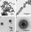
TEM images of nanocubes and pMCNPs. Notes: (A) TEM image of NCs; (B) NCs had a uniform cubic shape and were 22 nm in diameter; (C) TEM image of pMCNPs; (D) a single pMCNP had several NCs in the core. Abbreviations: TEM, transmission electron microscopy; NCs, nanocubes; pMCNPs, pCNPs loaded with 22 nm iron oxide NCs.
Figure 3
Figure 3
The hydrodynamic diameter of chitosan nanoparticles was measured in each step of conjugation and loading. Notes: CNPs had a diameter of 288.7 nm and polydispersity index of 0.2. The zeta-potential of CNPs was 9.7 mV. After conjugation to the bladder cancer-targeting peptide, CSNRDARRC (pCNPs), the diameter of pCNPs increased to 359.7 nm while the polydispersity and zeta-potential were similar. When we loaded the particles with nanocubes, the diameter increased to 481.8 nm (pMCNPs). The polydispersity index was similar, but the zeta-potential increased by more than three times (A). The size distribution of each chitosan particle was measured by DLS (B). Abbreviations: CNPs, chitosan nanoparticles; NCs, nanocubes; pCNPs, peptide-conjugated chitosan nanoparticles; pMCNPs, pCNPs loaded with 22 nm iron oxide NCs; SD, standard deviation.
Figure 4
Figure 4
We injected CNP and pCNP into athymic mice (four mice per group). Notes: We evaluated the biodistribution of nanoparticles at 24 and 48 h postinjection. At 24 and 48 h, CNPs showed high accumulation in the tumor and kidneys. At 48 h, higher nanoparticle accumulation was observed in kidneys and tumor (A). While we did not assess longer time points, we demonstrated that CNPs have a circulation time over 72 h and the signals decreased at 88 h. pCNP resulted in a higher NIRF signal than CNPs particularly in the liver, kidneys, and tumor at both 24 and 48 h. Similarly, the NIRF signal was higher at 48 than at 24 h indicating continuous accumulation and circulation (B). In comparing tumor accumulation at 24 and 48 h, pCNPs had more than twice the accumulation of CNPs. The difference was more significant at 48 h (P-value =2.24e−5) (C). Abbreviations: CNPs, chitosan nanoparticles; pCNPs, peptide-conjugated chitosan nanoparticles; NIRF, near infrared fluorescence.
Figure 5
Figure 5
pMCNPs were systemically injected to athymic mice through the tail-vein and in vivo and ex vivo NIRF imaging demonstrated tumor accumulation and bio-distribution of pMCNPs. Notes: At 24 h postinjection, we performed in vivo whole body and ex vivo NIRF imaging of major organs, including tumors, by IVIS. All images were normalized. In the whole body image, pMCNPs predominantly accumulated at the tumor site (A). The accumulation was also validated by ex vivo imaging of the organs and tumor. The nanoparticles specifically accumulated in even small solid tumors of 5 mm in diameter. The black dotted lines indicate the actual edge of the solid tumors. Without particle injection, we did not find high NIRF signals (B). pMCNP accumulation in the tumor clearly differed in liver, spleen, kidneys (*P<0.03), lung and heart (**P<0.0001). (C). Abbreviations: CNPs, chitosan nanoparticles; IVIS, in vivo imaging system; NIRF, near infrared fluorescence; NPs, nanoparticles; H, high; L, low; pMCNPs, pCNPs loaded with 22 nm iron oxide NCs.
Figure 6
Figure 6
pMCNPs were evaluated for NIRF imaging. Nanoparticle solutions were serially diluted in a 96-well plate. Notes: NCs were well dispersed in chloroform and stable in water after they were loaded into pMCNPs (A). The pMCNPs emitted a NIRF signal from Cy5.5 on the nanoparticles (B). pMCNPs were placed in a 96-well plate at serial two fold dilutions. NIRF images were observed with an IVIS NIRF machine. An increased number of particles resulted in brighter NIRF intensity (C). NIRF intensity was also measured as a function of the number of Fe ions in the pMCNP solution. A lower pMCNP concentration had a weaker NIRF intensity (D). Abbreviations: CNPs, chitosan nanoparticles; IVIS, in vivo imaging system; NCs, nanocubes; NIRF, near infrared fluorescence; pMCNPs, pCNPs loaded with 22 nm iron oxide NCs; H, high; L, low.
Figure 7
Figure 7
pMCNPs were evaluated for 3T GE MRI and ICP-MS. pMCNPs solutions were serially diluted in a 96-well plate and imaged by GE MRI. Notes: A lower number of pMCNPs had a brighter MR signal, which clearly demonstrated that T2 contrast effects depended on the pMCNP concentration (A). The total amount of Fe in pMCNPs directly influenced the MR signal (B). At longer echo times the hydrogen atoms were dephased by the particles faster, indicating the T2 contrast effects of pMCNPs (C). The r2 value (180.7 mM−1s−1) of pMCNP was calculated by ICP-MS and 3T GE MRI. The r2 value was higher than that of commercialized products, such as Feridex (120 mM−1s−1) (D). Abbreviations: CNPs, chitosan nanoparticles; ICP-MS, inductively coupled plasma mass spectrometry; NCs, nanocubes; pMCNPs, peptide-conjugated chitosan nanoparticles; MR, magnetic resonance; MRI, magnetic resonance imaging; TEs, echo times; GE, General Electric.
Figure 8
Figure 8
pMCNPs were incubated with K9TCC cells for different times. Notes: The cells were washed three times and imaged by confocal microscopy at different z-depths. XY, YZ, and XZ show the pMCNPs distribution in K9TCC cells (A). Internalization of pMCNPs in K9TCC cells was captured along multiple z-planes at every 1 µm after 4 h of incubation (B). Different incubation times (0, 0.5, 2, and 4 h) changed the NP internalization by K9TCC cells (C). The scale bar is 10 µm. Abbreviations: NCs, nanocubes; NP, nanoparticle; pMCNPs, pCNPs loaded with 22 nm iron oxide NCs.
Figure 9
Figure 9
The athymic mice were systemically injected with pMCNPs and imaged with 3T MRI. Notes: Axial view of a mouse before pMCNP administration (A). Axial view of a mouse at 24 hours after pMCNP administration (B). The knee coil was used for whole body imaging with the following conditions: TR =2,000 ms, TE =82 ms, 3D acquisition, and 512×512. The red dotted line 1 and 2 indicated the edge of tumor before/after pMCNP administration respectively. Yellow arrows indicate negative contrast effects of pMCNPs. The contrast intensity at the tumor sites was quantified with ImageJ. The tumors with pMCNPs had an average 67% lower signal, 37% darker minimum intensity, and 57% darker intensity per pixel (C). Abbreviations: MRI, magnetic resonance imaging; NCs, nanocubes; NP, nanoparticle; pMCNPs, pCNPs loaded with 22 nm iron oxide NCs.

References

    1. Xia W, Low PS. Folate-targeted therapies for cancer. J Med Chem. 2010;53(19):66–76. - PubMed
    1. Park J, Saravanakumar G, Kim K, Kwon I. Targeted delivery of low molecular drugs using chitosan and its derivatives. Adv Drug Deliv Rev. 2010;62(1):28–41. - PubMed
    1. Saravanakumar G, Min KH, Min DS, et al. Hydrotropic oligomer-conjugated glycol chitosan as a carrier of paclitaxel: synthesis, characterization, and in vivo biodistribution. J Control Release. 2009;140(3):210–217. - PubMed
    1. Min KH, Park K, Kim YS, et al. Hydrophobically modified glycol chitosan nanoparticles-encapsulated camptothecin enhance the drug stability and tumor targeting in cancer therapy. J Control Release. 2008;127(3):208–218. - PubMed
    1. Maeda H, Matsumura Y. EPR effects. Adv Drug Deliv Rev. 2011;63(3):161–169. - PubMed

MeSH terms