

Editorial
doi: 10.21037/jtd.2016.07.40.
Rare airway tumors: an update on current diagnostic and management strategies
Affiliations
- PMID: 27621844
- PMCID: PMC4999752
- DOI: 10.21037/jtd.2016.07.40
Item in Clipboard
Editorial
Rare airway tumors: an update on current diagnostic and management strategies
J Thorac Dis.
2016 Aug.
No abstract available
Conflict of interest statement
KH is a consultant for cook medical. The other authors have no conflicts of interest to declare.
Figures
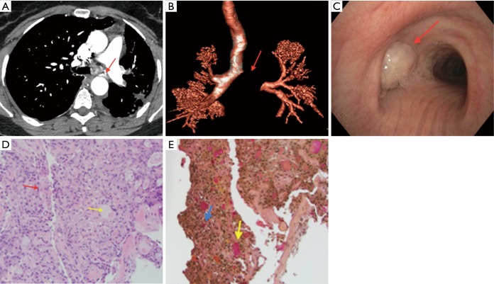
Mucoepidermoid carcinoma. (A) axial chest CT image showing the complete obstruction of the left main stem bronchus. The obstructive lesion is highly vascularized with a bronchial artery branch originating from the descending aorta (arrow); (B) chest CT three-dimensional reconstruction showing the complete obstruction of the left main stem (arrow); (C) bronchoscopic image showing the complete obstruction of the left main stem by a large tumor (arrow); (D) microscopic image of biopsy showing mucoepidermoid carcinoma. The image shows neoplastic tissue composed of round to oval epithelioid cells and occasional goblet cells (red arrow) punctuated by mucin containing cystic spaces (yellow arrow). Hematoxylin and Eosin stain (200×); (E) microscopic image showing mucicarmine staining of mucoepidermoid carcinoma. The image shows intracellular (blue arrow) and extracellular mucin (yellow arrow). Mucicarmine stain (200×).
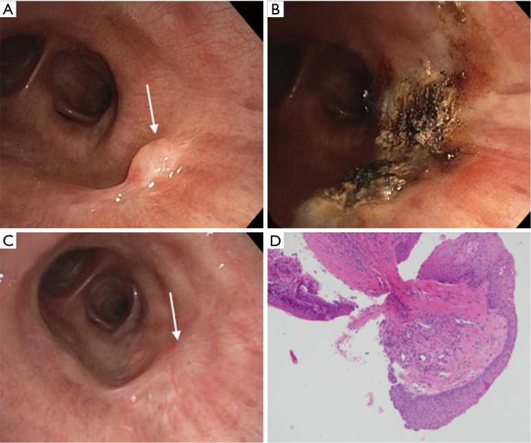
Endobronchial papilloma. (A) bronchoscopic view showing a small a polypoid endobronchial lesion (arrow) in the lateral wall of the bronchus intermedius; (B) ablation using argon plasma coagulation of the endobronchial lesion in the bronchus intermedius; (C) a six-month follow up showing endobronchial minimal scaring and no evidence of recurrence; (D) microscopic image of a papilloma. The image shows tissue fragments composed of fibrovascular core lined by benign squamous epithelium. Hematoxylin and Eosin stain (100×).
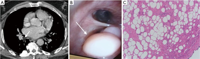
Endobronchial lipoma. (A) axial chest CT showing an endobronchial abnormality in the superior segment of the right lower lobe; (B) bronchoscopic image of the large endobronchial soft polypoid lesion originating from the superior segment of the right lower lobe; (C) microscopic image of an airway lipoma. The image shows airway epithelium on the right lower corner and submucosal tissue replaced by benign adipocytes. Hematoxylin and Eosin stain (200×).
References
Publication types
LinkOut - more resources
Full Text Sources
Other Literature Sources