

Giant thymoma successfully resected via hemiclamshell thoracotomy: a case report
- PMID: 27621898
- PMCID: PMC4999736
- DOI: 10.21037/jtd.2016.06.41
Giant thymoma successfully resected via hemiclamshell thoracotomy: a case report
Abstract
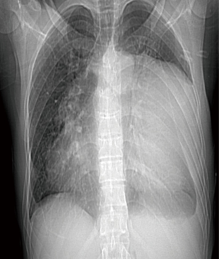
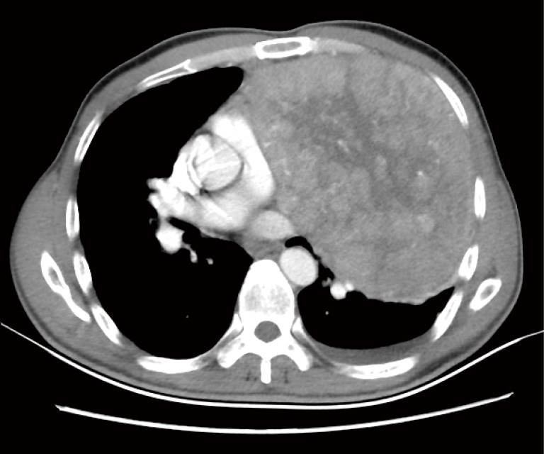
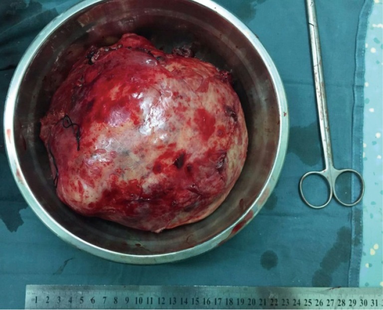
Thymoma is an epithelial neoplasm of the thymus. It commonly lies in the anterior mediastinum and represents 20-30% of mediastinal tumours in adults. In this report we present a case of giant thymoma locating in the anterior-inferior mediastinum. A 46-year-old male came to our institute with slight pectoralgia and dyspnea. Chest CT shows a giant tumor measuring 19 cm × 16 cm × 15 cm in the left thoracic cavity. After careful examination, we performed surgery. At surgery, we found the tumor was adherent to left upper lobe of the lung, mediastinal pleura, and parietal pleural. The tumor was completely resected with combined resection of part left upper lobe of lung. The weight of the tumor was 2,135 g. Pathological diagnosis indicated a type AB thymoma according to the World Health Organization classification and a diagnosis of Masaoka stage IIB was made.
Keywords: Giant thymoma; ectopic thymoma; hemiclamshell.
Conflict of interest statement
The authors have no conflicts of interest to declare.
Figures

Similar articles
-
Ectopic thymoma presenting as a giant intrathoracic mass: a case report.J Cardiothorac Surg. 2012 Jul 16;7:68. doi: 10.1186/1749-8090-7-68. J Cardiothorac Surg. 2012. PMID: 22800398 Free PMC article.
-
Giant thymoma presenting as a large bilateral intrathoracic mass: A case report and a comparison between median sternotomy and hemiclamshell approach.Ann Med Surg (Lond). 2021 Sep 13;70:102859. doi: 10.1016/j.amsu.2021.102859. eCollection 2021 Oct. Ann Med Surg (Lond). 2021. PMID: 34584686 Free PMC article.
-
Ectopic thymoma presenting as a large intrathoracic mass.J Cancer Res Ther. 2015 Oct-Dec;11(4):1033. doi: 10.4103/0973-1482.147388. J Cancer Res Ther. 2015. PMID: 26881626
-
[Thymoma with Hemorrhage and Necrosis Presenting with Fever and Chest Pain;Report of a Case].Kyobu Geka. 2015 Jun;68(6):476-9. Kyobu Geka. 2015. PMID: 26066884 Review. Japanese.
-
Ectopic thymoma in the paratracheal region of the middle mediastinum: a rare case report and literature review.BMC Res Notes. 2018 Apr 25;11(1):256. doi: 10.1186/s13104-018-3359-9. BMC Res Notes. 2018. PMID: 29695275 Free PMC article. Review.
Cited by
-
Perioperative strategies and management of giant anterior mediastinal tumors: a narrative review.Mediastinum. 2024 Feb 22;8:34. doi: 10.21037/med-23-40. eCollection 2024. Mediastinum. 2024. PMID: 38881815 Free PMC article. Review.
-
A Large Thymoma Resected via Left Antero-lateral Thoracotomy.Cancer Diagn Progn. 2021 Jul 3;1(4):363-370. doi: 10.21873/cdp.10048. eCollection 2021 Sep-Oct. Cancer Diagn Progn. 2021. PMID: 35403149 Free PMC article.
-
Trends, Symptoms, and Outcomes of Resectable Giant Mediastinal Tumors.Front Oncol. 2022 Feb 4;12:820720. doi: 10.3389/fonc.2022.820720. eCollection 2022. Front Oncol. 2022. PMID: 35186755 Free PMC article.
-
Analysis of giant thoracic neoplasms: Correlations between imaging, pathology and surgical management.Thorac Cancer. 2017 Sep;8(5):402-409. doi: 10.1111/1759-7714.12448. Epub 2017 Jun 13. Thorac Cancer. 2017. PMID: 28608450 Free PMC article.
-
Surgical treatment of giant mediastinal tumors.Turk Gogus Kalp Damar Cerrahisi Derg. 2021 Jan 13;29(1):52-60. doi: 10.5606/tgkdc.dergisi.2021.19586. eCollection 2021 Jan. Turk Gogus Kalp Damar Cerrahisi Derg. 2021. PMID: 33768981 Free PMC article.
References
Publication types
LinkOut - more resources
Full Text Sources
Other Literature Sources