. 2016 Sep 13;12(9):e1005880.
doi: 10.1371/journal.ppat.1005880. eCollection 2016 Sep.

The Matrix Protein of Nipah Virus Targets the E3-Ubiquitin Ligase TRIM6 to Inhibit the IKKε Kinase-Mediated Type-I IFN Antiviral Response

Affiliations

The Matrix Protein of Nipah Virus Targets the E3-Ubiquitin Ligase TRIM6 to Inhibit the IKKε Kinase-Mediated Type-I IFN Antiviral Response

Preeti Bharaj et al. PLoS Pathog. .

Abstract

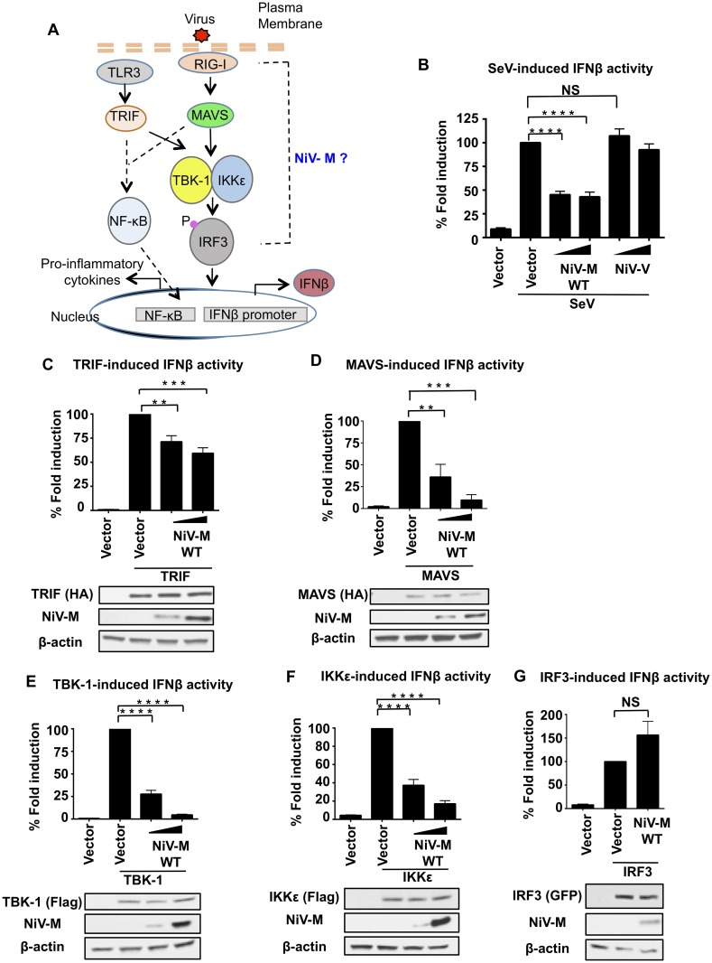
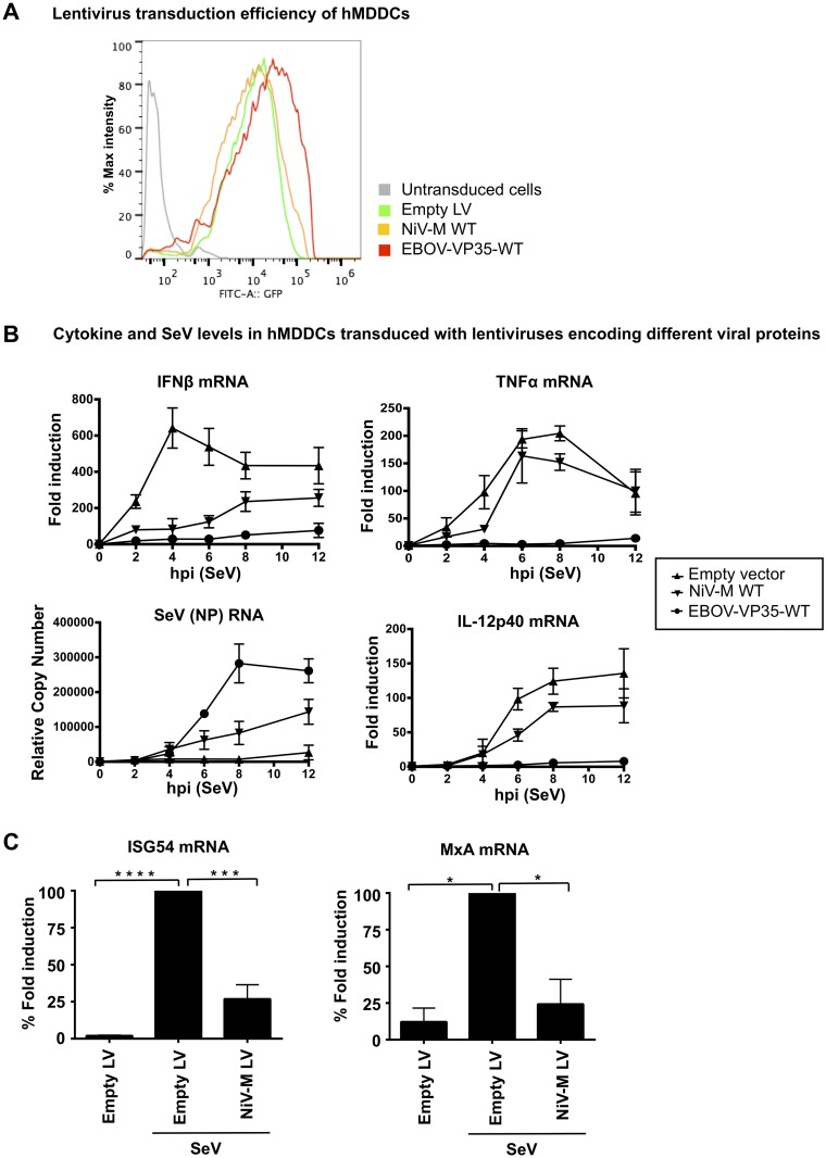
For efficient replication, viruses have developed mechanisms to evade innate immune responses, including the antiviral type-I interferon (IFN-I) system. Nipah virus (NiV), a highly pathogenic member of the Paramyxoviridae family (genus Henipavirus), is known to encode for four P gene-derived viral proteins (P/C/W/V) with IFN-I antagonist functions. Here we report that NiV matrix protein (NiV-M), which is important for virus assembly and budding, can also inhibit IFN-I responses. IFN-I production requires activation of multiple signaling components including the IκB kinase epsilon (IKKε). We previously showed that the E3-ubiquitin ligase TRIM6 catalyzes the synthesis of unanchored K48-linked polyubiquitin chains, which are not covalently attached to any protein, and activate IKKε for induction of IFN-I mediated antiviral responses. Using co-immunoprecipitation assays and confocal microscopy we show here that the NiV-M protein interacts with TRIM6 and promotes TRIM6 degradation. Consequently, NiV-M expression results in reduced levels of unanchored K48-linked polyubiquitin chains associated with IKKε leading to impaired IKKε oligomerization, IKKε autophosphorylation and reduced IFN-mediated responses. This IFN antagonist function of NiV-M requires a conserved lysine residue (K258) in the bipartite nuclear localization signal that is found in divergent henipaviruses. Consistent with this, the matrix proteins of Ghana, Hendra and Cedar viruses were also able to inhibit IFNβ induction. Live NiV infection, but not a recombinant NiV lacking the M protein, reduced the levels of endogenous TRIM6 protein expression. To our knowledge, matrix proteins of paramyxoviruses have never been reported to be involved in innate immune antagonism. We report here a novel mechanism of viral innate immune evasion by targeting TRIM6, IKKε and unanchored polyubiquitin chains. These findings expand the universe of viral IFN antagonism strategies and provide a new potential target for development of therapeutic interventions against NiV infections.


Conflict of interest statement

The authors have declared that no competing interests exist.

Figures

Fig 1
Fig 1. Nipah virus matrix protein inhibits IFNβ induction at the level of the TBK1/IKKε kinases.
A) Schematics of the signaling pathways investigated. (B-G) HEK293T cells were transfected with a Luciferase reporter plasmid under the control of the IFNβ promoter and a Renilla control plasmid, in the presence or absence of NiV-M and stimulated with SeV (B), or transfected with stimulating plasmids TRIF (C), MAVS (D), TBK-1 (E), IKKε (F) or IRF3 (G), followed by luciferase assay. Data were normalized first by none stimulated sample to obtain fold induction, and then stimulated samples were set to 100% to obtain percentage of fold induction. Data are from three independent experiments; each one in triplicate, and depicted is the mean ± SD (n = 9). *p < 0.05; **p < 0.01; ***p < 0.001, ****p < 0.0001, by Student’s t test. Immunoblots using lysates from transfected samples are shown as controls.
Fig 2
Fig 2. NiV-M protein inhibits IFNβ but not the pro-inflammatory cytokines TNFα and IL-12p40 in primary human MDDC.
A) Human MDDCs were either mock transduced, transduced with Vpx-VLPs only, or cotransduced with lentiviral (LV) vectors expressing NiV-M or Ebola virus VP35 and Vpx-VLPs. hMDDCs were harvested at day 5 post-transduction for GFP analysis by flow cytometry. B) hMDDCs where then stimulated with SeV and total RNA was isolated and analyzed by qRT-PCR to quantify IFNβ, SeV NP RNA, TNFα, and IL-12p40 levels or C) ISG54 and MxA. RPS11 was used as housekeeping gene and mRNA levels are shown as fold induction over mock treated samples. Error bars indicate standard deviations. Samples were collected from three independent donors. *p < 0.05; **p < 0.01; ***p < 0.001, ****p < 0.0001, by Student’s t test.
Fig 3
Fig 3. Lysine K258A on NiV matrix protein is important for inhibition of unanchored K48-linked polyubiquitin chains that associate with IKKε and required for IKKε activation.
(A-B) NiV-M requires membrane targeting for inhibition of IKKε/TBK-1 mediated IFNβ induction. HEK293T cells were transfected with IFNβ luciferase reporter in the presence of increasing concentrations of NiV-M WT or a K258A mutant and stimulating plasmid IKKε (A) or TBK-1 and the membrane targeting fusion proteins of M-K258A (S10-K258A or S15-K258A) (B). C) NiV-M-WT inhibits association of unanchored K48-linked polyubiquitin chains with IKKε leading to reduced IKKε-T501 phosphorylation and reduced IRF3 phosphorylation. HEK293T cells were transfected with NiV-M or NiV-M-K258A mutant in the presence or absence of Flag-IKKε. Cells were harvested and whole cell extracts (WCE) were used for IKKε immunoprecipitation using anti-Flag beads. A representative of at least 3 independent experiments is shown. Note that the WCE blot for K48-linked ubiquitin represents the levels of the total cellular pool of K48-linked ubiquitin (covalently ubiquitinated proteins and unanchored), which do not change in the presence of NiV-M. Only K48-linked ubiquitin that specifically associates with IKKε is reduced in the presence of NiV-M (shown in the IP panel). D) Schematics of IKKε activation by unanchored K48-linked polyubiquitin chains as described [13]. E) NiV-M-WT but not K258A inhibits oligomerization of IKKε. HEK293T cells were transfected with Flag-IKKε and Myc-IKKε in the presence of NiV-M, NiV-M-K258A or empty vector control. Oligomerization of IKKε was assessed by the ability of IKKε to interact with itself by immunoprecipitation. IKKε was immunoprecipitated from WCE with anti-Flag beads followed by immunoblot with Myc antibody. Representatives of at least 2 independent experiments are shown. *p < 0.05; **p < 0.01; ***p < 0.001, ****p < 0.0001, by Student’s t test.
Fig 4
Fig 4. Matrix proteins from henipaviruses with conserved Lysine K258A interact with IKKε and inhibit IKKε-mediated IFNβ induction.
A) Matrix proteins from henipaviruses inhibit IKKε-mediated IFNβ induction. HEK293T cells were transfected with IFNβ luciferase reporter and a Renilla control plasmid, in the presence or absence of NiV-M, HeV-M, GhV-M or CedV-M and IKKε, followed by luciferase assay. Data were normalized by none stimulated sample to obtain fold induction. Depicted is the mean ± SD (n = 3). B) Matrix proteins of henipaviruses interact with IKKε. HEK293T cells were transfected with Flag-NiV-M, Flag-HeV-M, Flag-GhV-M or Flag-CedV-M in the presence or absence of Myc-IKKε. Cells were harvested and whole cell extracts (WCE) were used for M immunoprecipitation using anti-Flag beads. C) Protein sequence alignment of the region mapping to the nuclear localization signal (NLS) of Nipah virus (NiV), Hendra Virus (HeV), Ghana virus (GhV) and Cedar virus (CedV). *p < 0.05; **p < 0.01; ***p < 0.001, ****p < 0.0001, by Student’s t test.
Fig 5
Fig 5. NiV-M interacts with TRIM6 and inhibits TRIM6-medited IFNβ induction and signaling.
A) NiV-M interacts with TRIM6. HEK293T cells were transfected with NiV-M, empty vector or HA-TRIM6. Cells were harvested and whole cell extracts (WCE) were used for TRIM6 immunoprecipitation using anti-HA-beads. (B-C) the C-terminal SPRY domain of TRIM6 interacts with NiV-M. B) TRIM6 deletion mutants used for coIP (GST-tagged). C) HEK293T cells were transfected with the GST-TRIM6 deletion mutants indicated together with NiV-M. Cells were harvested and WCE were used for TRIM6 immunoprecipitation using GST-beads. D) NiV-M co-localizes with TRIM6 in cytoplasmic bodies. HeLa cells were transfected with HA-TRIM6 and NiV-M-WT or NiV-M mutants K258A or K258R. Cells were fixed and stained with the indicated antibodies followed by confocal microscopy. Colocalization profiles are shown on the right. E) NiV-M inhibits the RIG-I induced IFNβ by targeting TRIM6. HEK293T were transfected with a non-targeting siRNA control (siControl) or aTRIM6-targeting siRNA (siTRIM6). After 24 hours, cells were transfected with IFNβ luciferase reporter and a Renilla control plasmid, in the presence or absence of NiV-M, or reconstitution with HA-TRIM6, followed by luciferase assay. Data were normalized by none stimulated sample to obtain fold induction. Depicted is the mean ± SD (n = 3). (F-G) NiV-M inhibits TRIM6-mediated IFNβ-induced ISG54 but not ISG15 reporter activity. HEK293T cells were transfected with ISG54-ISRE luciferase reporter (F) or ISG15 luciferase reporter (G) and a Renilla control plasmid and HA-TRIM6 in the presence of increasing concentrations of NiV-M-WT or NiV-MK258A, followed by luciferase assay. Data were normalized by none stimulated sample to obtain fold induction. Depicted is the mean ± SD (n = 3). Representatives of at least 2 independent experiments are shown. *p < 0.05; **p < 0.01; ***p < 0.001, ****p < 0.0001, by Student’s t test.
Fig 6
Fig 6. NiV-M targets TRIM6 for degradation.
A) HEK293T cells were transfected with increasing concentrations of NiV-M WT or a K258A mutant and the levels of endogenous TRIM6 were determined by immunoblot (representative of 3 experiments). For quantification, the levels of TRIM6 were normalized by actin, which were determined using ImageJ software. Data is combination of 3 independent experiments. *p < 0.05; **p < 0.01; ***p < 0.001, ****p < 0.0001, by Student’s t test. B) HEK293T cells were transfected with NiV-M-WT or NiV-M-K258A, empty vector and HA-TRIM6. Twenty-four hours post-transfection cells were treated with DMSO or the proteasome inhibitor MG132 for 4 hrs. Cells were harvested and whole cell extracts (WCE) were used for TRIM6 immunoprecipitation using anti-HA-beads.
Fig 7
Fig 7. NiV-M targets TRIM6 for degradation during NiV infection.
A) HeLa cells were infected with NiV-WT or NiV lacking the matrix protein (NiV-ΔM), expressing GFP, at MOI of 0.1. Forty-eight hours p.i. cells were fixed and stained with a TRIM6 antibody, followed by secondary anti-rabbit-555 (red) and immunofluorescence. Fluorescence intensity profiles are shown on the right. Mean Fluorescence Intensity (MFI) was quantified from three different images using ImageJ and graphs are shown below. TRIM6 MFI values were obtained from individual GFP+ or GFP- cells. For this quantification, individual cells were selected using the freehand selection tool in ImageJ software following the borders of each individual cell by following TRIM6 staining. Over 50 cells were selected for quantification in ImageJ software. B) Confocal microscopy of endogenous TRIM6 (red dots) in NiV-M-WT and NiV-ΔM infected cells. NiV-M-WT, GFP+ cells, have reduced levels of TRIM6 (MFI) and number of TRIM6 dots as compared to non-infected GFP- cells (quantification on the right). NiV-M-ΔM GFP+ infected cells, do not show significant differences in TRIM6 levels (MFI) or number of TRIM6 dots as compared to non-infected GFP- cells. A surface plot is shown, obtained using ImageJ based on pixels from the confocal images. C) NiV-M infected samples were stained with anti-M antibody (red) and anti-TRIM6 (for presentation TRIM6 is depicted in green). The region of high and low M expression was selected manually using the freehand tool in ImageJ. The region of high M expression correlates with very low TRIM6 staining as well as no visible TRIM6 dots. TRIM6 dots can clearly be seen in the cellular regions with low or no M expression. For quantification, the TRIM6 fluorescence intensity values in the low and high M areas where normalized by the MFI levels of M. *p < 0.05; **p < 0.01; ***p < 0.001, ****p < 0.0001, by Student’s t test.
Fig 8
Fig 8. Proposed model of NiV-M inhibition of IFN responses.
A) Upon virus recognition in Nipah virus infected cells lacking the matrix protein, PRR signaling promotes the synthesis of unanchored K48-linked polyubiquitin chains by the E3-ubiquitin ligase TRIM6. These polyubiquitin chains interact with IKKε and induce its oligomerization and T501 autophosphorylation. Consequently, activated IKKε phosphorylates IRF3 resulting in IFNβ induction and establishment of an antiviral response. IFNβ signaling through its receptor can also lead to TRIM6 and IKKε activation for induction of ISGs. B) The presence of NiV-M-WT promotes TRIM6 degradation resulting in reduced synthesis of K48-linked unanchored polyubiquitin chains, reduced IKKε oligomerization, IKKε-T501 autophosphorylation and reduced IRF3 phosphorylation with impaired antiviral responses.

References

    1. Medzhitov R, Preston-Hurlburt P, Janeway CA Jr. A human homologue of the Drosophila Toll protein signals activation of adaptive immunity. Nature. 1997;388(6640):394–7. - PubMed
    1. Meylan E, Tschopp J, Karin M. Intracellular pattern recognition receptors in the host response. Nature. 2006;442(7098):39–44. 10.1038/nature04946 . - DOI - PubMed
    1. Gitlin L, Barchet W, Gilfillan S, Cella M, Beutler B, Flavell RA, et al. Essential role of mda-5 in type I IFN responses to polyriboinosinic:polyribocytidylic acid and encephalomyocarditis picornavirus. Proc Natl Acad Sci U S A. 2006;103(22):8459–64. 10.1073/pnas.0603082103 - DOI - PMC - PubMed
    1. Yoneyama M, Kikuchi M, Natsukawa T, Shinobu N, Imaizumi T, Miyagishi M, et al. The RNA helicase RIG-I has an essential function in double-stranded RNA-induced innate antiviral responses. Nat Immunol. 2004;5(7):730–7. 10.1038/ni1087 . - DOI - PubMed
    1. Kawai T, Takahashi K, Sato S, Coban C, Kumar H, Kato H, et al. IPS-1, an adaptor triggering RIG-I- and Mda5-mediated type I interferon induction. Nat Immunol. 2005;6(10):981–8. 10.1038/ni1243 . - DOI - PubMed

MeSH terms

LinkOut - more resources