. 2016 Sep 13:6:33215.
doi: 10.1038/srep33215.

Cell wall degradation is required for normal starch mobilisation in barley endosperm

Affiliations

Cell wall degradation is required for normal starch mobilisation in barley endosperm

Vasilios M E Andriotis et al. Sci Rep. .

Abstract

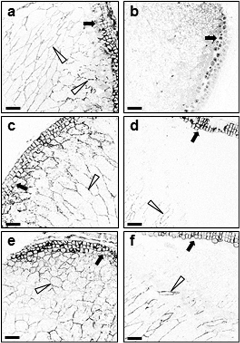
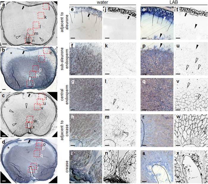
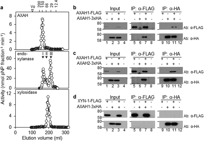
Starch degradation in barley endosperm provides carbon for early seedling growth, but the control of this process is poorly understood. We investigated whether endosperm cell wall degradation is an important determinant of the rate of starch degradation. We identified iminosugar inhibitors of enzymes that degrade the cell wall component arabinoxylan. The iminosugar 1,4-dideoxy-1, 4-imino-l-arabinitol (LAB) inhibits arabinoxylan arabinofuranohydrolase (AXAH) but does not inhibit the main starch-degrading enzymes α- and β-amylase and limit dextrinase. AXAH activity in the endosperm appears soon after the onset of germination and resides in dimers putatively containing two isoforms, AXAH1 and AXAH2. Upon grain imbibition, mobilisation of arabinoxylan and starch spreads across the endosperm from the aleurone towards the crease. The front of arabinoxylan degradation precedes that of starch degradation. Incubation of grains with LAB decreases the rate of loss of both arabinoxylan and starch, and retards the spread of both degradation processes across the endosperm. We propose that starch degradation in the endosperm is dependent on cell wall degradation, which permeabilises the walls and thus permits rapid diffusion of amylolytic enzymes. AXAH may be of particular importance in this respect. These results provide new insights into the mobilization of endosperm reserves to support early seedling growth.


Figures

Figure 1
Figure 1. The iminosugar 1, 4-dideoxy-1, 4-imino-l-arabinitol (LAB) is an inhibitor of barley endosperm arabinoxylan arabinofuranohydrolase (AXAH) activity.
(a) Structure of l-arabinofuranose (top) and LAB (bottom). (b) Structure of d-xylose (top) and of the iminosugar 1, 5-dideoxy-1, 5-imino-xylitol (xyloDNJ; bottom). (c–f) Chromatographic separation of barley endosperm AX degrading enzymes by FPLC. Soluble proteins from endosperm of seedlings at ten dpi were applied to a MonoQ anion exchange column (c–e). After washing, the column was eluted with a gradient of increasing NaCl concentration (dashed line). Proteins not binding on this column were applied to a MonoS cation exchange column (f) and eluted with a NaCl gradient (dashed line). Enzyme activities were assayed either in the absence (white circles) or in the presence of 500 μM LAB (black triangles) or xyloDNJ (grey squares). (c) Elution profile of endo-xylanase activity (assayed with 4-nitrophenyl xylotrioside). A single peak of endo-xylanase activity eluted from the MonoQ anion exchange column at about 200 mM NaCl. (d) Elution profile of AXAH activity (assayed with 4-nitrophenyl α-l-arabinofuranoside). AXAH eluted from the MonoQ anion exchange column at about 300 mM NaCl and was strongly inhibited by LAB. (e) Elution profile of xylosidase activity (assayed with 4-nitrophenyl xylanopyranoside). Xylosidase activity did not bind to the MonoQ anion exchange column. (f) Elution profile of xylosidase during MonoS cation exchange chromatography. Xylosidase activity that did not bind to a MonoQ anion exchange column (e) was applied to a MonoS cation exchange column and eluted as a single peak at about 150 mM NaCl.
Figure 2
Figure 2. Effects of LAB and xyloDNJ on seedling growth and endosperm starch content.
(a) Appearance of seedlings of cv NFC Tipple at ten dpi, grown in water alone or in the presence of 500 μM LAB or xyloDNJ. Bar = 1 cm. (b) Effect of inhibitors on coleoptiles (top panel) and roots (bottom panel) of seedlings at ten dpi grown in water alone or in the presence of 200 or 500 μM of each inhibitor. Values are means ± SE of measurements made on the numbers of seedlings indicated in the top part of the graph. (c) Endosperm starch content of barley seedlings at ten dpi grown in water alone or in the presence of the indicated levels (μM) of LAB or xyloDNJ. Values are means ± SE of measurements made on the number of seedlings indicated. (d) Effect of LAB on starch degradation in endosperm of embryo-less half-grains. Embryo-less half-grains were incubated with water or in the presence of 500 μM LAB, either supplemented with (closed bars) or lacking (open bars) gibberellin. Starch content was measured at eight dpi. Values are means ± SE (bars) of measurements on the number of half-grains indicated.
Figure 3
Figure 3. Effect of LAB on the activities of enzymes of starch degradation.
(a) Effect of inhibitors on α-amylase activity. Extracts of endosperm from seedlings at the dpi indicated were assayed for α-amylase activity in the absence (black bars) or in the presence of 500 μM (dark grey bars) or 100 μM (light grey bars) LAB, or acarbose, a known inhibitor of the enzyme (500 μM; white bars). Values are means ± SE of measurements on three independent extracts. (b) Effect of inhibitors on β-amylase activity. Extracts as in (a) were assayed for β-amylase activity either in the absence (black bars) or in the presence of 500 μM (dark grey bars) or 100 μM (light grey bars) LAB. Values are means ± SE of measurements on three independent extracts. (c) Effect of inhibitors on limit dextrinase activity. Activity of purified recombinant limit dextrinase was assayed against pullulan in the absence of inhibitors (black bar) or in the presence of 500 μM (dark grey bars), 100 μM (light grey bars), or 50 μM (white bars) of LAB, or β-cyclodextrin (βCD), a known inhibitor of the enzyme. Values are means ± SE of three independent measurements. (d) Effect of inhibitors on maltase (Agl97) activity. Purified recombinant Agl97 was assayed with pNPG in the presence of 100 μM (black bars), 10 μM (dark grey bars), 1 μM (grey bars), 0.1 μM (light grey bars), or 0.01 μM (white bars) of LAB, xyloDNJ or deoxynojirimycin (DNJ), a known inhibitor of the enzyme. Results are expressed as percent inhibition relative to control assays with no inhibitor. The enzyme preparation was as previously specified.
Figure 4
Figure 4. LAB reduces the rate of arabinoxylan degradation in endosperm cell walls.
A cell wall fraction prepared from pooled endosperm from 30–40 grains was treated with commercial endo-xylanase and the released oligosaccharides were analysed by HPAEC. (a) Representative elution profiles of AX-diagnostic peaks 1 and 2 (arrows). The full elution profile is shown in Fig. S4. (b) Quantification of peak area of AX-diagnostic peaks 1 and 2 from the elution profiles in (a). Values are means ± SE from three technical replicates. Differences between inhibitor-treated and water incubated grains at six dpi are statistically significant as indicated (Student’s t-test: *P < 0.05). (c) As in (a) but for AX-diagnostic peaks 3 and 4. Elution profiles are from the same chromatograms as in (a). The full elution profile is shown in Fig. S4. (d) Quantification of peak area of AX-diagnostic peaks 3 and 4 from the elution profiles in (c). Values are means ± SE from three technical replicates. Differences between inhibitor-treated and water incubated grains at six dpi are statistically significant as indicated (Student’s t-test: ***P < 0.001).
Figure 5
Figure 5. Treatment with LAB reduces the extent of arabinoxylan degradation in endosperm cell walls.
Transverse sections of endosperm of dry grains (a,b), or grains imbibed and grown in water (c,d), or LAB (500 μM; e,f) were incubated with a monoclonal antibody against AX (LM11) then with an Alexa Fluor® 633 conjugated secondary antibody, and subjected to confocal microscopy to visualise AX epitopes. All sections were prepared from approximately the same position of the grain. Open arrowheads point to fluorescence from AX epitopes in endosperm cell walls, closed arrows point to the aleurone layer. Original confocal images were edited such that colour was inverted in all panels at the same time, for optimal contrast between fluorescence signal and background. (a) Section through a dry grain, adjacent to the aleurone. The antibody decorates the cell walls of the endosperm and aleurone cells. (b) As in (a) but omitting the primary (LM11) antibody. No staining of cell walls is visible. (c) Section of barley grain grown in water, at two dpi. The distribution of fluorescence is the same as in dry grain (a). (d) Section of barley grain grown in water, at four dpi. Fluorescence is lower in endosperm cell walls adjacent to the aleurone than at two dpi. (e) Section of barley grain grown in LAB, at two dpi. The distribution of fluorescence is similar to that in dry grain (a). (f) Section of barley grain grown in water, at four dpi. Fluorescence is greater in the region adjacent to the aleurone layer than in grains at the same stage grown in water (d). Scale bars are 100 μm throughout.
Figure 6
Figure 6. Treatment with LAB reduces the extent of both arabinoxylan and starch degradation in the endosperm.
Transverse sections of endosperm of grains incubated for six dpi in water (a,b,e–n), or LAB (c,d,o–x) were either incubated with the LM11 antibody and processed as in Fig. 5, to visualise AX epitopes (a,c,j–n,t–x), or stained with toluidine blue and iodine solution (b,d,e–i,o–s) to visualise cellular structures and starch deposits. All sections were prepared from approximately the same position of the grain. Open arrowheads: fluorescence from AX epitopes in endosperm cell walls; closed arrowheads: interface between the sub-aleurone zone and endosperm tissue in which starch degradation has been initiated; closed arrows: aleurone layer; dashed lines (a–d,h,i,m,n,r,s,w,x) edge of the crease; asterisks (b,d): sub-aleurone zone. Results were reproducible in two separate incubations. In (a–d) letters next to the red boxes correspond to panels (e–x) as indicated. (a) Section through a grain grown in water, incubated with the LM11 antibody and viewed under a confocal microscope. The antibody decorates the cell walls of the endosperm cells around the crease of the grain, and the aleurone. (b) Section consecutive to that shown in (a), stained with toluidine blue and iodine solution. (c) As in (a) but for a grain grown in LAB. The LM11 antibody decorates cell walls in a much larger area of the endosperm surrounding the crease, compared to (a). (d) As in (b) but for a grain grown in LAB. Loss of starch from the sub-aleurone zone has not progressed to the same extent as in (b). (e–n) Close-up views of a section through a grain grown in water. The sections correspond to the five boxed areas of the grain shown in (a),(b). (e–i) were stained with toluidine blue and iodine to reveal starch; (j–n) were stained with LM11 antibody to reveal AX epitopes. (o–x) As in (e–n) but for a grain grown with LAB. (o–s) were stained with toluidine blue and iodine to reveal starch; (t–x) were stained with LM11 antibody to reveal AX epitopes. Scale bars are 100 μm (a–d) and 50 μm (e–x).
Figure 7
Figure 7. Size fractionation of AXAH, endo-xylanase and β-d-xylosidase from endosperm extracts, and detection of oligomeric protein complexes.
(a) Size fractionation of AX-degrading enzymes from endosperm extracts of barley seedlings at ten dpi were applied to a Sephacryl S200 column by FPLC. Fractions were assayed for AXAH (top panel), endo-xylanase (middle panel) and β-d-xylosidase (bottom panel) activity. Molecular masses of eluted activities were estimated by comparison to the elution profile of proteins of known size (numbers at the top, in kDa). (b) AXAH1 can homodimerise. AXAH1-FLAG and/or AXAH1-3xHA proteins were transiently expressed in N. benthamiana leaves. +Above a lane indicates that the protein indicated at the left was expressed in the leaf from which extracts were made. Input samples are extracts prior to immunoprecipitation; IP: α-FLAG and IP: α-HA are proteins precipitated with the FLAG and HA antisera respectively. Ab: α-FLAG and Ab: α-HA indicate the antiserum used for immunodetection of proteins following SDS-PAGE. Numbers on the left are positions of molecular mass markers (in kDa). Numbers below each panel indicate lane numbers. Samples were subjected to electrophoresis on the same gel and to immunoblot analysis at the same time. Asterisk: position of the IgG heavy chain occasionally detected by the antisera. (c) AXAH1 interacts with AXAH2. AXAH1-FLAG and/or AXAH2-3xHA were transiently expressed in N. benthamiana leaves. Annotation is as for (b). (d) AXAH1 does not interact with XYN-1. AXAH1-3xHA and/or XYN-1-FLAG were transiently expressed in N. benthamiana leaves. Annotation is as for (b).

References

    1. Ritchie S., Swanson S. J. & Gilroy S. From common signalling components to cell specific responses: insights from the cereal aleurone. Physiol. Plant. 15, 342–351 (2002). - PubMed
    1. Clarke S. E., Hayes P. M. & Henson C. A. Effects of single nucleotide polymorphisms in β-amylase1 alleles from barley on functional properties of the enzymes. Plant Physiol. Biochem. 41, 798–804 (2003).
    1. Muralikrishna G. & Nimala M. Cereal α-amylases - an overview. Carbohydr. Polym. 60, 163–173 (2005).
    1. Xie Z. et al.. Interactions of two abscisic-acid induced WRKY genes in repressing gibberellin signalling in aleurone cells. Plant J. 46, 231–242 (2006). - PubMed
    1. Moreno-Risueno M. A. et al.. The HvDOF19 transcription factor mediates the abscisic acid-dependent repression of hydrolase genes in germinating barley aleurone. Plant J. 51, 352–365 (2007). - PubMed

Publication types

MeSH terms

LinkOut - more resources