. 2016 Sep 13;11(9):e0162698.
doi: 10.1371/journal.pone.0162698. eCollection 2016.

SPARC Regulates Transforming Growth Factor Beta Induced (TGFBI) Extracellular Matrix Deposition and Paclitaxel Response in Ovarian Cancer Cells

Affiliations

SPARC Regulates Transforming Growth Factor Beta Induced (TGFBI) Extracellular Matrix Deposition and Paclitaxel Response in Ovarian Cancer Cells

David A Tumbarello et al. PLoS One. .

Abstract

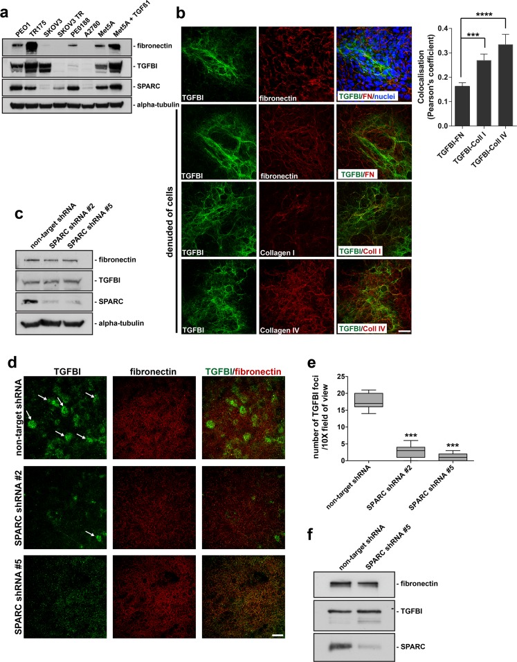
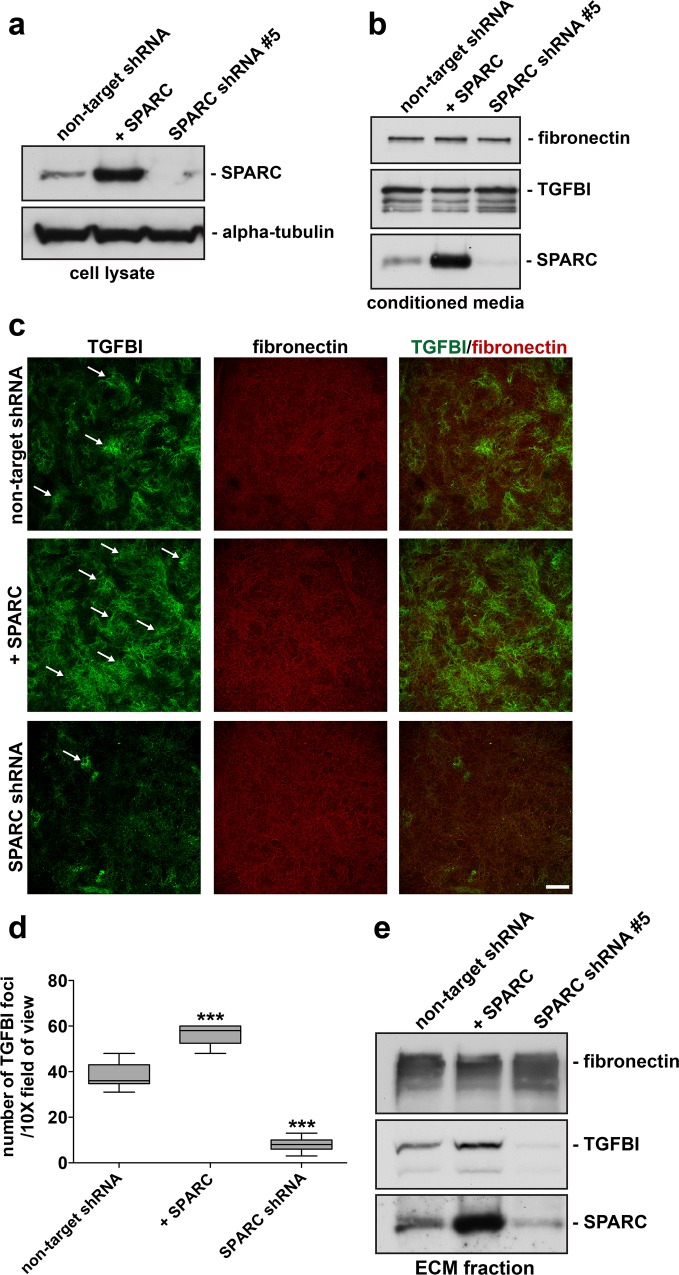
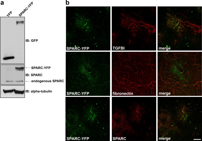
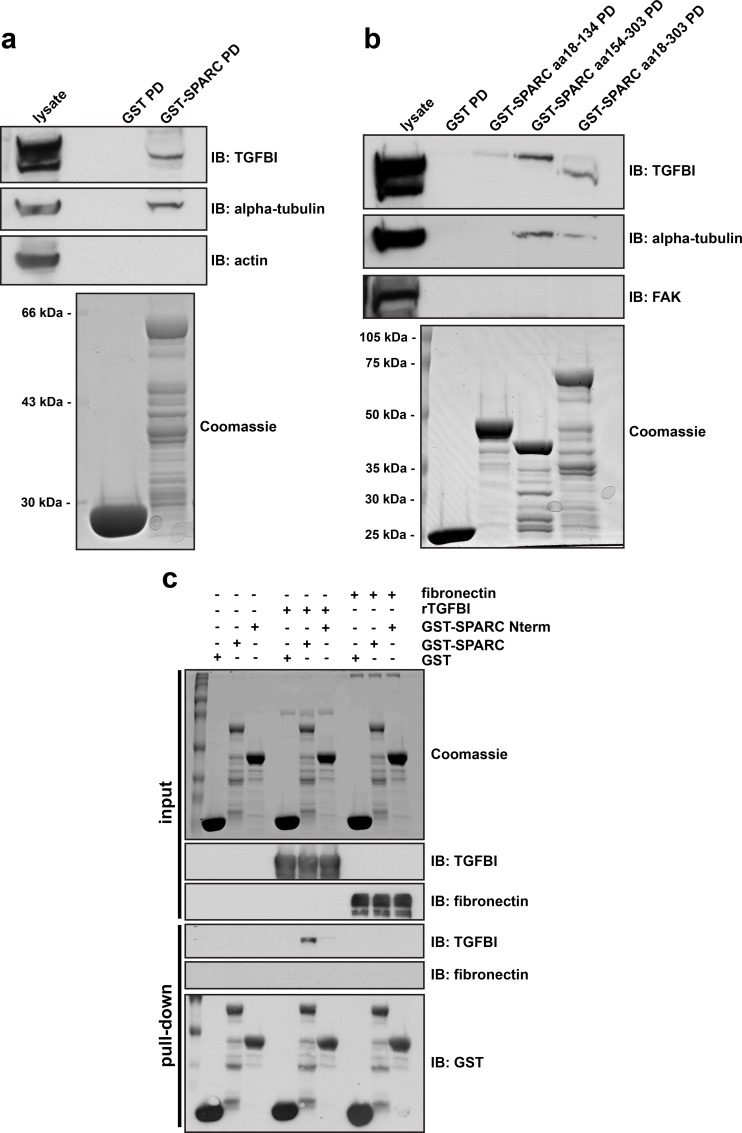
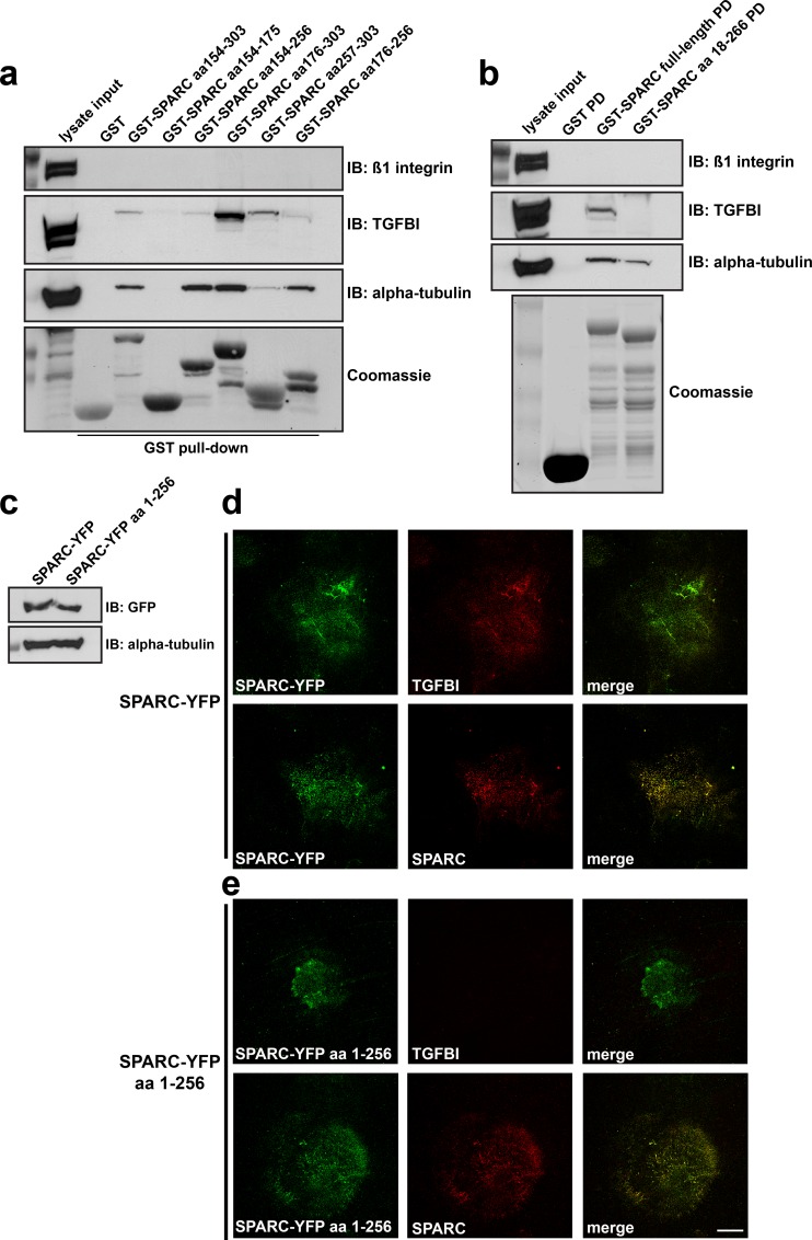
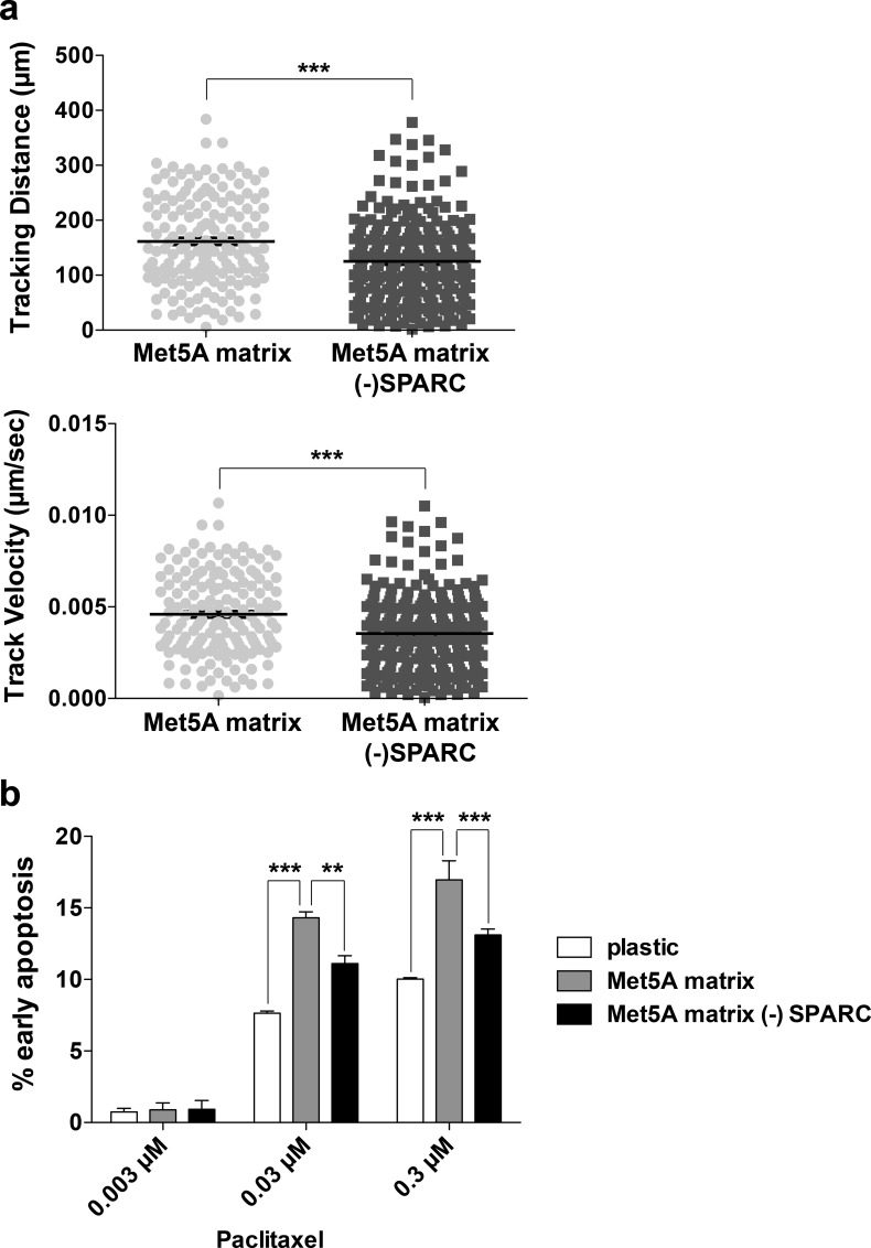
TGFBI has been shown to sensitize ovarian cancer cells to the cytotoxic effects of paclitaxel via an integrin receptor-mediated mechanism that modulates microtubule stability. Herein, we determine that TGFBI localizes within organized fibrillar structures in mesothelial-derived ECM. We determined that suppression of SPARC expression by shRNA decreased the deposition of TGFBI in mesothelial-derived ECM, without affecting its overall protein expression or secretion. Conversely, overexpression of SPARC increased TGFBI deposition. A SPARC-YFP fusion construct expressed by the Met5a cell line co-localized with TGFBI in the cell-derived ECM. Interestingly, in vitro produced SPARC was capable of precipitating TGFBI from cell lysates dependent on an intact SPARC carboxy-terminus with in vitro binding assays verifying a direct interaction. The last 37 amino acids of SPARC were shown to be required for the TGFBI interaction while expression of a SPARC-YFP construct lacking this region (aa 1-256) did not interact and co-localize with TGFBI in the ECM. Furthermore, ovarian cancer cells have a reduced motility and decreased response to the chemotherapeutic agent paclitaxel when plated on ECM derived from mesothelial cells lacking SPARC compared to control mesothelial-derived ECM. In conclusion, SPARC regulates the fibrillar ECM deposition of TGFBI through a novel interaction, subsequently influencing cancer cell behavior.


Conflict of interest statement

The authors have declared that no competing interests exist.

Figures

Fig 1
Fig 1. TGFBI produced by mesothelial cells forms a fibrillar matrix distinct from fibronectin and loss of SPARC expression disrupts TGFBI deposition in mesothelial-derived ECM.
(a) Western blot analysis of RIPA soluble lysates derived from a panel of ovarian cancer cell lines and the Met5a mesothelial cell line either untreated or treated with TGFß1. The membrane was probed with antibodies specific to the indicated proteins. (b) Confocal microscopy of Met5a cells and extracellular matrix preparations denuded of Met5a cells cultured for 9 days, was performed following immunostaining for TGFBI, fibronectin, Collagen type I, and Collagen type IV. Hoechst dye was utilized to visualize nuclei and merged images are indicated. Scale bar 40 μm. Quantitation of colocalisation from >3 fields of view were performed and represented as a Pearson’s coefficient. Error bars indicate s.d., ****p<0.0001, ***p<0.001. (c) RIPA soluble lysates harvested from Met5a cells stably expressing either control non-target shRNA or SPARC shRNA target #2 and #5, while cultured under 3D matrix conditions. Western blot analysis was performed utilizing antibodies specific to the indicated proteins. (d) Confocal microscopy of matrix preparations from Met5a cells stably expressing non-target shRNA, SPARC shRNA #2, or SPARC shRNA #5 immunostained for TGFBI and fibronectin. Merged images are indicated. Arrows indicate individual foci. Scale bar 200 μm. (e) Quantitation of results from immunostained matrices. The number of enriched TGFBI immunostained foci were counted from each group and represented as number of TGFBI foci/10X field of view. ***represents significance from control of p<0.001, ANOVA. (f) Conditioned media was harvested from Met5a cells stably expressing either control non-target shRNA or SPARC shRNA #5, while cultured under 3D matrix conditions. Western blot was performed utilizing antibodies specific to the indicated proteins.
Fig 2
Fig 2. Modulation of SPARC expression influences TGFBI deposition in mesothelial-derived ECM.
RIPA soluble lysates (a) and conditioned media (b) were harvested from Met5a cells stably expressing either control non-target shRNA, overexpressing SPARC cDNA, or SPARC shRNA #5, while cultured under 3D matrix conditions. Western blot analysis was performed using antibodies specific to the indicated proteins. (c) Confocal microscopy was performed on ECM preparations from Met5a cells stably expressing non-target shRNA, SPARC shRNA #5, or overexpressing SPARC cDNA immunostained for TGFBI and fibronectin as indicated. Merged images are indicated. Arrows indicate individual foci. Scale bar 200 μm. (d) Level of TGFBI immunostaining was quantified by counting TGFBI enriched immunostained foci from each group and represented by number of TGFBI foci/10X field of view. ***represents significance from control of p<0.001, ANOVA. (e) Matrix denuded of Met5a cells was solubilised in SDS-sample buffer and Western blot analysis was performed utilizing antibodies to the indicated proteins.
Fig 3
Fig 3. SPARC colocalizes with TGFBI in mesothelial-derived ECM.
(a) Western blot analysis was performed on RIPA soluble lysates harvested from Met5a cells transfected with either YFP alone or SPARC-YFP. Immunoblotting with anti-GFP antibody recognizes YFP fusion constructs. (b) Extracellular matrix preparation was carried out from Met5a cells transfected with SPARC-YFP. Confocal microscopy was performed following immunostaining for YFP, TGFBI, fibronectin, and SPARC as indicated. Merged images are indicated. Scale bar 40 μm.
Fig 4
Fig 4. SPARC directly interacts with TGFBI via its carboxy-terminus.
(a) In vitro GST-binding assays. Coomassie stained gel of purified GST and GST-SPARC (aa 18–303) expressed in bacteria. Western blot analysis was performed following GST pull-down from SKOV3 lysates, probed with antibodies specific to the indicated proteins. (b) Coomassie stained gel of bacterially expressed and purified GST, GST-SPARC (aa 18–303), GST-SPARC Nterm (aa 18–134), and GST-SPARC Cterm (aa 154–303) fusion proteins. Western blot analysis was performed following GST pull-down assay from SKOV3 lysates, probed with antibodies specific to the indicated proteins. (c) In vitro binding of purified GST-SPARC to bacterially expressed and purified recombinant TGFBI. GST, GST-SPARC (aa 18–303), or GST-SPARC Nterm (aa 18–134) fusion proteins were incubated with rTGFBI or fibronectin, followed by pull-down with Glutathione sepharose 4B beads. Subsequently, Western blot analysis was performed with antibodies specific to the indicated proteins. Coomassie stained gel represents experimental input.
Fig 5
Fig 5. Interaction of SPARC with TGFBI is necessary for TGFBI extracellular matrix deposition.
(a) GST pull-down assay from SKOV3 cell lysates utilizing truncated GST-fusion proteins derived from the Carboxy-terminus of SPARC. Following GST-pull down, Western blot analysis was performed utilizing antibodies specific to the indicated proteins. (b) GST pull-down assay from SKOV3 cell lysates using either full-length SPARC or full-length SPARC lacking the carboxy-terminal 37 amino acids (aa 18–266). Following GST-pull-down (PD), Western blot analysis was performed to the indicated proteins. Coomassie brilliant blue staining of SDS-PAGE confirms expression and purification of GST fusion proteins. (c) Full-length SPARC-YFP or SPARC-YFP lacking the carboxy-terminal 47 amino acids (SPARC-YFP aa 1–256) were transiently transfected into Met5a cells and Western blot analysis was performed to confirm their expression. (d) and (e) Extracellular matrix preparation derived from these cells following 6-day culture was subsequently processed for confocal immunofluorescence microscopy. Cell derived ECM was immunostained for YFP, TGFBI, and SPARC as indicated. Merged images are indicated. Scale bar 40 μm.
Fig 6
Fig 6. Mesothelial-derived ECM influences cancer cell motility and response to the chemotherapeutic agent paclitaxel.
(a) Time lapse video microscopy was performed of SKOV3 cells plated on Met5A derived ECM derived from cells expressing either control shRNA (Met5A matrix) or SPARC shRNA (Met5A matrix—SPARC). Images were collected for 10 hours and cell centroids were tracked using Volocity software. Circles represent tracking distance and velocity of each individual cell and black bars represent the mean ±S.E.M. (b) SKOV3 cells were plated on either plastic or Met5A derived ECM derived from cells expressing either control shRNA or SPARC shRNA (- SPARC). Cells were treated with 0.003 μM, 0.03 μM, or 0.3 μM paclitaxel for 30 hours prior to staining with FITC-Annexin V and propidium iodide before analyzing by flow cytometry. Three independent experiments were performed and the results are represented by percent of cells in early apoptosis (Annexin V +, PI -). ** represents significance of p<0.01 and *** represents significance of p<0.001.

References

    1. Ahmed AA, Mills AD, Ibrahim AE, Temple J, Blenkiron C, Vias M, et al. (2007) The extracellular matrix protein TGFBI induces microtubule stabilization and sensitizes ovarian cancers to paclitaxel. Cancer Cell 12: 514–527. - PMC - PubMed
    1. Zutter MM (2007) Integrin-mediated adhesion: tipping the balance between chemosensitivity and chemoresistance. Adv Exp Med Biol 608: 87–100. - PubMed
    1. Tumbarello DA, Temple J, Brenton JD (2012) Beta3 integrin modulates transforming growth factor beta induced (TGFBI) function and paclitaxel response in ovarian cancer cells. Mol Cancer 11: 36 10.1186/1476-4598-11-36 - DOI - PMC - PubMed
    1. Denys H, Braems G, Lambein K, Pauwels P, Hendrix A, De Boeck A, et al. (2009) The extracellular matrix regulates cancer progression and therapy response: implications for prognosis and treatment. Curr Pharm Des 15: 1373–1384. - PubMed
    1. Sangaletti S, Di Carlo E, Gariboldi S, Miotti S, Cappetti B, Parenza M, et al. (2008) Macrophage-derived SPARC bridges tumor cell-extracellular matrix interactions toward metastasis. Cancer Res 68: 9050–9059. 10.1158/0008-5472.CAN-08-1327 - DOI - PubMed

MeSH terms