. 2016 Nov:100:146-158.
doi: 10.1016/j.neuint.2016.09.008. Epub 2016 Sep 10.

PKCδ knockout mice are protected from para-methoxymethamphetamine-induced mitochondrial stress and associated neurotoxicity in the striatum of mice

Affiliations

PKCδ knockout mice are protected from para-methoxymethamphetamine-induced mitochondrial stress and associated neurotoxicity in the striatum of mice

Eun-Joo Shin et al. Neurochem Int. 2016 Nov.

Abstract

Para-methoxymethamphetamine (PMMA) is a para-ring-substituted amphetamine derivative sold worldwide as an illegal psychotropic drug. Although PMMA use has been reported to lead to severe intoxication and even death, little is known about the mechanism(s) by which PMMA exerts its neurotoxic effects. Here we found that PMMA treatment resulted in phosphorylation of protein kinase Cδ (PKCδ) and subsequent mitochondrial translocation of cleaved PKCδ. PMMA-induced oxidative stress was more pronounced in mitochondria than in the cytosol. Moreover, treatment with PMMA consistently resulted in significant reductions in mitochondrial membrane potential, mitochondrial complex I activity, and mitochondrial Mn superoxide dismutase-immunoreactivity. In contrast, PMMA treatment led to a significant increase in intramitochondrial Ca2+ level. Treatment with PMMA also significantly increased ionized calcium binding adaptor molecule 1 (Iba-1)-labeled microglial activation and upregulated tumor necrosis factor alpha (TNF-α) gene expression. PKCδ knockout attenuated these mitochondrial effects and dampened the neurotoxic effects of PMMA. Importantly, TNF-α knockout mice were significantly protected from PMMA-induced increases in phospho-PKCδ expression, mitochondrial translocation of cleaved PKCδ, and Iba-1-labeled microgliosis. Both rottlerin, a pharmacological inhibitor of PKCδ, and etanercept, a pharmacological inhibitor of TNF-α, significantly protected against PMMA-mediated induction of apoptosis, as assessed by terminal deoxynucleotidyl transferase dUDP nick end labeling (TUNEL) assays. In addition, PKCδ knockout and TNF-α knockout both resulted in decreased PMMA-mediated induction of dopaminergic loss. Therefore, our results suggest that PKCδ mediates PMMA-induced neurotoxicity by facilitating oxidative stress (mitochondria > cytosol), mitochondrial dysfunction, microglial activation, and pro-apoptotic signaling. Our results also indicate that PMMA-induced PKCδ activation requires the proinflammatory cytokine TNF-α.

Keywords: Microglia; Mitochondria; Oxidative stress; Para-methoxymethamphetamine; Protein kinase Cδ; Tumor necrosis factor-α.


Conflict of interest statement

Conflict of interest disclosure

There is no conflict of interest

Figures
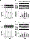
Fig. 1.
Fig. 1.
Time-dependent alterations in phosphorylation of PKCδ (A and C) and mitochondrial translocation of cleaved PKCδ (B and D) in the striata of wild type (WT) mice induced by multiple doses (A and B) or a single dose (C and D) of PMMA. Sal = Saline. Each value is the mean ± S.E.M. of 6 animals. One-way ANOVA showed a significant effect of PMMA time-point on phosphorylation of PKCδ (A and C) and mitochondrial translocation of cleaved PKCδ (B and D) activation (Supplemental Table 1). *p < 0.05, **p < 0.01 vs. Sal (one-way ANOVA followed by Fisher’s LSD pairwise comparisons).
Fig. 2.
Fig. 2.
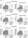
PMMA-induced time-dependent alterations in cytosolic and mitochondrial protein oxidation (A), lipid peroxidation (B), and reactive oxygen species (ROS; C) in the striata of wild type and PKCδ KO mice. Sal = Saline. WT = wild type. Each value is the mean ± S.E.M. of 6 animals. Two-way ANOVA showed significant effects of PMMA time-point (A-C) and PKCδ KO (A-C). In addition, two-way ANOVA revealed a significant interaction between PMMA time-point and PKCδ KO (mitochondrial protein oxidation and mitochondrial lipid peroxidation) (Supplemental Table 1). *p < 0.05, **p < 0.01 vs. WT with saline. #p < 0.05, ##p < 0.01 vs. the corresponding time-point of WT with PMMA (two-way ANOVA followed by Fisher’s LSD pairwise comparisons).
Fig. 3.
Fig. 3.
PMMA-induced alterations in mitochondrial membrane potential (A), intramitochondrial Ca2+ level (B), mitochondrial complex I activity (C), mitochondrial Mn-superoxide dismutase (MnSOD) expression (D), and MnSOD immunoreactivity (E) in the striata of wild type (WT) and PKCδ KO mice. Sal = Saline. WT = wild type. Each value is the mean ± S.E.M. of 6 animals. Two-way ANOVA showed significant effects of PMMA (A-E) and PKCδ KO (A-E). Two-way ANOVA also showed a significant interaction between PMMA and PKCδ KO (B) (Supplemental Table 2). *p < 0.05, **p < 0.01 vs. WT with saline. #p < 0.05 vs. WT with PMMA (two-way ANOVA followed by Fisher’s LSD pairwise comparisons). Scale bar = 0.5 mm.
Fig. 4.
Fig. 4.
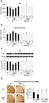
PMMA-induced alterations in dopamine (A), dopamine turnover rate (B), tyrosine hydroxylase (TH) expression (C), and TH-immunoreactivity (D) in the striata of wild type and PKCδ KO mice. Sal = Saline. WT = wild type. Each value is the mean ± S.E.M. of 6 animals. Two-way ANOVA showed significant effects of PMMA time points (C) or PMMA (D) and PKCδ KO (B-D), and a significant interaction between PMMA time points and PKCδ KO (B). Additional statistical analysis with two-way ANOVA revealed significant effects of PMMA (A and B) and PKCδ KO (A and B), and a significant interaction between PMMA and PKCδ KO (A and B) at 12 h post-PMMA (Supplemental Table 2). *p < 0.05 vs. WT with saline. #p < 0.05 vs. the corresponding time-point of WT with PMMA (two-way ANOVA followed by Fisher’s LSD pairwise comparisons). Scale bar = 1 mm.
Fig. 5.
Fig. 5.
PMMA-induced alterations in Iba-1-expression (A) and Iba-1 immunoreactivity (B) in the striata of wild type and PKCδ KO mice. Sal = Saline. WT = wild type. Each value is the mean ± S.E.M. of 6 animals. One-way ANOVA showed a significant effect of PMMA time-point on the Iba-1 expression (A). In addition, two-way ANOVA showed significant effects of PMMA (B) and PKCδ KO (B), and a significant interaction between PMMA and PKCδ KO (B) (Supplemental Table 3). *p < 0.01 vs. Sal or WT with saline. #p < 0.01 vs. WT with PMMA [one-way ANOVA (A) or two-way ANOVA (B) followed by Fisher’s LSD pairwise comparisons]. Scale bar = 100 μm.
Fig. 6.
Fig. 6.
PMMA-induced alterations in mRNA and protein levels of IL-6 (A), INF-γ (B), TNF-α (C), TNFR1 (D) and TNFR2 (E) in the striata of wild type mice. Each value is the mean ± S.E.M. of 6 animals. One-way ANOVA showed a significant effect of PMMA time-point on the mRNA and protein levels of IL-6 (A), INF-γ (B), TNF-α (C), TNFR1 (D) and TNFR2 (E) in the striata of wild type mice (Supplemental Table 3). *p < 0.05, **p < 0.01 vs. Sal (one-way ANOVA followed by Fisher’s LSD pairwise comparisons).
Fig. 7.
Fig. 7.
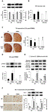
PMMA-induced alteration in dopamine level and TH expression (A), dopamine turnover rate (B), TH-immunoreactivity (C), phospho-PKCδ expression (D), mitochondrial translocation of cleaved PKCδ (E), and Iba-1-immunoreactivity and expression (F) in the striata of wild type (WT) and TNF-α KO mice. Each value is the mean ± S.E.M. of 6 animals. Two-way ANOVA showed significant effects of PMMA (A-F) and PKCδ KO (A-F), and a significant interaction between PMMA and PKCδ KO (B and D-F) (Supplemental Table 4). *p < 0.05, **p < 0.01 vs. WT with saline. #p < 0.01 vs. WT with PMMA (two-way ANOVA followed by Fisher’s LSD pairwise comparisons). Scale bar = 1 mm (C) or 100 μm (F).
Fig. 8.
Fig. 8.
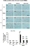
Effect of rottlerin (Rot), or etanercept (Eta) on PMMA-induced apoptotic cell death. Apoptotic cell death was observed as an increase in TUNEL-positive cells in the striata of Taconic ICR mice. Sal = Saline. Veh = vehicle [10% (v/v) DMSO] for rottlerin. Rot = rottlerin (3μg, i.c.v.). Eta = etanercept (50μg, i.c.v.). Each value is the mean ± S.E.M. of 6 animals. Two-way ANOVA showed significant effects of PMMA and pretreatment with rottlerin or etanercept, and a significant interaction between PMMA and pretreatment (Supplemental Table 4). *p < 0.01 vs. vehicle + saline. #p < 0.01 vs. vehicle + PMMA (two-way ANOVA followed by Fisher’s LSD pairwise comparisons). Scale bar = 100 μm.

References

    1. Bach MV, Coutts RT, Baker GB, 1999. Involvement of CYP2D6 in the in vitro metabolism of amphetamine, two N-alkylamphetamines and their 4-methoxylated derivatives. Xenobiotica 29, 719–732. - PubMed
    1. Bankson MG, Cunningham KA, 2002. Pharmacological studies of the acute effects of (+)-3,4-methylenedioxymethamphetamine on locomotor activity: role of 5-HT(1B/1D) and 5-HT(2) receptors. Neuropsychopharmacology 26, 40–52. - PubMed
    1. Block ML, Hong JS, 2005. Microglia and inflammation-mediated neurodegeneration: multiple triggers with a common mechanism. Prog. Neurobiol 76, 77–98. - PubMed
    1. Block ML, Zecca L, Hong JS, 2007. Microglia-mediated neurotoxcity: uncovering the molecular mechanisms. Nat. Rev. Neurosci 8, 57–69. - PubMed
    1. Bubar MJ, Pack KM, Frankel PS, Cunningham KA, 2004. Effects of dopamine D1- or D2-like receptor antagonists on the hypermotive and discriminative stimulus effects of (+)-MDMA. Psychopharmacology (Berl) 173, 326–336. - PubMed

Publication types

MeSH terms