. 2016 Sep 13;5(9):e362.
doi: 10.1038/mtna.2016.67.

Reprogramming the Dynamin 2 mRNA by Spliceosome-mediated RNA Trans-splicing

Affiliations

Reprogramming the Dynamin 2 mRNA by Spliceosome-mediated RNA Trans-splicing

Delphine Trochet et al. Mol Ther Nucleic Acids. .

Abstract

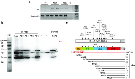
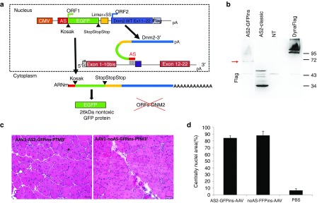
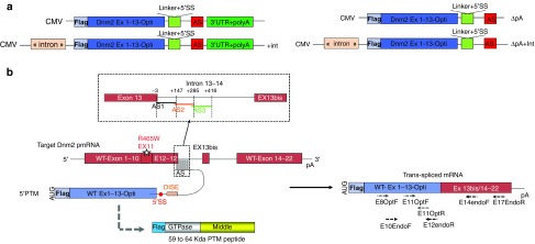
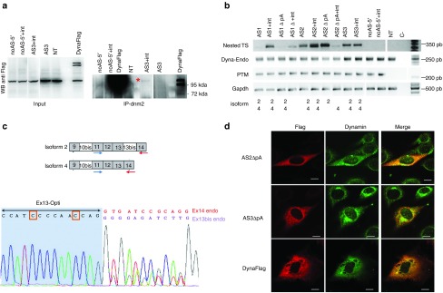
Dynamin 2 (DNM2) is a large GTPase, ubiquitously expressed, involved in membrane trafficking and regulation of actin and microtubule cytoskeletons. DNM2 mutations cause autosomal dominant centronuclear myopathy which is a rare congenital myopathy characterized by skeletal muscle weakness and histopathological features including nuclear centralization in absence of regeneration. No curative treatment is currently available for the DNM2-related autosomal dominant centronuclear myopathy. In order to develop therapeutic strategy, we evaluated here the potential of Spliceosome-Mediated RNA Trans-splicing technology to reprogram the Dnm2-mRNA in vitro and in vivo in mice. We show that classical 3'-trans-splicing strategy cannot be considered as accurate therapeutic strategy regarding toxicity of the pre-trans-splicing molecules leading to low rate of trans-splicing in vivo. Thus, we tested alternative strategies devoted to prevent this toxicity and enhance frequency of trans-splicing events. We succeeded to overcome the toxicity through a 5'-trans-splicing strategy which also allows detection of trans-splicing events at mRNA and protein levels in vitro and in vivo. These results suggest that the Spliceosome-Mediated RNA Trans-splicing strategy may be used to reprogram mutated Dnm2-mRNA but highlight the potential toxicity linked to the molecular tools which have to be carefully investigated during preclinical development.


Figures

Figure 1
Figure 1
The 3′ trans-splicing strategy. (a) Schematic illustration of the 3′-PTM constructs containing CMV promoter, the AS sequence, the 3′ splice site, the wild-type Dnm2 cDNA (optimized sequence of exons 11 to 22 with exon 13 bis) in frame with Flag epitope, and the polyadenylation site. (b) Schematic illustration of 3′ trans-splicing reaction between the endogenous Dnm2 mRNA and the 3′-PTM correcting the mutation in exon 11. Locations of AS sequences are shown in the dotted line square. Primers used for trans-spliced mRNA amplification by RT-PCR are depicted by arrows on the trans-spliced mRNA. CMV, cytomegalovirus promoter; AS, antisense sequence; SS, splice site; pA, polyadenylation site; Dnm2, Dynamin 2; PTM, pre-trans-splicing molecules; RT-PCR, reverse transcription-polymerase chain reaction.
Figure 2
Figure 2
Expression of the 3′-PTM in vitro. (a) RT-PCR detection of trans-spliced Dnm2 transcripts in 3T3 transfected cells. Trans-spliced mRNA were detected after two rounds of PCR (E8-F/E14Opt-R) in cells transfected with AS2- and AS3-3-PTMs. Total Dnm2 mRNAs (endogenous and trans-spliced mRNAs) are amplified using E8-F/E11-R primers. TS, trans-spliced; endo, endogenous. (b) AntiFlag western blot on total extracts from 3T3 transfected cells. The red arrow indicated the expected molecular weight for trans-spliced protein. A 43 kDa nonspecific band (asterisk) associated with the antiFlag antibody observed in both NT and PTM-transfected cells is used as a loading control. AS, antisense sequence; NT, nontransfected cells. 3T3 cells were transfected three times in duplicates. (c) Location of the “Flagged” ORFs relative to the PTM sequence. Eleven ATG are in frame with the Flag sequence, their Kozak score (Ks) obtained on the ATGPR software are indicated. Their positioning relative to the Dnm2 domains and the predicted polypeptide weight are indicated. Possible cryptic CUG start codons are shown in red. M, methionine; L, leucine; MD, middle domain; PH: Plekstrin homology domain; GED, GTPase effector domain, PRD, proline rich domain; Dnm2, Dynamin 2; ORF, open reading frame; PTM, pre-trans-splicing molecules; RT-PCR, reverse transcription-polymerase chain reaction.
Figure 3
Figure 3
Evaluation of Dnm2 3′ trans-splicing strategy in transduced muscles. (a) Muscle integrity after AAV-AS5-3′-PTM injection in TA muscle. HE staining shows the extent of the necrosis process 15 days after injection (top) and the regenerated areas 1.5 months postinjection (bottom). Scale bar = 500 µm (left panel) and 50 µm (right panel). (b) Quantification of AAV viral genomes (vg) by quantitative PCR (qPCR) showing the loss of viral particles during the necrosis/regeneration process. AAV genome copy number is expressed as an absolute value per ng of DNA (mean + SD). (c) RT-PCR detection of trans-spliced Dnm2 transcripts in muscles transduced with AAV-3′-PTM. PCR1-TS/PCR2-TS: First and second RT-PCR using primers Ex8-F/E14OptR amplifying the trans-spliced mRNA. Dyna-Endo: RT-PCR using primers Ex8-F/E14EndoR amplifying the endogenous dynamin 2. TS, trans-spliced; Endo, endogenous. (d) Trans-spliced sequence showing endogenous Dnm2 ex10 bis followed by PTM exon 11 and optimized exon 14 sequence (detected after AAV-AS2-3′-PTM injection). Two TAs were analyzed for each AAV-PTM. AAV, adeno-associated viruses; DNM2, dynamin 2; HE, Hematoxylin-Eosin; PTM, pre-trans-splicing molecules; RT-PCR, reverse transcription-polymerase chain reaction; TA, Tibialis anterior.
Figure 4
Figure 4
The GFP-inserted-3′ trans-splicing strategy. (a) Schematic illustration of the “GFP-inserted” PTM containing the AS sequence, the GFP ORF, the 3′ splice site (SS), and the Dnm2 wild-type sequence (exons 11 to 22) fused to the Flag. CMV, cytomegalovirus promoter; ORF, Open Reading Frame; AS, antisense sequence; SS, Splice site. 3T3 cells were transfected three times in duplicates. (b). Flag western blot on 3T3 transfected cells showing the lack of PTM expression in cells transfected with the “GFP-inserted” constructs, while a ~65 kDa protein is expressed. (c) HE staining obtained 1 month after TA injection with AAV-AS2-GFP-ins-3′-PTM and the AAV-noAS-GFP-ins-3′-PTM. Scale bar = 50 µm. (d) Histogram showing quantification of the regeneration extent on muscles section. The area containing fibres with internal nuclei was measured and divided by the total area of the muscle section (mean + SD, n = 3 sections counted for each condition). GFP, Green Fluorescent Protein; AAV, adeno-associated viruses; DNM2, dynamin 2; HE, Hematoxylin-Eosin; PTM, pre-trans-splicing molecules; RT-PCR, reverse transcription-polymerase chain reaction; TA, Tibialis anterior.
Figure 5
Figure 5
The 5′ trans-splicing strategy. (a) Schematic illustration the 5′-PTM constructs containing a CMV promoter, the wild-type Dnm2 cDNA (optimized sequence of exons 1 to 13 including exon 10 bis), a 5′ splice donor site followed by the AS and with or without polyadenylation site and intron. (b) Schematic illustration of the 5′ trans-splicing reaction between the 5′-PTM and the endogenous Dnm2 mRNA correcting the mutation in exon 11. Location of AS sequences is shown above the dotted line square. The predicted translation of the PTM (i.e., a peptide containing the Flag, the Dnm2 GTPase and Middle domain (525 aa) plus few amino acids encoded by the linker and a part of the AS sequence (59 to 64  kDa) is shown below the PTM. Primers used for RT-PCR mRNA amplification are depicted by arrows below the TS mRNA. CMV, cytomegalovirus promoter; AS, antisense sequence; SS, splices site; DISE, Downstream intronic splice enhancer; pA, polyadenylation site. PTM, pre-trans-splicing molecules; RT-PCR, reverse transcription-polymerase chain reaction; TA, Tibialis anterior.
Figure 6
Figure 6
Detection of 5′ trans-splicing in vitro. (a) Left: Anti-Flag Western Blot on total proteins from 3T3 cells transfected with 5′-PTM or a plasmid expressing full length Dnm2-Flag fusion protein as control (Dynaflag). NT: nontransfected. Right: Dnm2 immunoprecipitation followed by Flag western blot on 3T3 cells transfected with 5′-PTM. The image is overexposed and over contrasted to enhance the detection of the band indicated by an asterisk. (b) RT-PCR analysis on mRNA extracted from 3T3 cells transfected with 5′-PTM. Dyna-endo: endogenous Dnm2 transcripts amplified using E10EndoF/E12endoR primers, TS: trans-spliced RNA amplified using E8OptiF/Ex17endoR and Ex11OptiF/Ex14endoR primers; PTM are amplified using E8OptF/Ex11OptiR primers. C−: negative PCR control. The isoform of Dnm2 identified after sequencing for each band is indicated below (i.e., isoform 2 or 4 or both). 3T3 cells were transfected three times. (c) Top: Schematic representation of Dnm2 alternative splicing leading to isoforms 2 and 4, locations of primers used for PCR amplification are depicted. Bottom: Example of heterozygous trans-spliced sequence showing Ex13 optimized sequence followed by ex13 bis and ex14 endogenous sequence. The orange squares show the optimized nucleotides. (d) Anti-Flag and anti-DNM2 immunofluorescence on 3T3 cells 48 hours after transfection with AS2-, AS3-, noAS-▵pA-5′-PTM and full length Dnm2-Flag fusion constructs (scale bar = 10 µm). PTM, pre-trans-splicing molecules; RT-PCR, reverse transcription-polymerase chain reaction.
Figure 7
Figure 7
Detection of 5′ trans-spliced Dynamin 2 mRNA and protein in muscles. (a) Muscle integrity after AAV-mediated AS1-5′-PTM transduction; Histogram showing the area of muscle sections with internal or central nuclei (%). The area of fibres with internal nuclei was normalized by the total area of the muscle section (Mean + SD, n = 3 sections counted for each condition). (b) RT-PCR analysis on mRNA extracted from transduced muscles using specific primers for trans-spliced and cis-spliced mRNA TS: Trans-spliced PCR amplicon, Endo: endogenous. (c) Electropherogram of trans-spliced amplicons showing Ex13 optimized sequence followed by ex13 bis (isoform 2, top) or ex14 endogenous sequence (isoform 4, bottom). The orange squares show the optimized nucleotides (d). Immunofluorescence analysis on TA transverse sections from mice injected of AAV-AS1-5′-PTM. Fibres were double-stained with anti-FLAG (red) and anti-DNM2 (green) antibodies. Scale bar = 50 µm. AAV, adeno-associated viruses; DNM2, dynamin 2; PTM, pre-trans-splicing molecules; RT-PCR, reverse transcription-polymerase chain reaction; TA, Tibialis anterior.

References

    1. Durieux, AC, Prudhon, B, Guicheney, P and Bitoun, M (2010). Dynamin 2 and human diseases. J Mol Med (Berl) 88: 339–350. - PubMed
    1. Diatloff-Zito, C, Gordon, AJ, Duchaud, E and Merlin, G (1995). Isolation of an ubiquitously expressed cDNA encoding human dynamin II, a member of the large GTP-binding protein family. Gene 163: 301–306. - PubMed
    1. Warnock, DE, Baba, T and Schmid, SL (1997). Ubiquitously expressed dynamin-II has a higher intrinsic GTPase activity and a greater propensity for self-assembly than neuronal dynamin-I. Mol Biol Cell 8: 2553–2562. - PMC - PubMed
    1. Henley, JR, Krueger, EW, Oswald, BJ and McNiven, MA (1998). Dynamin-mediated internalization of caveolae. J Cell Biol 141: 85–99. - PMC - PubMed
    1. Jones, SM, Howell, KE, Henley, JR, Cao, H and McNiven, MA (1998). Role of dynamin in the formation of transport vesicles from the trans-Golgi network. Science 279: 573–577. - PubMed

LinkOut - more resources