. 2016 Nov 1;311(5):E859-E868.
doi: 10.1152/ajpendo.00515.2015. Epub 2016 Sep 13.

Development of a reliable automated screening system to identify small molecules and biologics that promote human β-cell regeneration

Affiliations

Development of a reliable automated screening system to identify small molecules and biologics that promote human β-cell regeneration

Kristie I Aamodt et al. Am J Physiol Endocrinol Metab. .

Abstract

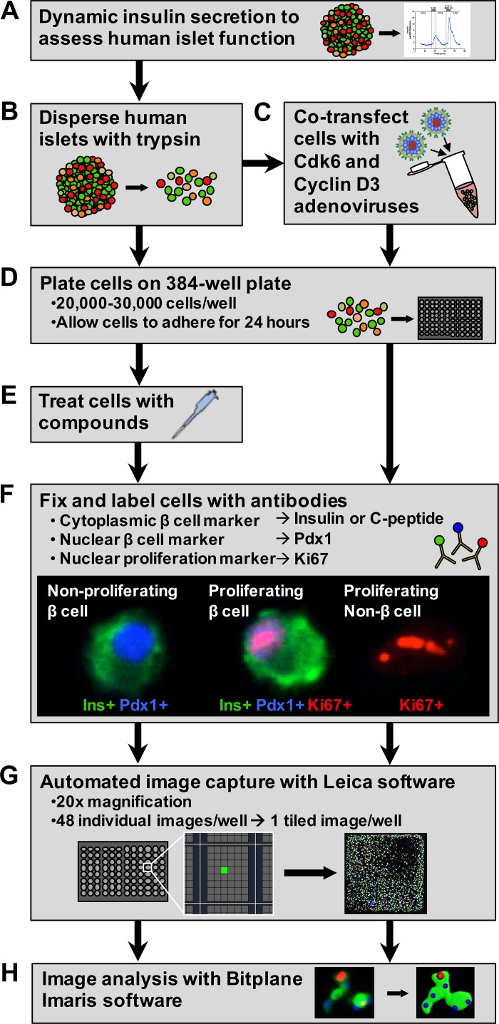
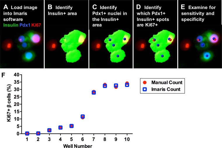
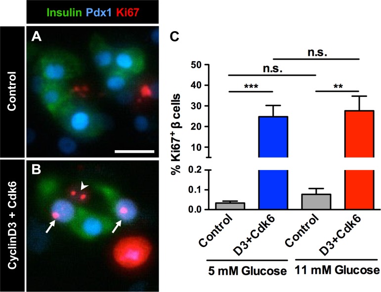
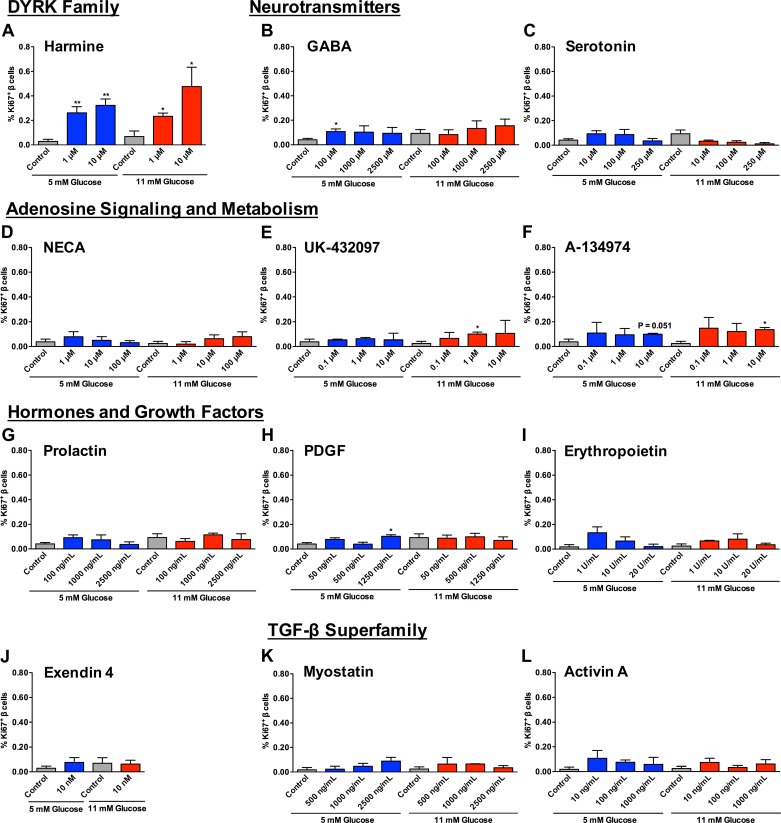
Numerous compounds stimulate rodent β-cell proliferation; however, translating these findings to human β-cells remains a challenge. To examine human β-cell proliferation in response to such compounds, we developed a medium-throughput in vitro method of quantifying adult human β-cell proliferation markers. This method is based on high-content imaging of dispersed islet cells seeded in 384-well plates and automated cell counting that identifies fluorescently labeled β-cells with high specificity using both nuclear and cytoplasmic markers. β-Cells from each donor were assessed for their function and ability to enter the cell cycle by cotransduction with adenoviruses encoding cell cycle regulators cdk6 and cyclin D3. Using this approach, we tested 12 previously identified mitogens, including neurotransmitters, hormones, growth factors, and molecules, involved in adenosine and Tgf-1β signaling. Each compound was tested in a wide concentration range either in the presence of basal (5 mM) or high (11 mM) glucose. Treatment with the control compound harmine, a Dyrk1a inhibitor, led to a significant increase in Ki-67+ β-cells, whereas treatment with other compounds had limited to no effect on human β-cell proliferation. This new scalable approach reduces the time and effort required for sensitive and specific evaluation of human β-cell proliferation, thus allowing for increased testing of candidate human β-cell mitogens.

Keywords: human islet; β-cell proliferation.


Figures

Fig. 1.
Fig. 1.
Workflow for semiautomated evaluation of human β-cell proliferation. A and B: human islets from each donor were evaluated for β-cell function (A) and then dispersed into a single cell suspension (B). C and D: an aliquot of dispersed cells from each donor was cotransfected with cdk6 and cyclin D3 adenoviruses to evaluate their proliferative potential (C) before being plated on 384-well collagen I-coated plates (D) to evaluate β-cell proliferative potential. E: nontransfected cells were also plated, allowed to adhere for 24 h, and then treated with compounds for 72 h. F: following the treatment period, all cells were fixed and labeled with antibodies to identify β-cells using both cytoplasmic (insulin or C-peptide) and nuclear (Pdx1) markers and to identify proliferating cells (Ki-67). G: cells were imaged using Leica LAS AF Matrix Developer software, which automates the capture of 48 images/well at ×20 magnification and tiles them to produce a single merged image of each well. H: these images were then analyzed for β-cell proliferation using Bitplane Imaris software.
Fig. 2.
Fig. 2.
Validation of the Imaris analysis method for quantifying human β-cell proliferation. A and B: images were loaded in Imaris software (A), and then insulin+ area was identified using the Surfaces function (B). C: the Spots function was then used to identify Pdx1+ nuclei that colocalized with Pdx1+ staining masked by the insulin+ surface. These Pdx1+insulin+ spots represent the total number of β-cells in the well. D: Ki-67+ nuclei were then identified using the Spots function and colocalized with the Pdx1+insulin+ spots to determine the number of Pdx1+insulin+Ki-67+ proliferating β-cells. E: the image with Surface and Spot markups was then examined for sensitivity and specificity of the analysis. F: quantification of human β-cell proliferation using Imaris software was validated using images from 10 different wells with varying β-cell proliferation rates due to different levels of cyclin D3 and cdk6 [wells 1 and 2, no transduction; wells 3-10, transduced with 50–250 multiplicity of infection (MOI)] plated at basal (5 mM) glucose. Each well was evaluated by both manual counting and the Imaris algorithm (A-E) to determine the number of proliferating β-cells. The average coefficient of variation between manual and Imaris counts for each well was 3.2% (range 0.6–10.7%), indicating that there is no significant difference between manual and Imaris analysis methods.
Fig. 3.
Fig. 3.
Adenoviral expression of cyclin D3 and cdk6 induces human β-cell proliferation. Dispersed islet cells from all human donors were plated following dispersion in basal (5 mM) or high (11 mM) glucose. An aliquot of cells at each glucose level was cotransduced with adenoviruses encoding cell cycle regulators cyclin D3 and cdk6 (D3+Cdk6) before plating (100–250 MOI). A and B: labeling for cytoplasmic β-cell marker insulin (Ins, green), nuclear β-cell marker Pdx1 (blue), and proliferation marker Ki-67 (red) in control (A) and transduced (B) cells at 5 mM glucose. Arrows mark proliferating β-cells, and arrowhead marks a proliferating non-β-cell. Scale bar in A represents 20 μm and also applies to B. C: adenoviral expression of cyclin D3 and cdk6 induces human β-cell proliferation in cells from all donors at both basal (5 mM) and high (11 mM) glucose; n = 6–9 donors/treatment. **P < 0.01, 5 mM glucose control vs. D3+Cdk6. ***P < 0.001, 11 mM glucose control vs. D3+Cdk6. Comparisons between controls or transfected cells at 5 vs. 11 mM glucose were not statistically significant (ns).
Fig. 4.
Fig. 4.
Human β-cell proliferation is induced by treatment with some, but not all, compounds tested. A: treatment of human islet cells with harmine at 1 and 10 μM in 5 or 11 mM glucose increased β-cell proliferation. **P < 0.01, 5 mM glucose control vs. 1 μM harmine or 10 μM harmine. *P < 0.01, 5 mM glucose control vs. 1 μM harmine or 10 μM harmine. B and C: neurotransmitters GABA (B) and serotonin (C) had a limited effect on β-cell proliferation, and only treatment with 100 μM GABA at 5 mM glucose was statistically significant *P < 0.05, 5 mM glucose control vs. 100 μM GABA. D–F: compounds involved in adenosine signaling and metabolism [NECA (D), UK-432097 (E), and A-134974 (F)] had a modest effect, with 1 μM UK-432097 and 10 μM A-134974 at 11 mM glucose causing a small but statistically significant increase in β-cell proliferation. *P < 0.05, 11 mM glucose control vs. 1 μM UK-432097 and control vs. 10 μM A-134974. G–J: hormones prolactin (G), PDGF (H), erythropoietin (I), and exendin-4 (J) had no effect on β-cell proliferation other than 1,250 ng/ml PDGF at 5 mM glucose, which caused a small but significant increase in proliferation. *P < 0.05, 5 mM glucose control vs. 1,250 ng/ml PDGF. K and L: treatment with members of the TGF-β superfamily myostatin (K) and activin A (L) had no significant effect on β-cell proliferation. Comparisons between controls or compound treatments at 5 vs. 11 mM glucose were not statistically significant in any compound or control tested, and there was no difference between vehicle controls (P = 0.13–0.62); n = 3–6 donors/treatment condition.

References

    1. Andersson O, Adams BA, Yoo D, Ellis GC, Gut P, Anderson RM, German MS, Stainier DYR. Adenosine signaling promotes regeneration of pancreatic β cells in vivo. Cell Metab 15: 885–894, 2012. - PMC - PubMed
    1. Annes JP, Ryu JH, Lam K, Carolan PJ, Utz K, Hollister-Lock J, Arvanites AC, Rubin LL, Weir G, Melton DA. Adenosine kinase inhibition selectively promotes rodent and porcine islet β-cell replication. Proc Natl Acad Sci USA 109: 3915–3920, 2012. - PMC - PubMed
    1. Beattie GM, Cirulli V, Lopez AD, Hayek A. Ex vivo expansion of human pancreatic endocrine cells. J Clin Endocrinol Metab 82: 1852–1856, 1997. - PubMed
    1. Brelje TC, Stout LE, Bhagroo NV, Sorenson RL. Distinctive roles for prolactin and growth hormone in the activation of signal transducer and activator of transcription 5 in pancreatic islets of langerhans. Endocrinology 145: 4162–4175, 2004. - PubMed
    1. Brissova M, Fowler MJ, Nicholson WE, Chu A, Hirshberg B, Harlan DM, Powers AC. Assessment of human pancreatic islet architecture and composition by laser scanning confocal microscopy. J Histochem Cytochem 53: 1087–1097, 2005. - PubMed

MeSH terms

Substances