Case Reports
. 2016 Oct;17(4):227-232.
doi: 10.1007/s10048-016-0493-1. Epub 2016 Sep 13.

PARP10 deficiency manifests by severe developmental delay and DNA repair defect

Affiliations
Case Reports

PARP10 deficiency manifests by severe developmental delay and DNA repair defect

Maher Awni Shahrour et al. Neurogenetics. 2016 Oct.

Erratum in

Abstract

DNA repair mechanisms such as nucleotide excision repair (NER) and translesion synthesis (TLS) are dependent on proliferating cell nuclear antigen (PCNA), a DNA polymerase accessory protein. Recently, homozygosity for p.Ser228Ile mutation in the PCNA gene was reported in patients with neurodegeneration and impaired NER. Using exome sequencing, we identified a homozygous deleterious mutation, c.648delAG, in the PARP10 gene, in a patient suffering from severe developmental delay. In agreement, PARP10 protein was absent from the patient cells. We have previously shown that PARP10 is recruited by PCNA to DNA damage sites and is required for DNA damage resistance. The patient cells were significantly more sensitive to hydroxyurea and UV-induced DNA damage than control cells, resulting in increased apoptosis, indicating DNA repair impairment in the patient cells. PARP10 deficiency joins the long list of DNA repair defects associated with neurodegenerative disorders, including ataxia telangiectasia, xeroderma pigmentosum, Cockayne syndrome, and the recently reported PCNA mutation.

Keywords: DNA repair; Neurodegeneration.


Figures

Figure 1
Figure 1
Family pedigree, PARP10 genotype and patient photos. A-Family pedigree with genotyping of the c.648delAG mutation in the PARP10 gene. B-the chromatogram of the mutation, of a patient (upper panel), a carrier (middle panel) and a healthy control (lower panel) (deleted nucleotides are marked by asterisk in the lower panel). C, D – patient photos at 3 years of age. Note microcephaly, long philtrum, micrognathia and wide alveolar ridge.
Figure 2
Figure 2
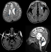
Brain MRI at 3 years of age. T2-weighted axial (A), midsagittal (D) and T1-weighted (B, C) axial images. Note cortical atrophy (arrows) and delayed myelination (four-pointed stars) with paucity of white matter with both cerebral and cerebellar involvement (chevrons).
Figure 3
Figure 3
Patient-derived PARP10 mutant lymphoblast cells show sensitivity to DNA damage. A. Western blot showing that PARP10 protein is absent in Patient-derived lymphoblasts. As controls, PARP10 is detectable in control LCL721 cells. PARP10 was also detected in 293T cells, but not after transfecting these cells with PARP10-targeting siRNA. B. Patient-derive PARP10 mutant lymphoblasts are sensitive to DNA damage compared to control LCL721 cells. Patient and control LCL721 lymphoblasts were treated with Hydroxyurea (HU; 5mM) and Mytomycin C (MMC; 0.1µg/ml) for 6 days. Cellular viability was assayed using trypan blue staining and viable cells were counted using an automated cell counter. The average of 4 independent experiments, with standard errors, is shown. Statistical significance was calculated using the TTEST (two-tailed, equal variance). C. Increased apoptosis in DNA damage–treated patient cells. Cells were exposed to HU (5mM) and MMC (0.1µg/ml) for 24h, and Annexin V staining was performed. Flow cytometry was used to quantify the mean Annexin V staining. The average of 3 independent experiments, with standard deviations, is shown. Statistical significance was calculated using the TTEST (two-tailed, equal variance). D. Increased PARP1 cleavage, indicating increased apoptosis, in DNA damage–treated patient cells. Patient and LCL cells were treated with HU (2mM) and MMC (0.1µg/ml) for 24h. Cells were lysed and extracts were analyzed by western blot with the indicated antibodies. E. Increased DNA damage and reduced mitotic progression in PARP10 mutant cells exposed to HU and MMC. Patient and LCL cells were treated with HU (2mM) and MMC (0.1µg/ml) for 24h. Cells were lysed and extracts were analyzed by western blot with antibodies against γH2AX (DNA damage marker) and phospho-H3 (mitotic marker). F. Alkaline comet assay showing increased DNA damage in PARP10-mutant cells exposed to UV radiation. Cells were analyzed 4h after exposure to 15J/m2. In total, 78 and respectively 79 comet tails were measured, pooled from two independent experiments. Error bars indicate standard errors. Statistical significance was calculated using the TTEST (two-tailed, equal variance).

References

    1. Rass U, Ahel I, West SC. Defective DNA repair and neurodegenerative disease. Cell. 2007;130:991–1004. - PubMed
    1. McKinnon PJ. DNA repair deficiency and neurological disease. Nat Rev Neurosci. 2009;10:100–112. - PMC - PubMed
    1. O'Driscoll M. Diseases associated with defective responses to DNA damage. Cold Spring Harb Perspect Biol. 2012;4(12):a012773. - PMC - PubMed
    1. Hedglin M, Benkovic SJ. Regulation of Rad6/Rad18 Activity During DNA Damage Tolerance. Annu Rev Biophys. 2015;44:207–228. - PMC - PubMed
    1. Chang DJ, Cimprich KA. DNA damage tolerance: when it’s OK to make mistakes. Nat Chem Biol. 2009;5:82–90. - PMC - PubMed

Publication types

Substances