. 2016 Oct 17;26(11):4282-4298.
doi: 10.1093/cercor/bhw216.

Reelin Regulates the Maturation of Dendritic Spines, Synaptogenesis and Glial Ensheathment of Newborn Granule Cells

Affiliations

Reelin Regulates the Maturation of Dendritic Spines, Synaptogenesis and Glial Ensheathment of Newborn Granule Cells

Carles Bosch et al. Cereb Cortex. .

Abstract

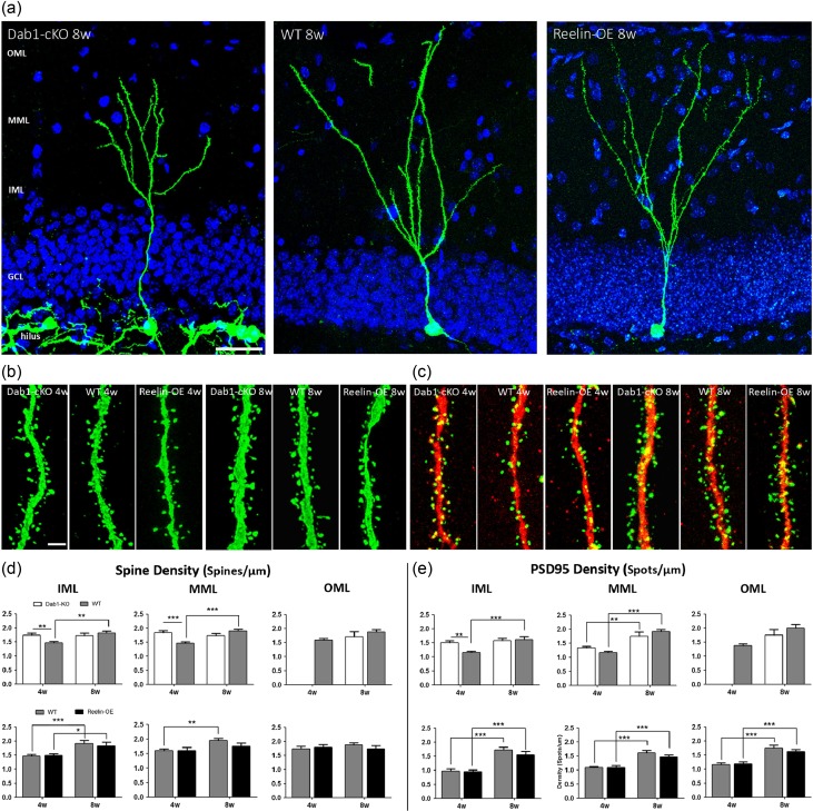
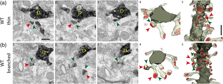
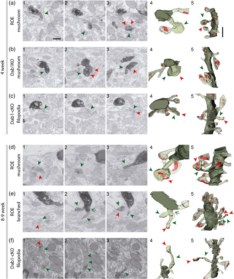
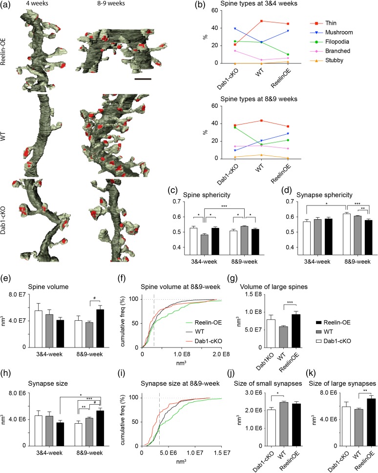
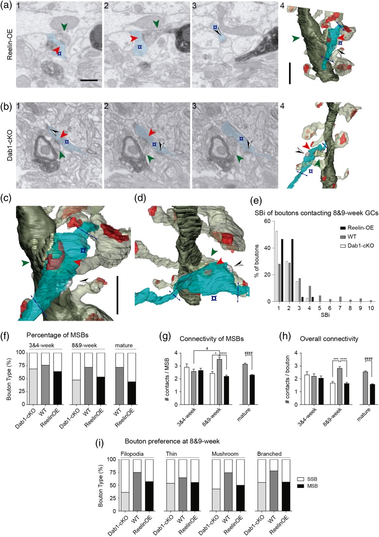
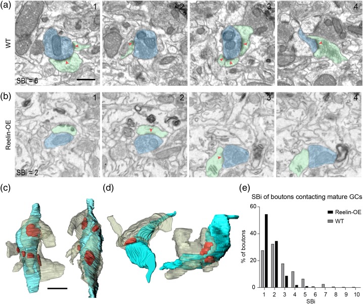
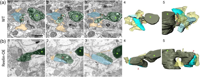
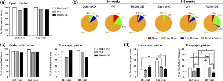
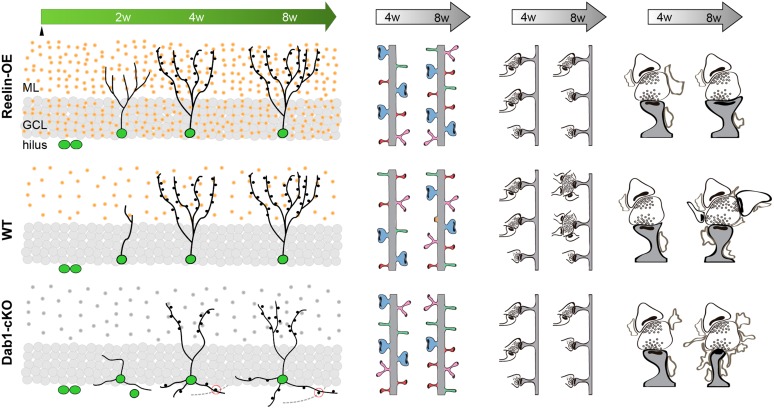
The extracellular protein Reelin has an important role in neurological diseases, including epilepsy, Alzheimer's disease and psychiatric diseases, targeting hippocampal circuits. Here we address the role of Reelin in the development of synaptic contacts in adult-generated granule cells (GCs), a neuronal population that is crucial for learning and memory and implicated in neurological and psychiatric diseases. We found that the Reelin pathway controls the shapes, sizes, and types of dendritic spines, the complexity of multisynaptic innervations and the degree of the perisynaptic astroglial ensheathment that controls synaptic homeostasis. These findings show a pivotal role of Reelin in GC synaptogenesis and provide a foundation for structural circuit alterations caused by Reelin deregulation that may occur in neurological and psychiatric disorders.

Keywords: 3D-electron microscopy; FIB/SEM; adult neurogenesis; axospinous synapses; glia.


Figures

Figure 1.
Figure 1.
Density of spines and postsynaptic densities remain largely unaltered in Reelin-OE and Dab1-cKO newborn GCs. (a) Representative images of GFP-labeled young-adult-born neurons from Dab1-cKO, WT, and Reelin-OE mice at 8 weeks. (b and c) Representative examples of dendritic segments analyzed from adult-born neurons expressing GFP (b) and PSD95-GFP (c) from Dab1-cKO, WT, and Reelin-OE mice at 4 and 8 weeks. (d and e) Quantifications of the spine density (d) and PSD95-GFP spot density (e) in dendritic segments of newborn neurons in the 3 ML layers. Scale bar = 50 µm (a) and 2 µm (b and c). Abbreviations: GCL, Granule Cell Layer. Data represented as mean ± SEM; *P < 0.05; **P < 0.01; ***P < 0.001.
Figure 2.
Figure 2.
Spines of 8-week-old WT GFP/DAB-labeled GCs reconstructed with FIB/SEM microscopy. Examples of a thin (a) and a branched (b) spine arising from their parent dendrite (D). The left images (1–3) show selected serial planes of the spines depicting the head (green arrowheads), neck (green arrow) and synaptic contact (red arrowheads). The right 3D reconstructions (4–5) show the labeled spines in 2 orthogonal orientations. The dendritic shaft is shown in solid dark green, the spine of interest in solid pale green, and its synapse in solid red. Neighboring spines and synapses are indicated in light pale green and red, respectively. Scale bar in a1 is 0.5 µm and applies to a-b 1–4. Scale bar in a5 is 1 µm and applies to a-b5. Abbreviations: D, dendrite.
Figure 3.
Figure 3.
Spines of 8- to 9-week-old Reelin-OE and Dab1-cKO GFP/DAB-labeled GCs reconstructed with FIB/SEM microscopy. Examples of spines of the different reported types arising from dendrites of 4-week (a–c) and 8- to 9-week-old (d–f) Reelin-OE (a, d and e) and Dab1-cKO (b and c, f) GCs. Mushroom, branched, and filopodial spines are shown. The left images (1–3) show selected serial planes of the spines depicting the head (green arrowheads), neck and synaptic contact (red arrowheads). The right 3D reconstructions (4–5) show the labeled spines in 2 orthogonal orientations. The dendritic shaft is shown in solid dark green, the spine of interest in solid pale green, and its synapse in solid red. Neighboring spines and synapses are indicated in light pale green and red, respectively. Scale bar in a1 is 0.5 µm and applies to a–f 1–4. Scale bar in a5 is 1 µm and applies to a–f5.
Figure 4.
Figure 4.
The Reelin/Dab1 pathway alters newborn GC dendritic spine morphology and types. (a) 3D reconstructions allowing comparison of dendritic segments and spines at 3–4 and 8–9 weeks. The color code is the same as that described in Fig. 3. (b) Plots show the percentages of the different types of spines at 3–4 and 8–9 weeks. (c and d) Sphericities of fully reconstructed spines (c) and synapses (d) at the different time points and genotypes. (e) Spine volumes of fully reconstructed spines at the different time points and genotypes. (f) Cumulative distribution of spine volumes at 8–9 weeks. Notice the threshold (dashed gray line) above which Reelin-OE spine volumes distribute differently from WT. This population of large spines is analyzed in detail in (g). (h) Synapse sizes of fully reconstructed synapses at the different time points and genotypes. (i) Cumulative distribution of synapse sizes at 8–9 weeks. Notice the threshold (dashed gray line) above which Reelin-OE synapses show larger sizes than WT. This threshold was used to split all synapses into 2 groups, the smaller synapses being analyzed in (j) and the larger ones in (k). Scale bar in a is 1 µm. Data represent mean ± SEM. *P < 0.05, **P < 0.01, ***P < 0.001; Kruskal–Wallis test and post hoc Dunn's. #P < 0.05; Mann–Whitney test.
Figure 5.
Figure 5.
The Reelin/Dab1 pathway strongly regulates presynaptic innervation of newborn GC spines. (a–d) Two examples depicting multiple synaptic boutons establishing synaptic contacts with Reelin-OE (a, c) and Dab1-cKO (b, d) GCs. The left FIB/SEM images (1–3) show selected serial planes of the labeled dendritic spine heads (green arrowhead) and the presynaptic bouton (colored in blue) establishing a synapse onto them (red arrowhead). The boutons establish additional synapses (black and white arrowheads) on non-labeled spines. The right 3D reconstructions (4) show the same elements in a different orientation. Tilted larger magnifications of a4, b4 are shown in c, d, respectively. Note that only varicosities presynaptic to the labeled spine were analyzed (delimited by blue dashed lines in the 3D panels). The color code is as described in Fig. 3; additionally, the axon is shown in light blue, and synapses established by the axon on non-labeled spines in solid gray. (e) Histogram showing the frequency of synaptic contacts (SBi, number of synapses per bouton) established by axon terminals at 8–9 weeks. (f) Percentage of SSB and MSB that make synapses onto dendritic spines aged 3–4 and 8–9 weeks. (g and h) Average number of synaptic contacts established by MSBs (g) and by any bouton (h) on 3–4 and 8- to 9-week-old GCs. Values obtained for neighboring “mature” axons are also shown. (i) Percentage of SSBs and MSBs establishing contacts on each spine type at 8- to 9-week-old WT, Reelin-OE and Dab1-cKO GCs. Scale bar in a1 is 0.5 µm and applies to a and b 1–3. Scale bar in a4 is 1 µm and applies to a and b4. Scale bar in c is 1 µm and applies to c and d. Data represent mean ± SEM. *P < 0.05, ***P < 0.001, ****P < 0.0001; Kruskal–Wallis test and post hoc Dunn's. #P < 0.05, ####P < 0.0001; Mann–Whitney test.
Figure 6.
Figure 6.
Presynaptic innervation of mature GCs in WT and Reelin-OE neurons. Two examples depicting synaptic boutons establishing multiple synaptic contacts (total number of contacts, SBi, indicated in a and b1) with mature WT (a) and Reelin-OE (b) GCs. The EM panels (a and b 1–4) show selected serial planes of multisynaptic boutons (blue) establishing synapses (red arrowhead) with non-labeled spines (green). (c and d) 3D reconstructions of the bouton shown in a (c) and b (d) in different orientations. (e) Histogram showing the frequency of synaptic contacts established by axon terminals on mature GCs. Scale bar in a is 0.5 µm and applies to a and b. Scale bar in c is 0.5 µm and applies to c and d.
Figure 7.
Figure 7.
Astrocytic ensheathment of GC synapses in WT and Reelin-OE genotypes. Examples of synaptic couples enwrapped by PAPs in WT (a) and Reelin-OE (b) mice. The left images (1–3) show selected serial planes of PAPs (colored in yellow) surrounding the spines from 8- to 9-week GCs (colored in green, asterisk indicates the dendritic shaft) that establish synaptic contacts (red arrowheads) with presynaptic boutons (colored in blue). The right 3D reconstructions (4–5) show the astrocytic ensheathment in different orientations (yellow). Scale bar in a1 is 0.5 µm and applies for all panels.
Figure 8.
Figure 8.
The Reelin/Dab1 pathway regulates the astroglial ensheathment of newborn GC synapses. (a) Proportion of synapses (spines+boutons) ensheathed by PAPs in 3–4 and 8- to 9-week-old WT, Reelin-OE, and Dab1-KO genotypes. (b) Round graphs showing the frequency of the different types of astrocytic ensheathment on synapses (only on the postsynaptic spine, only on the presynaptic bouton, or on both) of newborn WT, Reelin-OE, and Dab1-KO GCs at 3–4 and 8–9 weeks. (c) Proportion of postsynaptic spines and presynaptic boutons ensheathed by PAPs at 3–4 and 8–9-week-old WT, Reelin-OE, and Dab1-KO genotypes. (d) Percentage of the perimeter covered by astrocytic processes on spines and boutons at 3–4 and 8- to 9-week-old WT, Reelin-OE, and Dab1-KO genotypes. Data represent mean ± SEM. *P < 0.05, **P < 0.01, ***P < 0.001; Kruskal–Wallis test and post hoc Dunn's. #P < 0.05; Mann–Whitney test.
Figure 9.
Figure 9.
The Reelin/Dab1 pathway regulates dendritic, spine, and presynaptic development in young-adult-generated GCs. Representative diagram of adult neurogenesis in WT DG (center, left) shows Reelin presence in the ML of the DG (orange dots) and how new neurons increase their dendritic complexity and spine density over time. Gain of function of Reelin (top left) accelerates the growth of the dendritic tree of new GCs (green) without affecting their final morphology. Loss of Reelin signaling (gray dots) leads to the formation of aberrant granular cells, which possess a less complex dendritic tree in the ML and project several basal dendrites into the hilus, where they receive excitatory inputs (bottom left). Spines in WT experience changes in density and morphology from 4 to 8 w, and maturation occurs along with an increase in the presence of branched spines (central panels). Overexpression of Reelin does not change their density but it triggers a larger presence of mushroom spines at both 4 and 8 w (top). Dab1-cKO neurons exhibit a transient increase in dendritic spines at 4 w, but at 8 w no differences are present compared with WT. Loss of Dab1 leads to major changes in spine morphology, including a transient increase in mushroom and branched spines at 4 w, followed by a marked decrease in mushroom spines at 8 weeks and an increase in the filopodial type (bottom). Regarding the presynaptic bouton, the increase in synaptic multi-innervation seen in WT from 4 to 8 weeks is severely disrupted upon both Reelin overexpression and Dab1 loss of function. Finally, downregulation or overactivation of the Reelin/Dab1 pathway leads to severe alterations in the perisynaptic astroglial ensheathment (right). Abbreviations: GCL, Granule Cell Layer.

References

    1. Abdolmaleky HM, Cheng KH, Russo A, Smith CL, Faraone SV, Wilcox M, Shafa R, Glatt SJ, Nguyen G, Ponte JF, et al. . 2005. Hypermethylation of the reelin (RELN) promoter in the brain of schizophrenic patients: a preliminary report. Am J Med Genet B Neuropsychiatr Genet. 134B:60–66. - PubMed
    1. Aimone JB, Deng W, Gage FH. 2011. Resolving new memories: a critical look at the dentate gyrus, adult neurogenesis, and pattern separation. Neuron. 70:589–596. - PMC - PubMed
    1. Akers KG, Martinez-Canabal A, Restivo L, Yiu AP, De Cristofaro A, Hsiang HL, Wheeler AL, Guskjolen A, Niibori Y, Shoji H, et al. . 2014. Hippocampal neurogenesis regulates forgetting during adulthood and infancy. Science. 344:598–602. - PubMed
    1. Alcantara S, Ruiz M, D'Arcangelo G, Ezan F, de Lecea L, Curran T, Sotelo C, Soriano E. 1998. Regional and cellular patterns of reelin mRNA expression in the forebrain of the developing and adult mouse. J Neurosci. 18:7779–7799. - PMC - PubMed
    1. Althaus AL, Sagher O, Parent JM, Murphy GG. 2015. Intrinsic neurophysiological properties of hilar ectopic and normotopic dentate granule cells in human temporal lobe epilepsy and a rat model. J Neurophysiol. 113:1184–1194. - PMC - PubMed

MeSH terms