. 2016 Oct 21;291(43):22583-22593.
doi: 10.1074/jbc.M116.742304. Epub 2016 Sep 13.

Transcriptional Regulation of Brain-derived Neurotrophic Factor Coding Exon IX: ROLE OF NUCLEAR RESPIRATORY FACTOR 2

Affiliations

Transcriptional Regulation of Brain-derived Neurotrophic Factor Coding Exon IX: ROLE OF NUCLEAR RESPIRATORY FACTOR 2

Bindu Nair et al. J Biol Chem. .

Abstract

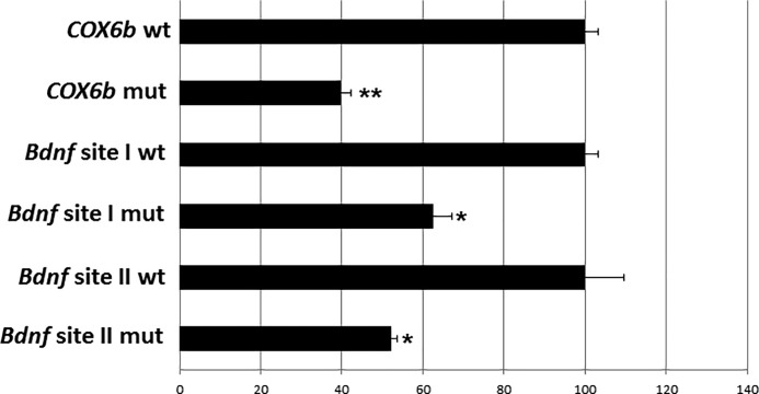
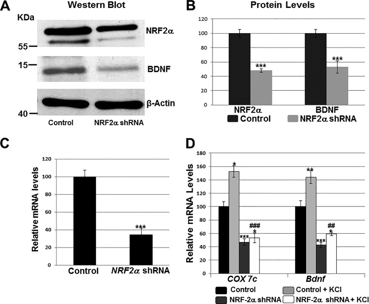
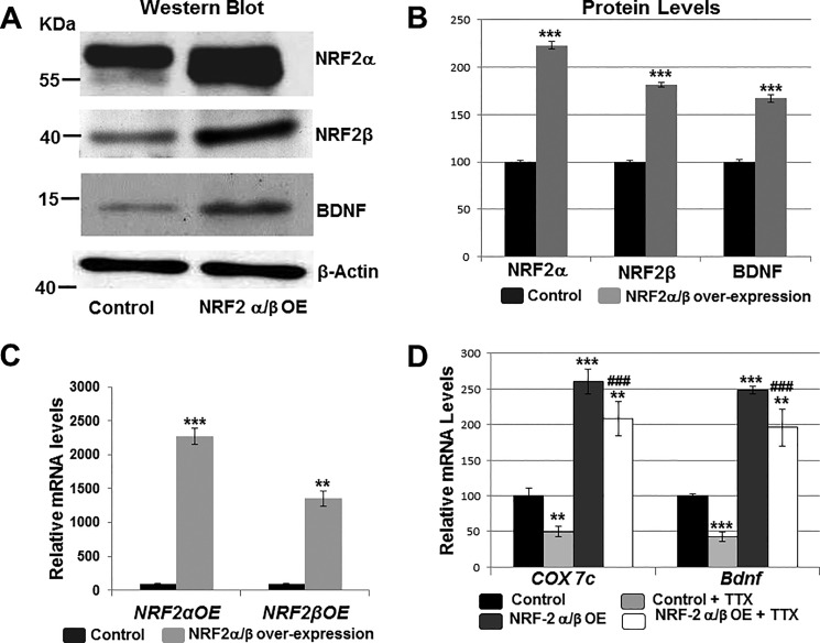
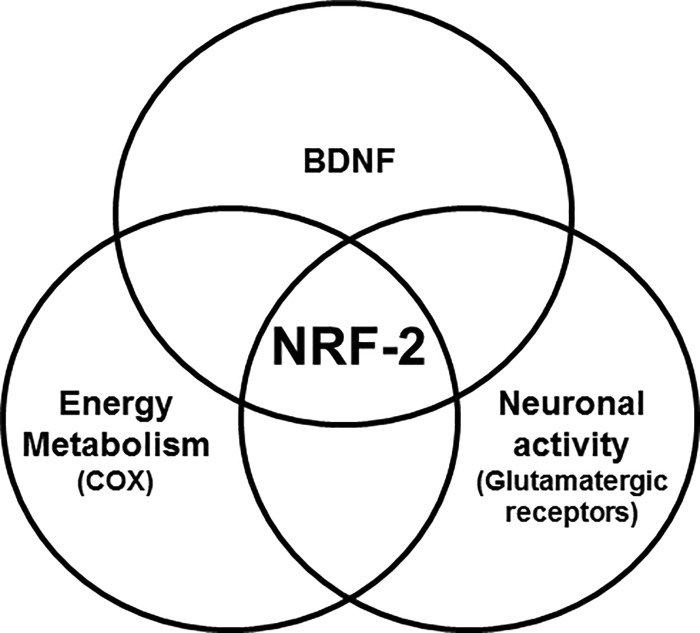
Brain-derived neurotrophic factor (BDNF) is an active neurotrophin abundantly expressed throughout the nervous system. It plays an important role in synaptic transmission, plasticity, neuronal proliferation, differentiation, survival, and death. The Bdnf gene in rodents has eight non-coding exons and only a single coding exon (IX). Despite its recognized regulation by neuronal activity, relatively little is known about its transcriptional regulation, and even less about the transcription factor candidates that may play such a role. The goal of the present study was to probe for such a candidate that may regulate exon IX in the rat Bdnf gene. Our in silico analysis revealed tandem binding sites for nuclear respiratory factor 2 (NRF-2) on the promoter of exon IX. NRF-2 is of special significance because it co-regulates the expressions of mediators of energy metabolism (cytochrome c oxidase) and mediators of neuronal activity (glutamatergic receptors). To test our hypothesis that NRF-2 also regulates the Bdnf gene, we performed electrophoretic mobility shift assay (EMSA), chromatin immunoprecipitation (ChIP), promoter cloning, and site-directed mutagenesis, real-time quantitative PCR (RT-qPCR), and Western blotting analysis. Results indicate that NRF-2 functionally regulates exon IX of the rat Bdnf gene. The binding sites of NRF-2 are conserved between rats and mice. Overexpressing NRF-2 up-regulated the expression of Bdnf exon IX, whereas knocking down NRF-2 down-regulated such expression. These findings are consistent with our hypothesis that NRF-2, in addition to regulating the coupling between neuronal activity and energy metabolism, also regulates the expression of BDNF, which is intimately associated with energy-demanding neuronal activity.

Keywords: brain-derived neurotrophic factor (BDNF); chromatin immunoprecipitation (ChiP); short hairpin RNA (shRNA); transcription factor; transcription regulation.


Figures

FIGURE 1.
FIGURE 1.
Analysis of all Bdnf exons' mRNA levels in the rat visual cortex. Values given are mean ± S.E. The expression level of exon IX is represented as 100%, and those of all other exons are depicted as percentages in comparison to exon IX (n for each exon = 6).
FIGURE 2.
FIGURE 2.
Schematic representation of the rat Bdnf gene showing its eight non-coding exons and the single, coding exon IX, with IXA providing the precursor portion of the pro-BDNF (modified from Ref. , not drawn to scale). The enclosed portion at the top right for exons IXA and IX is enlarged to indicate the four putative NRF-2 binding sites tested. Each binding site contains a tandem repeat of GGAA or TTCC. The translation start site (ATG) of exon IX is located 21 nucleotides downstream of the transcription start point of exon IX. The sequences for sites I and II used for EMSA are indicated below, with the NRF-2 binding motifs underlined. There is high homology between rat and mouse sequences at these two sites. Nucleotide sequences for sites III and IV are also given.
FIGURE 3.
FIGURE 3.
In vitro EMSA and supershift assays to show the binding of NRF-2 to putative sites II and I on the promoter of exon IX in the Bdnf gene. All lanes contain 32P-labeled oligonucleotides containing either site II or site I, and they are indicated by a ”+“ or a ”−“ sign depending on whether they also contain rat brain nuclear extract, excess unlabeled oligos, unlabeled mutant oligos, or NRF-2 antibody. NRF-2 shift, supershift, and nonspecific complexes are indicated by arrows. The positive control for NRF-2 binding is COX6b. Specific NRF-2 shift bands are revealed upon incubation with cortical nuclear extract (lanes 1 and 3). Excess unlabeled competitor competed out the shift band (lane 2). The addition of NRF-2 antibody yielded a strong supershift band (lane 3). Incubation of cortical nuclear extract with the Bdnf probes yielded a specific NRF-2 shift band (lanes 4 and 6 for site II, lanes 12 and 14 for site I) that was competed out with the addition of excess cold probe (lanes 5 and 13). The addition of NRF-2 antibody yielded specific supershift band (lanes 6 and 14). The addition of excess unlabeled mutant Bdnf probes did not compete out the shift reaction (lanes 7 and 15). Labeled Bdnf probe with NRF-2 antibody without any nuclear extract was used as a control to check for any antibody-oligo interaction, and no shift or supershift bands are evident (lanes 8 and 16). Labeled Bdnf probes with mutant NRF-2 sites did not yield prominent specific NRF-2 shift or supershift bands (lanes 9–11 and 17–19).
FIGURE 4.
FIGURE 4.
In vivo chromatin immunoprecipitation assay using rat visual cortical nuclear extract shows that NRF-2 interacts with the promoter of Bdnf exon at both sites I and II, but not at sites III and IV. Immunoprecipitation was carried out with anti-NRF-2α antibody (NRF-2 lane), anti-nerve growth factor receptor p75 antibody (negative control, NGFR lane), or no antibody (negative control, blank lane). 0.5% of input chromatin (input DNA lane) served as the control for PCR. The positive control for NRF-2 binding was COX6b, whereas exon 8 of NRF-1 was the negative control.
FIGURE 5.
FIGURE 5.
Promoter mutational analysis shows relative luciferase activity of wild type (wt) and mutant (mut) promoters of Bdnf exon IX. COX6b served as the positive control. Mutating the NRF-2 binding site in COX6b, and sites I and II in Bdnf resulted in a significant decrease in luciferase activity as compared with the wild type controls (down to 40% for COX6b mutant and to 62.44 and 52% for Bdnf sites I and II mutants, respectively). n = 3 for each construct. **, p < 0.01; *, p < 0.05 as compared with the respective wild type controls.
FIGURE 6.
FIGURE 6.
Effect of silencing NRF-2α with shRNA and KCl depolarizing stimulation. Scrambled shRNA vectors served as the control. A and B, Western blots revealed a down-regulation of NRF-2α and BDNF protein levels in shRNA-transfected primary visual cortical neurons. β-Actin served as a loading control (n = 4 for each). The molecular masses of NRF-2α, BDNF, and β-actin are ∼56, ∼14, and ∼42 kDa, respectively. ***, p < 0.001 when compared with controls. C and D, RT-qPCR revealed a down-regulation of NRF-2α, COX7c, and Bdnf transcripts in neurons transfected with NRF-2α shRNA (n = 4 for each) as compared with those transfected with scrambled shRNA controls. ***, p < 0.001 when compared with controls. Primary neurons exposed to KCl up-regulated their mRNA levels for COX7c and Bdnf (n = 4 for each). *, p < 0.05; **, p < 0.01 when compared with unstimulated controls. In the presence of NRF-2α shRNA, KCl could no longer up-regulate COX7c and Bdnf transcripts to control + KCl levels (n = 4 for each). ##, p < 0.01; ###, p < 0.001 when compared with control + KCl. There were no statistically significant differences in transcript levels of Bdnf and COX7c in neurons treated with NRF-2α shRNA with or without KCl.
FIGURE 7.
FIGURE 7.
Effect of overexpressing NRF-2 on BDNF levels and TTX impulse blockade. Empty vectors served as the control. A and B, Western blots showed that primary neurons transfected with NRF-2α and NRF-2β overexpression vectors up-regulated their NRF-2α, NRF-2β, and BDNF protein levels (n = 4 for each). ***, p < 0.001 when compared with empty vector controls. C and D, overexpressing NRF-2α and -β in primary neurons increased mRNA levels of NRF-2α, NRF-2β, COX7c, and Bdnf in primary neurons (n = 4 for each). **, p < 0.01; ***, p < 0.001 when compared with empty vector controls. Transcript levels of COX7c and Bdnf were reduced with 3 days of 0.4 μm TTX treatment (n = 4 for each). **, p < 0.01; ***, p < 0.001 when compared with no-TTX controls. NRF-2α/β overexpression rescued the TTX-mediated down-regulation of COX7c and Bdnf transcripts (n = 4). **, p < 0.01 when compared with controls. ###, p < 0.001 when compared with control + TTX. There were no statistically significant differences in transcript levels of Bdnf and COX7c in neurons treated with NRF-2 overexpression vectors with or without TTX.
FIGURE 8.
FIGURE 8.
Venn diagram shows how NRF-2 transcriptionally regulates the coupling of energy metabolism (represented by COX), neuronal activity (represented by glutamatergic receptor subunit genes), and BDNF, which is intimately associated with both energy metabolism and neuronal activity.

References

    1. Ernfors P., Wetmore C., Olson L., and Persson H. (1990) Identification of cells in rat brain and peripheral tissues expressing mRNA for members of the nerve growth factor family. Neuron 5, 511–526 - PubMed
    1. Conner J. M., Lauterborn J. C., Yan Q., Gall C. M., and Varon S. (1997) Distribution of brain-derived neurotrophic factor (BDNF) protein and mRNA in the normal adult rat CNS: evidence for anterograde axonal transport. J. Neurosci. 17, 2295–2313 - PMC - PubMed
    1. Katoh-Semba R., Takeuchi I. K., Semba R., and Kato K. (1997) Distribution of brain-derived neurotrophic factor in rats and its changes with development in the brain. J. Neurochem. 69, 34–42 - PubMed
    1. McCarthy D. M., Brown A. N., and Bhide P. G. (2012) Regulation of BDNF expression by cocaine. Yale J. Biol. Med. 85, 437–446 - PMC - PubMed
    1. Yoshii A., and Constantine-Paton M. (2010) Postsynaptic BDNF-TrkB signaling in synapse maturation, plasticity, and disease. Dev. Neurobiol. 70, 304–322 - PMC - PubMed

MeSH terms

Substances

LinkOut - more resources