. 2016 Nov 4;291(45):23557-23568.
doi: 10.1074/jbc.M116.752766. Epub 2016 Sep 13.

Novel Insights into the PKCβ-dependent Regulation of the Oxidoreductase p66Shc

Affiliations

Novel Insights into the PKCβ-dependent Regulation of the Oxidoreductase p66Shc

Martina Haller et al. J Biol Chem. .

Abstract

Dysfunctional mitochondria contribute to the development of many diseases and pathological conditions through the excessive production of reactive oxygen species (ROS), and, where studied, ablation of p66Shc (p66) was beneficial. p66 translocates to the mitochondria and oxidizes cytochrome c to yield H2O2, which in turn initiates cell death. PKCβ-mediated phosphorylation of serine 36 in p66 has been implicated as a key regulatory step preceding mitochondrial translocation, ROS production, and cell death, and PKCβ thus may provide a target for therapeutic intervention. We performed a reassessment of PKCβ regulation of the oxidoreductase activity of p66. Although our experiments did not substantiate Ser36 phosphorylation by PKCβ, they instead provided evidence for Ser139 and Ser213 as PKCβ phosphorylation sites regulating the pro-oxidant and pro-apoptotic function of p66. Mutation of another predicted PKCβ phosphorylation site also located in the phosphotyrosine binding domain, threonine 206, had no phenotype. Intriguingly, p66 with Thr206 and Ser213 mutated to glutamic acid showed a gain-of-function phenotype with significantly increased ROS production and cell death induction. Taken together, these data argue for a complex mechanism of PKCβ-dependent regulation of p66 activation involving Ser139 and a motif surrounding Ser213.

Keywords: PKC; cell death; p66shc; phosphorylation; phosphotyrosine binding (PTB) domain; reactive oxygen species (ROS); redox signaling.


Figures

FIGURE 1.
FIGURE 1.
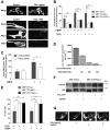
Regulation of p66 redox activity by PKC. A, HEK293 cells were transfected with pECFP-PKCα, β, or θ, and fluorescence images were acquired before and after treatment with TPA (1 μg) or an oxidative challenge (H2O2 1 mm). Before stress application, all PKC isoforms showed a predominantly cytosolic distribution as described previously (31). Upon TPA stimulation or H2O2 treatment, distinct membrane fluorescence was detected (n ≥ 3). All images were acquired using a ×63 oil immersion objective, and for better visualization, the images were adjusted, and representative cells are shown. B, MEF 3T3 WT, p66−/−, and PKCβ−/− cells were treated with t-BHP or H2O2 (1 mm, 30 min) (n ≥ 4). C, effect of PKCβ knockdown on mitochondrial ROS levels in MEF 3T3 WT cells after t-BHP exposure (1 mm, 30 min) (n = 4). D, PKCβ expression was determined by real-time quantitative PCR after transfecting cells with PKCβ or control siRNA and normalized to the housekeeping gene RSP-29. E, MEF 3T3 WT, p66−/−, and PKCβ−/− cells were treated for 24 h with 800 μm H2O2, 30 μm t-BHP. The percentage of vital cells was determined after Annexin V/propidium iodide staining (n ≥ 3). F, H2O2 (1 mm, 30 min) induced p66 phosphorylation on Ser36 in both PKCβ 3T3 WT and PKCβ−/− MEFs, whereas preincubation with Gö6976 (500 nm, 1 h) did not abrogate p66 Ser36 phosphorylation (n ≥ 3). G, pretreatment of PKCβ-CFP-transfected HEK293 cells with Gö6976 (500 nm) prevents PKCβ plasma membrane translocation. Statistics were done using ANOVA (*, p < 0.05; ***, p < 0.001).
FIGURE 2.
FIGURE 2.
PKCβ regulatory sites in p66. A, PyMOL was used to visualize and inspect the structure of the PTB domain of the Shc1 protein harboring Ser139, Thr206, and Ser213. Structural information obtained from the PDB (1SHC) was obtained for the smaller p52Shc isoform, and the numbering of amino acids, therefore, had to be adjusted accordingly. Thus, Ser139, Thr206, and Ser213 correspond to Ser29, Thr96, and Ser103, respectively. B, recombinant PKCβ was used to phosphorylate peptides containing Ser36, Ser139, Thr206, or Ser213. Pos, positive; Neg, negative; CPM, counts per minute.
FIGURE 3.
FIGURE 3.
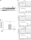
Ser139, Thr206, and Ser213 are PKCβ-regulatory sites. A, for mass spectrometry, an in vitro kinase assay was performed with recombinant kinases and p66 as substrate and subjected to SDS-PAGE. The gel was stained with Coomassie or immunoblotted for p66 Ser36. B, the protein bands were excised from Coomassie-stained gel and digested with trypsin or Lys C. Both Tryptic (C) and Lys C (D) digests were analyzed by nano-HPLC coupled via an electrospray ionization interface to a Q Exactive HF mass spectrometer. Data analysis and peak area calculation were performed using Proteome Discoverer 1.4.1.14.
FIGURE 4.
FIGURE 4.
Cellular phosphorylation of Ser139 by PKCβ. A, p66 overexpressed in HEK293 cells and stressed with 1 mm H2O2 for 15 min either alone or in the presence of Gö6976. Cells lysates were analyzed for p66 Ser36 phosphorylation by Western blotting or immunoprecipitated for mass spectrometry. B and C, proteins were digested on beads with trypsin and analyzed by nano-LC coupled via an electrospray ionization interface to a Velos mass spectrometer. The amounts of phosphorylated and non-phosphorylated peptide were calculated by the peak heights of the extracted ion chromatograms provided. A summary graph of more than three individual biological experiments and mass spectrometry analyses is provided. Statistical significance was determined by using ANOVA (*, p < 0.05; **, p < 0.01).
FIGURE 5.
FIGURE 5.
Ser139 in p66 contributes to ROS and cell death regulation upon stress. A, wild-type p66 or the mutants p66S36A and p66S139A were transfected in p66Shc−/− 3T3 MEFs in triplicates and selected for 2 weeks with puromycin (4 μg/ml). Cells were lysed and checked for protein expression with immunoblotting, and cells showing equal expression were scaled up for further experiments. B, cells were treated for 30 min with 1 mm H2O2. Mitochondrial ROS levels were detected by staining the cells with MitoTracker Red CM-H2XROS and visualized by fluorescent microscope (n ≥ 3) or (C) by staining the cells with DCF-DA, which was measured via FACS. D, cell death of MEFs expressing WT p66 or p66 mutated in Ser139 after 24-h treatment with 500 μm H2O2 (n ≥ 5). Statistical significance was determined using t test or ANOVA (**, p < 0.01; ***, p ≤ 0.001; n.s., not significant).
FIGURE 6.
FIGURE 6.
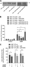
Ser213 in p66 is critical for ROS production. Wild-type p66 or the mutants T206A, S213A, T206/S213A, and T206E/S213E were transfected in p66Shc−/− 3T3 MEFs in triplicates and selected for 2 weeks with puromycin (4 μg/ml). A, cells were lysed and checked for protein expression with immunoblotting, and cells showing equal expression were scaled up for further experiments. The results shown in individual panels were run on the same blot and cropped for clarity. B, ROS levels of p66−/− MEFs expressing wild-type p66 or p66 mutated in Thr206 and/or Ser213 in full-serum medium (control) after 30 min of t-BHP treatment (1 mm) (n ≥ 4). C, mild uncoupling with carbonyl cyanide p-trifluoromethoxyphenylhydrazone (FCCP) (5 μm, 15 min preincubation) decreased mitochondrial ROS levels in MEFs expressing wild-type or Thr206-mutated p66 (n ≥ 5). Statistical analysis was performed using either t test or analysis of variance (*, p < 0.05; **, p < 0.01; ***, p < 0.001).
FIGURE 7.
FIGURE 7.
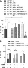
Ser213 in p66 regulates ROS-induced apoptosis. A, survival of MEFs expressing WT p66 or p66 mutated in Thr206 and/or Ser213 after 24-h treatment with 30 μm t-BHP (n ≥ 5). B, pretreatment with NAC (10 mm, 1 h) rescued MEFs expressing wild-type or Thr206-mutated p66 from apoptosis induced by t-BHP (30 μm, 24 h) (n ≥ 5). Statistical significances were determined using t test or analysis of variance (*, p < 0.05; **, p < 0.01; ***, p < 0.001).

References

    1. Kulkarni A. C., Kuppusamy P., and Parinandi N. (2007) Oxygen, the lead actor in the pathophysiologic drama: enactment of the trinity of normoxia, hypoxia, and hyperoxia in disease and therapy. Antioxid. Redox Signal. 9, 1717–1730 - PubMed
    1. McKay A., Cassidy D., Sutherland F., and Dixon E. (2008) Clinical results of N-acetylcysteine after major hepatic surgery: a review. J. Hepatobiliary Pancreat. Surg. 15, 473–478 - PubMed
    1. Fuller T. F., Serkova N., Niemann C. U., and Freise C. E. (2004) Influence of donor pretreatment with N-acetylcysteine on ischemia/reperfusion injury in rat kidney grafts. J. Urol. 171, 1296–1300 - PubMed
    1. Jegatheeswaran S., and Siriwardena A. K. (2011) Experimental and clinical evidence for modification of hepatic ischaemia-reperfusion injury by N-acetylcysteine during major liver surgery. HPB 13, 71–78 - PMC - PubMed
    1. Kozower B. D., Christofidou-Solomidou M., Sweitzer T. D., Muro S., Buerk D. G., Solomides C. C., Albelda S. M., Patterson G. A., and Muzykantov V. R. (2003) Immunotargeting of catalase to the pulmonary endothelium alleviates oxidative stress and reduces acute lung transplantation injury. Nat. Biotechnol. 21, 392–398 - PubMed

MeSH terms

LinkOut - more resources