. 2016 Sep 14:6:33420.
doi: 10.1038/srep33420.

The PCP pathway regulates Baz planar distribution in epithelial cells

Affiliations

The PCP pathway regulates Baz planar distribution in epithelial cells

Benoit Aigouy et al. Sci Rep. .

Abstract

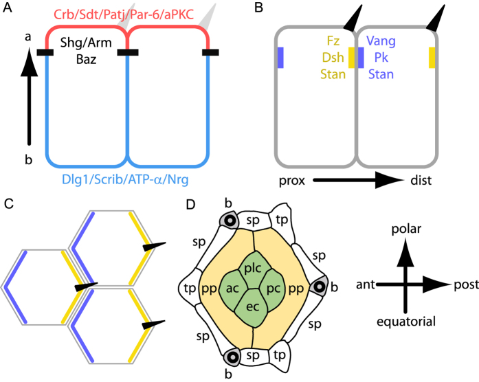
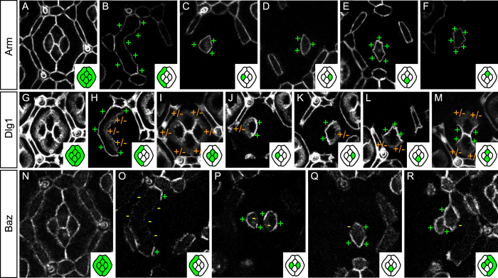
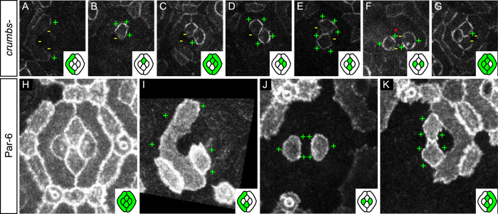
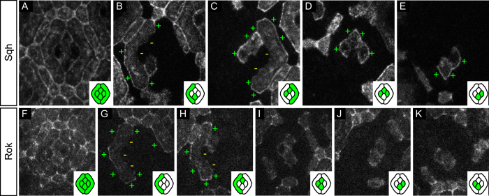
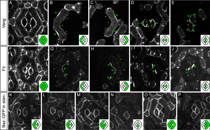
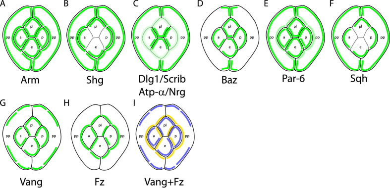
The localisation of apico-basal polarity proteins along the Z-axis of epithelial cells is well understood while their distribution in the plane of the epithelium is poorly characterised. Here we provide a systematic description of the planar localisation of apico-basal polarity proteins in the Drosophila ommatidial epithelium. We show that the adherens junction proteins Shotgun and Armadillo, as well as the baso-lateral complexes, are bilateral, i.e. present on both sides of cell interfaces. In contrast, we report that other key adherens junction proteins, Bazooka and the myosin regulatory light chain (Spaghetti squash) are unilateral, i.e. present on one side of cell interfaces. Furthermore, we demonstrate that planar cell polarity (PCP) and not the apical determinants Crumbs and Par-6 control Bazooka unilaterality in cone cells. Altogether, our work unravels an unexpected organisation and combination of apico-basal, cytoskeletal and planar polarity proteins that is different on either side of cell-cell interfaces and unique for the different contacts of the same cell.


Figures

Figure 1
Figure 1. Planar and transversal distributions of apico-basal and planar polarity proteins.
(A,B) transverse section showing the distribution of apico-basal (A) and planar polarity proteins (B) in fly wing epithelial cells. (A) The apical most region of the cell is shown in red, adherens junction are indicated in black and baso-lateral domains in blue. (B) Proximal (prox) PCP domains containing Vang, Pk and Stan are indicated in blue. Distal (dist) PCP domains containing Fz, Dsh, Dgo and Stan are indicated in yellow. Hairs (dark triangles) grow specifically from the distal side of cells. (C) Top view of the cells shown in (B). (D) Scheme of a 32 h APF fly ommatidium. pp, sp and tp indicate the primary, secondary and tertiary pigment cells, respectively. ac, pc, ec and plc indicate the anterior, posterior, equatorial and polar cone cells, respectively. Wild type ommatidia contain three bristles (b). Here and wherever applicable, the ommatidium is oriented with anterior left and polar up.
Figure 2
Figure 2. Planar distribution of apico-basal polarity proteins.
(A–F) Arm::GFP mosaics. (A) Characteristic distribution of Arm::GFP in a 32 h APF ommatidial epithelium. (B) GFP-labeled primary pigment cell. Note the even Arm::GFP distribution around the primary pigment cell cortex (+). (C–F) Even distribution of Arm::GFP around the cortices of the anterior (C), posterior (D), equatorial (E), and polar (F) cone cells. (G–M) Dlg1::GFP mosaics. (G) Characteristic distribution of Dlg1::GFP in the 32 hAPF ommatidial epithelium. (H) Dlg1::GFP is enriched on the outer interface of the primary pigment cell (+) while its inner interface shows a diffuse Dlg1::GFP signal (+/−). (I) Outer cone cell interfaces show diffuse Dlg1::GFP signal (+/−) while all cone-cone interfaces (J–M) show a strong and sharp Dlg1::GFP signal (+). (N–R) Baz::GFP mosaics. (N) Every interface of the 32 h APF ommatidial epithelium carries Baz::GFP. (O) Baz distribution in primary pigment cells. Baz is devoid from outer and inner primary pigment cells interfaces (−). Baz is specifically enriched at the zone of contact between adjacent primary pigment cells (+). (P) In anterior cone cells (left) Baz::GFP is specifically depleted from the interface shared with the polar cone cell (−) and present elsewhere (+). In posterior cone cells (right), Baz::GFP is specifically depleted on the interface with the equatorial cone cell (−). (Q) In equatorial cone cells, Baz::GFP is excluded from the interface with the anterior cone cell (−). (R) In polar cone cells, Baz::GFP is excluded from the interface with the posterior cone cell. In this figure and the following, insets contain a cartoon representation of the ommatidia where GFP positive cells are shown in green and GFP negative cells in white. To gain space, the posterior primary pigment cell is not shown. Note however that the protein distribution in posterior primary pigment cell is mirror symmetric to that in the anterior primary pigment cell (data not shown).
Figure 3
Figure 3. Crumbs and Par-6 do not regulate the planar distribution of Baz.
(A–G) Baz::GFP mosaics in crb null mutant cells (wild type cone and pigment cells are indicated by a red asterisk). (A,C,G) Inner primary pigment cell interfaces are devoid of Baz signal. (B–G) Baz remains unilateral in crb null mutant cone cells. (H–K) Par-6::GFP mosaics. (H) Characteristic distribution of Par-6 in 32 h APF eyes. (I) Par-6 is enriched on outer (+) primary pigment cell interfaces. (J,K) Par-6 is evenly distributed around the cortex of cone cells (+).
Figure 4
Figure 4. Planar distribution of Sqh and its kinase Rok.
(A–E) Sqh::GFP mosaics. (A) Characteristic distribution of Sqh::GFP in the 32 h APF ommatidial epithelium. (B) Sqh::GFP is enriched on the outer primary pigment cell interfaces (+) and depleted from their inner interfaces (−). (C–E) Outer cone cell interfaces are positive for Sqh::GFP (+). All cone-cone interfaces carry low amounts of Sqh::GFP proteins, preventing us from drawing strong conclusions on the uni- or bilaterality of the protein there. (F–K) Rok::GFP mosaics. (F) Characteristic distribution of Rok::GFP in the 32 h APF ommatidial epithelium. (G,H) Rok::GFP is enriched on the outer primary pigment cell interfaces (+) and depleted from their inner interfaces (−). (H–K) Planar distribution of Rok in cone cells is highly variable, four random samples are presented here.
Figure 5
Figure 5. Planar organisation and function of PCP proteins in the eye.
(A–E) Vang::YFP mosaics. (A) Characteristic distribution of Vang::YFP in 32 h APF ommatidia. (B,C) Vang::YFP is enriched on outer interfaces of primary pigment cells (+) and depleted on their inner interfaces (−). (D,E) Vang::YFP in cone cells. (D) Vang::YFP is depleted (−) in anterior cone cells (left) at their interface with polar cone cells and in posterior cone cells (right) at their interface with equatorial cone cells. (E) Polar cone cells (top) are devoid of Vang::YFP signal (−) at their interface with the posterior cone cells and equatorial cone cells (bottom) are devoid of Vang::YFP signal (−) at their interface with anterior cone cells. (F–J) Fz::YFP mosaics. (F) Characteristic distribution of Fz::YFP in ommatidia. (G,H) In primary pigment cells, Fz is enriched on inner interfaces in contact with cone cells (+) and depleted elsewhere (−). (I,J) Fz is enriched on one interface per cone cell. (I) Fz is enriched in anterior cone cells (left) at the interface with polar cone cells (+). In posterior cone cells (right) Fz is enriched on the interface with equatorial cone cells (+). (J) Fz is enriched in polar cone cells (top) at the interface with posterior cone cells (+). In equatorial cone cells (bottom) Fz is loaded at the interface with anterior cone cells (+). Altogether, the Fz pattern is the negative of the Vang pattern. (K–P) Baz::GFP mosaics in stan null mutant cells (remaining wild type cells are indicated by asterisks). (K–P) Improper ommatidial rotation and misplaced bristles in stan mutants prevent us from determining the antero-posterior and the polar-equatorial axes. Therefore, ommatidia are oriented using the long axis of primary pigment cells in (K–P). (K–M) Polar/equatorial cells express Baz::GFP on their contacts with anterior/posterior cone cells, i.e. no Baz::GFP depletion is observed (compare with Fig. 2P). Baz::GFP shows an even distribution around the cortex of isolated anterior/posterior (L) and polar/equatorial (N) cone cells. (O,P) Polar/equatorial cells express Baz::GFP on their shared interface with anterior/posterior cone cells (compare with Fig. 2Q,R).
Figure 6
Figure 6. Schematic representation of the planar distribution of apico-basal and planar polarity proteins in the ommatidial epithelium.
(A–I) Planar distribution of (A) Arm, (B) Shg, (C) Dlg1/Scrib/ATP-α/Nrg, (D) Baz, (E) Par-6, (F) Sqh, (G) Vang and (H) Fz. (I) Combined planar distribution of Fz (yellow) and Vang (blue). Note the complementary distributions of Fz and Vang proteins. Due to the weakness of the signal on the interfaces between cone cells, Shg (B) and Sqh (F) planar localisation is not represented in (B,F). Similarly the weak PCP signal for Fz and Vang on the interface between equatorial and polar cone cells prevents us from drawing strong conclusions on the planar distribution of PCP proteins on this interface.

References

    1. St Johnston D. & Ahringer J. Cell polarity in eggs and epithelia: parallels and diversity. Cell 141, 757–774, doi: 10.1016/j.cell.2010.05.011 (2010). - DOI - PubMed
    1. Tepass U. The apical polarity protein network in Drosophila epithelial cells: regulation of polarity, junctions, morphogenesis, cell growth, and survival. Annu Rev Cell Dev Biol 28, 655–685, doi: 10.1146/annurev-cellbio-092910-154033 (2012). - DOI - PubMed
    1. Assemat E., Bazellieres E., Pallesi-Pocachard E., Le Bivic A. & Massey-Harroche D. Polarity complex proteins. Biochim Biophys Acta 1778, 614–630, doi: 10.1016/j.bbamem.2007.08.029 (2008). - DOI - PubMed
    1. Tepass U., Theres C. & Knust E. crumbs encodes an EGF-like protein expressed on apical membranes of Drosophila epithelial cells and required for organization of epithelia. Cell 61, 787–799 (1990). - PubMed
    1. Medina E. et al.. Crumbs interacts with moesin and beta(Heavy)-spectrin in the apical membrane skeleton of Drosophila. J Cell Biol 158, 941–951, doi: 10.1083/jcb.200203080 (2002). - DOI - PMC - PubMed

Publication types

MeSH terms

Substances

LinkOut - more resources