. 2016 Sep 13;9(445):rs10.
doi: 10.1126/scisignal.aaf7694.

Chemical proteomic map of dimethyl fumarate-sensitive cysteines in primary human T cells

Affiliations

Chemical proteomic map of dimethyl fumarate-sensitive cysteines in primary human T cells

Megan M Blewett et al. Sci Signal. .

Abstract

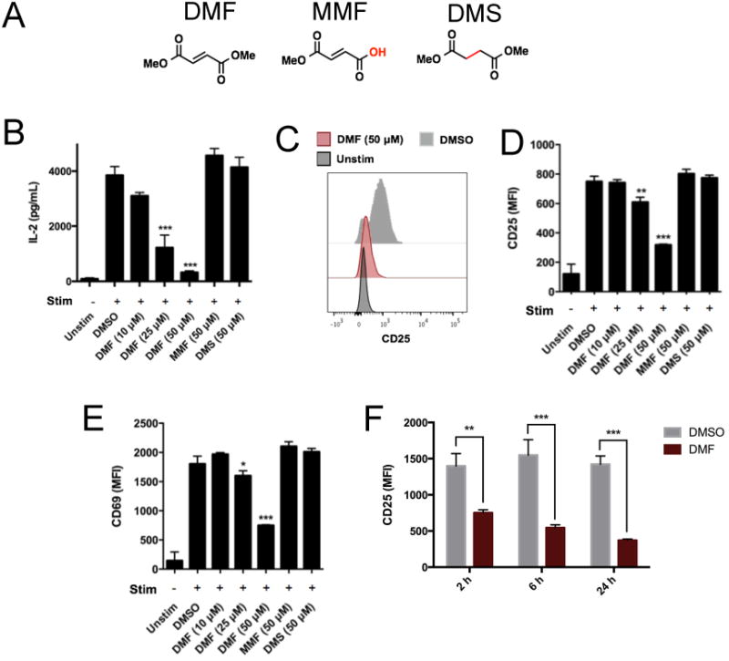
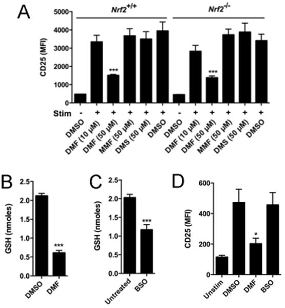
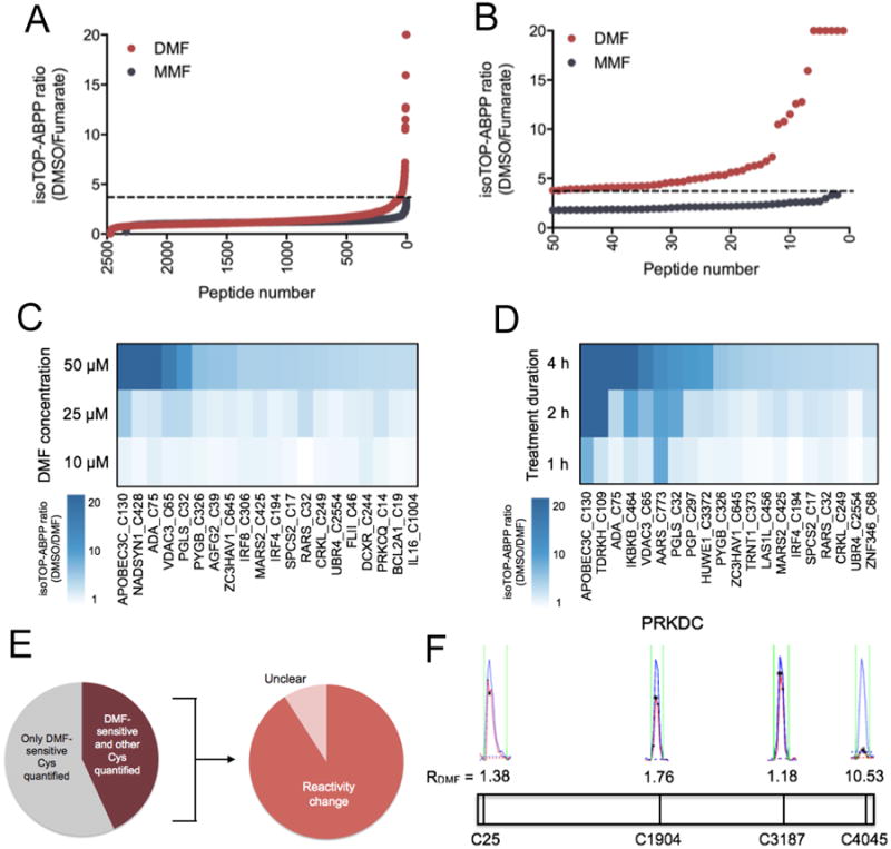
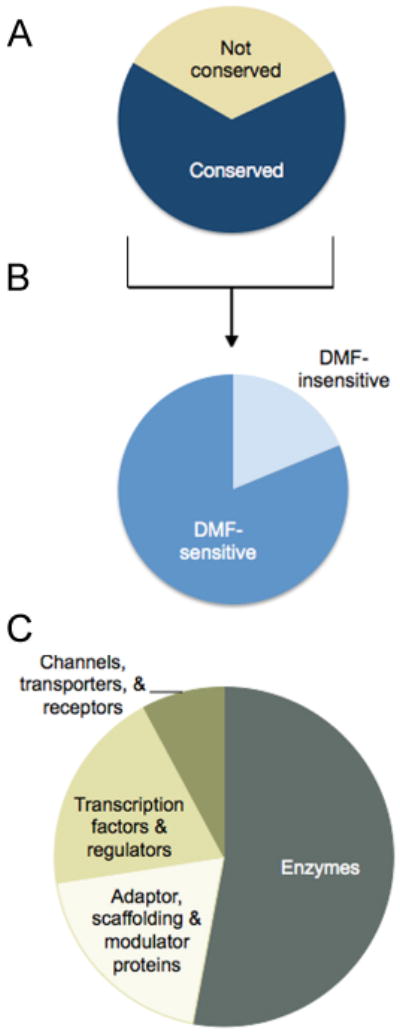
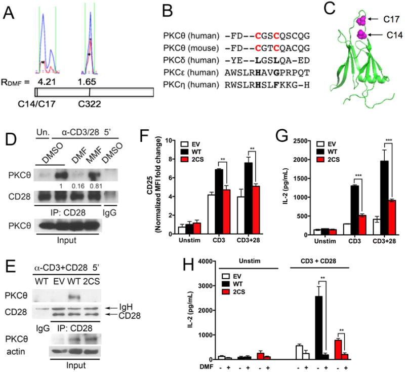
Dimethyl fumarate (DMF) is an electrophilic drug that is used to treat autoimmune conditions, including multiple sclerosis and psoriasis. The mechanism of action of DMF is unclear but may involve the covalent modification of proteins or DMF serving as a prodrug that is converted to monomethyl fumarate (MMF). We found that DMF, but not MMF, blocked the activation of primary human and mouse T cells. Using a quantitative, site-specific chemical proteomic platform, we determined the DMF sensitivity of >2400 cysteine residues in human T cells. Cysteines sensitive to DMF, but not MMF, were identified in several proteins with established biochemical or genetic links to T cell function, including protein kinase Cθ (PKCθ). DMF blocked the association of PKCθ with the costimulatory receptor CD28 by perturbing a CXXC motif in the C2 domain of this kinase. Mutation of these DMF-sensitive cysteines also impaired PKCθ-CD28 interactions and T cell activation, designating the C2 domain of PKCθ as a key functional, electrophile-sensing module important for T cell biology.


Figures

Fig. 1
Fig. 1. DMF inhibits the activation of primary human T cells
(A) The chemical structures of DMF, MMF, and DMS. (B to E) Primary human T cells were left unstimulated (Unstim) or were stimulated (Stim) with anti-CD3 and anti-CD28 (anti-CD3/CD28) antibodies in the presence of DMSO or the indicated concentrations of DMF, MMF, or DMS for 8 hours. The cells were then analyzed by ELISA to determine the amount of IL-2 secreted (B) or were subjected to flow cytometric analysis of the cell surface abundance of CD25 (C and D) and CD69 (E). Flow cytometry data are presented as the mean fluorescence intensity (MFI) of the indicated markers. (F) Time-course analysis of the effects of DMF on CD25 abundance. Primary human T cells were stimulated with anti-CD3/CD28 antibodies in the presence of DMSO or DMF for the indicated times before the DMF-containing medium was replaced with DMF-free medium. The cells were harvested 24 hours after T cell stimulation was begun. The cell-surface abundance of CD25 was measured on gated CD4+ T cells. Data in (B) and (D) to (F) are means ± SEM of four to six technical replicates from each of two experiments. *P < 0.05, **P < 0.01, ***P < 0.001 by two-tailed, unpaired t test in comparison to the DMSO-treated cells.
Fig. 2
Fig. 2. The inhibitory effects of DMF are equivalent in Nrf2+/+ and Nrf2–/– T cells and are not caused by reductions in cellular GSH abundance
(A) Total splenic T cells from Nrf2+/+ and Nrf2–/– mice were left unstimulated or were stimulated with anti-CD3/CD28 antibodies in the presence of the indicated compounds for 24 hours. The cells were then analyzed by flow cytometry to determine the cell surface abundance of CD25. (B and C) DMF and BSO markedly reduce the amount of GSH in human T cells. Primary human T cells were stimulated with anti-CD3/CD28 antibodies in the presence of 50 μM DMF for 2 hours (B) or 2.5 mM BSO for 4 hours (C), after which intracellular GSH concentrations were measured. (D) BSO does not alter T cell activation. Primary human T cells were left unstimulated or were stimulated with anti-CD3/CD28 antibodies in the presence of DMSO, 50 μM DMF, or 2.5 mM BSO for 8 hours. The cells were then analyzed by flow cytometry to measure the cell-surface abundance of CD25. Data are means ± SEM of three or four technical replicates from each of two experiments. *P < 0.05, **P < 0.01, ***P < 0.001 by two-tailed, unpaired t test, in comparison to the DMSO-treated cells.
Fig. 3
Fig. 3. Analysis of DMF-treated primary human T cells by isoTOP-ABPP
(A) isoTOP-ABPP ratios, or R values, for > 2400 cysteine residues in primary human T cells that were activated with anti-CD3/CD28 antibodies and treated for four hours with DMSO, 50 μM DMF, or 50 μM MMF. (B) Expanded profile for DMF-sensitive cysteine residues, where sensitivity is defined as a > four-fold reduction in IA-alkyne reactivity in DMF-treated vs. DMSO-treated T cells (DMSO/DMF R value > 4). For (A) and (B), data represent aggregate quantified cysteine residues from five biological replicates. For the cysteine residues quantified in more than one replicate, average ratios are reported. The dashed line designates R values > 4. (C and D) Concentration-dependent (C) and time-dependent (D) R values for DMF-sensitive cysteine residues in primary human T cells. Each column corresponds to an individual DMF-sensitive cysteine residue, and each tile represents the average R value for that cysteine residue in isoTOP-ABPP experiments performed at the indicated concentration of DMF (concentration-dependence treatments were performed for 4 hours) or duration of treatment (time course treatments were performed with 50 μM DMF). (E) Left: Fraction of proteins for which both a DMF-sensitive cysteine residue and at least one additional cysteine residue was quantified. Right: Fraction of these proteins in which the additional cysteine residue was unchanged (DMSO/DMF R value < 2.0). Unclear calls mark proteins with DMF-sensitive cysteine residues and for which the R value for the second cysteine showed marginal evidence of a potential change (R values between 2.0 and 3.9). (F) Representative MS1 profiles for quantified cysteine residues in PRKDC, one of which (Cys4045) showed sensitivity to DMF. Blue indicates the DMSO-treated sample (heavy isotopic tag); red indicates the DMF-treated sample (light isotopic tag). Asterisks represent MS2 events that were recorded by the MS instrument.
Fig. 4
Fig. 4. Conservation and functional analysis of DMF-sensitive cysteines
(A) Fraction of the DMF-sensitive cysteine residues in the human T cell proteome that are conserved in mice. (B) Fraction of conserved DMF-sensitive cysteine residues in human T cells that were quantified and also sensitive to DMF in mouse T cells. (C) Distribution of proteins in primary human T cells harboring DMF-sensitive cysteine residues by functional class.
Fig. 5
Fig. 5. The DMF-sensitive residues Cys14 and Cys17 in PKC0 are required for its interactions with CD28 and for T cell activation
(A) Representative MS1 profiles for DMF-sensitive (Cys14 and Cys17) and -insensitive (Cys322) cysteine residues in PKCθ. Blue indicates the DMSO-treated sample (heavy isotopic tag); red indicates the DMF-treated sample (light isotopic tag). Asterisks represent MS2 events recorded by the MS instrument. (B) Sequence conservation analysis of human and mouse PKCθ, human PKCδ, human PKCε, and human PKCη. Cys14 and Cys17 are in red. (C) Location of the DMF-sensitive residues Cys14 and Cys17 in the C2 domain of human PKCθ (PDB accession number 2ENJ). (D) DMF, but not MMF, blocks the association of PKCθ with CD28. Peripheral CD4+ T cells from C57BL/6 mice were pre-incubated with DMSO, 50 μM DMF, or 50 μM MMF and then were left unstimulated or were stimulated with anti-CD3/CD28 antibodies for 5 min. The cells were washed, lysed, subjected to immunoprecipitation (IP) with anti-CD28 or control IgG antibodies, and then analyzed by Western blotting with antibodies specific for CD28 and PKCθ. Whole-cell lysates (Input) were also analyzed. Western blots are representative of four experiments. Quantification of band intensities from multiple experiments is shown in fig. S7A. (E) PKCθ–/– T cells were reconstituted with control retrovirus (EV), retrovirus encoding wild-type (WT) PKCθ, or retrovirus encoding the 2CS PKCθ mutant. The cells were then stimulated with anti-CD3/CD28 antibodies for 5 min before being lysed and subjected to immunoprecipitation with anti-CD28 or IgG antibodies. Immunoprecipitated samples and whole-cell lysates (Input) were then analyzed by Western blotting with antibodies against the indicated targets. Western blots are representative of three experiments. Quantification of band intensities from multiple experiments is shown in fig. S7B. (F and G) PKCθ−/− T cells reconstituted with WT PKCθ or the 2CS mutant PKCθ were left unstimulated or were stimulated with anti-CD3 antibody alone or in the presence of anti-CD28 antibody. The cells were then analyzed by flow cytometry to determine the cell-surface abundance of CD25 (F) or by ELISA to measure the amount of secreted IL-2 (G). (H) PKCθ−/− T cells reconstituted with empty vector, WT PKCθ, or the 2CS mutant PKCθ were left unstimulated or were stimulated with anti-CD3/CD28 antibodies overnight in the presence or absence of 50 μM DMF. Cell culture medium was then analyzed by ELISA to measure the amount of secreted IL-2. For (E) to (H), PKCθ−/− T cells were pre-activated with plate-coated anti-CD3/CD28 antibodies for 24 hours before undergoing retroviral transduction. The cells were rested in culture medium without stimulation for 48 hours and then were incubated with or without plate-coated anti-CD3/CD28 antibodies (1 μg/ml) overnight (F) or for 48 hours (G and H), or with soluble anti-CD3/CD28 antibodies (10 μg/ml) for 5 min before being subjected to immunoprecipitation (E). Data in (F) to (H) are means ± SEM of three biological replicates. *P < 0.05, **P < 0.01, ***P < 0.001 by two-tailed, unpaired t test.

Similar articles

Cited by

References

    1. Ropper AH. The “;poison chair”; treatment for multiple sclerosis. The New England journal of medicine. 2012;367:1149–1150. - PubMed
    1. Schimrigk S, Brune N, Hellwig K, Lukas C, Bellenberg B, Rieks M, Hoffmann V, Pohlau D, Przuntek H. Oral fumaric acid esters for the treatment of active multiple sclerosis: an open-label, baseline-controlled pilot study. European journal of neurology. 2006;13:604–610. - PubMed
    1. Gold R, Kappos L, Arnold DL, Bar-Or A, Giovannoni G, Selmaj K, Tornatore C, Sweetser MT, Yang M, Sheikh SI, Dawson KT, Investigators DS. Placebo-controlled phase 3 study of oral BG-12 for relapsing multiple sclerosis. The New England journal of medicine. 2012;367:1098–1107. - PubMed
    1. Fox RJ, Miller DH, Phillips JT, Hutchinson M, Havrdova E, Kita M, Yang M, Raghupathi K, Novas M, Sweetser MT, Viglietta V, Dawson KT, Investigators CS. Placebo-controlled phase 3 study of oral BG-12 or glatiramer in multiple sclerosis. The New England journal of medicine. 2012;367:1087–1097. - PubMed
    1. Linker RA, Gold R. Dimethyl fumarate for treatment of multiple sclerosis: mechanism of action, effectiveness, and side effects. Current neurology and neuroscience reports. 2013;13:394. - PubMed

Publication types