Review
. 2016 Aug 30:10:75.
doi: 10.3389/fnsys.2016.00075. eCollection 2016.

Cortical Plasticity and Olfactory Function in Early Blindness

Affiliations
Review

Cortical Plasticity and Olfactory Function in Early Blindness

Rodrigo Araneda et al. Front Syst Neurosci. .

Abstract

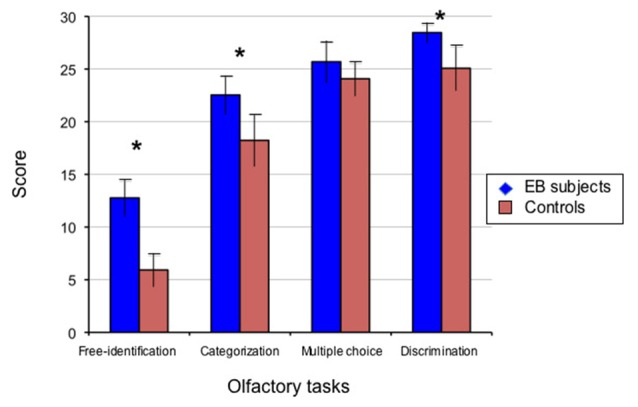
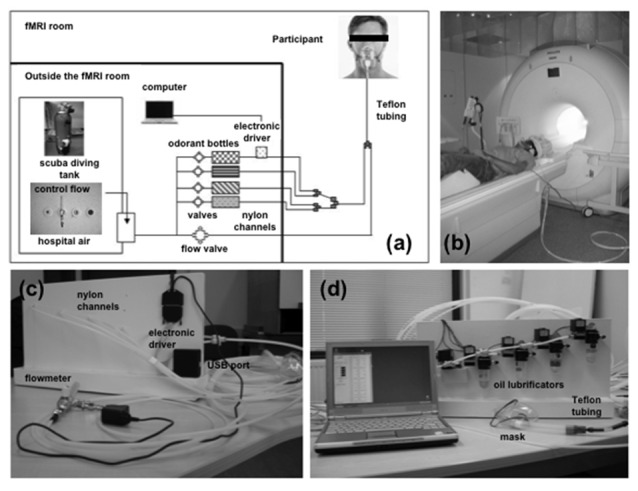
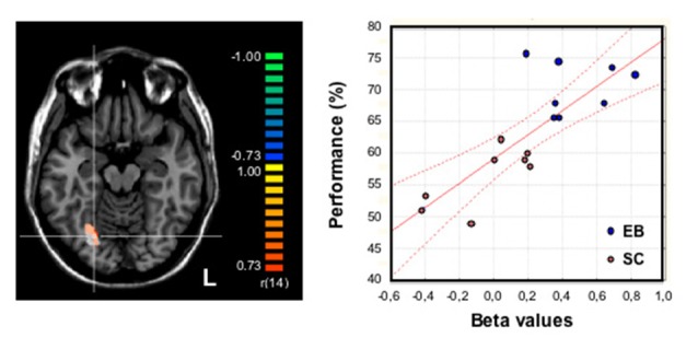
Over the last decade, functional brain imaging has provided insight to the maturation processes and has helped elucidate the pathophysiological mechanisms involved in brain plasticity in the absence of vision. In case of congenital blindness, drastic changes occur within the deafferented "visual" cortex that starts receiving and processing non visual inputs, including olfactory stimuli. This functional reorganization of the occipital cortex gives rise to compensatory perceptual and cognitive mechanisms that help blind persons achieve perceptual tasks, leading to superior olfactory abilities in these subjects. This view receives support from psychophysical testing, volumetric measurements and functional brain imaging studies in humans, which are presented here.

Keywords: congenital blindness; cross-modal plasticity; functional neuroimaging; olfaction; olfactory perception; visual deprivation.


Figures

Figure 1
Figure 1
Performance of early blind (EB) subjects and controls in psychophysical olfactory testing. The perceptual aspects of olfaction were investigated in 13 subjects with early-onset blindness (EB) and 13 sighted controls (SCs) studied blindfolded, using a set of 30 commercially available bottles that contained microencapsulated granules of odorants selected by a perfumer (http://www.sentosphere.fr). Odorants (flower, fruit, plant or domestic elements) were presented orthonasally in one single session. A discrimination task and an identification task with three levels of cuing, i.e., free identification (no cue), categorization (semantic cue), multiple choice (semantic and phonological cues) were used to assess the olfactory abilities. In each task, the quotation was made on a 0/1 basis (for each trial in the related task, 0: wrong; 1: correct), with the total number of correct responses providing the score (maximum: 30/30). Histograms display the mean values and standard deviations of these scores for EB subjects and matched SCs as indicated. The group difference was significant (*p < 0.05) in all conditions except multiple choice identification. EB subjects significantly outperformed the SC participants in odor discrimination (p < 0.0002), free-identification (p < 0.0001) and categorization (p < 0.0004). The multiple choice identification scores showed a trend to a slightly better performance in the EB subjects compared to the blindfolded controls, though not significant (p = 0.063). Adapted from Cuevas et al. (2009).
Figure 2
Figure 2
Results from measurements of olfactory bulb (OB) volume in EB subjects and controls. Using a 3-Tesla MRI and a T2-weighted fast spin-echo sequence in 10 male subjects with early blindness and 10 matched controls, individual OB volume was calculated by plannimetric manual contouring on 23 coronal slices (1.5 mm thickness) perpendicular to the cribriform plane and covering the middle segment of the basifrontal area. Measurements were taken twice by two observers and the mean of these measurements was included as the definite volume, according to a validated protocol for OB analysis (Rombaux et al., and references therein). The OB volume (right + left) in mm3 is plotted as a function of age in EB subjects (blue diamonds, r = 0.30) and controls (orange squares, r = −0.62). Coronal T2 sequence MRI scans of a representative EB subject (top) and a control (bottom) are displayed at the level of OB (indicated by the white arrow). OB: occipital bulb; EB: early blind. Adapted from Rombaux et al. (2010).
Figure 3
Figure 3
Images of the experimental setup for fMRI study of olfactory processing. (A) Schematic representation of the computer-controlled, MRI-compatible odor delivery system. Outside and partly inside the fMRI room, the five nylon channels transmit odorless pulsed air and odorants in separate ways, until they reach the last 30-cm segment nearest to the participant; these channels converge into a single Teflon tube connected to a mask. Outside of the fMRI room, compressed air—either from a scuba-diving tank or a hospital air-care delivery (constant flow)—provides a clean air supply for the stimulator. Bottles containing the odorants (lemon, banana, lavender, rose) are kept in the odor delivery system. An electronic driver is located in the back of the stimulator device (represented schematically in the figure). The computer that controls the stimulator device is located outside the fMRI room. (B) Image of a volunteer participating in a fMRI experiment using the odor delivery system. Auditory signals that allow synchronization of breathing with odor stimulations are delivered via headphones. (C,D) Overall view of the computer-controlled stimulator device, showing nylon channels, fittings, and Teflon tube that deliver the switched air streams to the participant via a removable medical mask; panel (C) shows the view from the back, showing the flowmeter, the start of the five nylon channels, the main power, and the electronic driver, which is equipped with a USB port; panel (D) gives a detailed front view of the device, showing the solenoid valves and oil lubrificators containing the odors in solution. The main part of the device and the computer remain outside the fMRI room, whereas the five nylon channels are passed to the fMRI room through a conventional security hole. Adapted from Cuevas et al. (2010a).
Figure 4
Figure 4
Relationship between olfactory performance and brain activity during odor processing (odor discrimination and categorization). Activation maps were obtained from an analysis of covariance on olfactory conditions plotted together in 16 (8 EB) subjects using their averaged performance in odor free-identification, categorization and discrimination as the covariate (the ability to discriminate, categorize and identify 30 odor samples was assessed before the fMRI study and further averaged to provide a global odor recognition performance expressed in percentage). Brain regions with a positive covariation (p = 0.001 uncorrected, with a cluster size threshold correction of p = 0.05 based on Monte Carlo simulation) were superimposed on the transversal view of a normalized MRI brain of a representative subject. An activation focus was found in the right fusiform gyrus [FG, in orange-yellow: 380 mm3 (center of gravity: x: 24, y: −64, z: −13)] that largely overlapped a brain area that had been identified in the group comparison (EB − SC) for olfactory processing and displayed here in white color as a reference. The crosshairs intersect on a voxel in the right FG (x: 24, y: −67, z: −13). The graph at the right part of the figure shows the strong correlation between brain activity (beta weights) in the right FG during odor processing (white region) and the individual performance (averaged score, %) in the whole group of subjects (n = 16): r = 0.80; p = 0.001. The red lines indicate the confidence interval (CI, 95%). EB, early blind; SC, sighted control (studied blindfolded); L, left hemisphere. Adapted from Renier et al. (2013).

Comment in

References

    1. Afarid M., Torabi-Nami M., Nemati A., Khosravi A., Malekzadeh M. (2015). Brain-derived neurotrophic factor in patients with advanced age-related macular degeneration. Int. J. Ophthalmol. 8, 991–995. 10.3980/j.issn.2222-3959.2015.05.25 - DOI - PMC - PubMed
    1. Amedi A., Malach R., Hendler T., Peled S., Zohary E. (2001). Visuo-haptic object-related activation in the ventral visual pathway. Nat. Neurosci. 4, 324–330. 10.1038/85201 - DOI - PubMed
    1. Amedi A., Raz N., Pianka P., Malach R., Zohary E. (2003). Early visual cortex activation correlates with superior verbal memory performance in the blind. Nat. Neurosci. 6, 758–766. 10.1038/nn1072 - DOI - PubMed
    1. Arno P., De Volder A. G., Vanlierde A., Wanet-Defalque M. C., Streel E., Robert A., et al. . (2001). Occipital activation by pattern recognition in the early blind using auditory substitution of vision. Neuroimage 13, 632–645. 10.1006/nimg.2000.0731 - DOI - PubMed
    1. Bavelier D., Neville H. J. (2002). Cross-modal plasticity: where and how? Nat. Rev. Neurosci. 3, 443–452. 10.1038/nrn848 - DOI - PubMed

LinkOut - more resources