

Traumatic brain injury and subsequent glioblastoma development: Review of the literature and case reports
- PMID: 27625888
- PMCID: PMC5009580
- DOI: 10.4103/2152-7806.189296
Traumatic brain injury and subsequent glioblastoma development: Review of the literature and case reports
Abstract
Background: Previous reports have proposed an association between traumatic brain injury (TBI) and subsequent glioblastoma (GBM) formation.
Methods: We used literature searches and radiographic evidence from two patients to assess the possibility of a link between TBI and GBM.
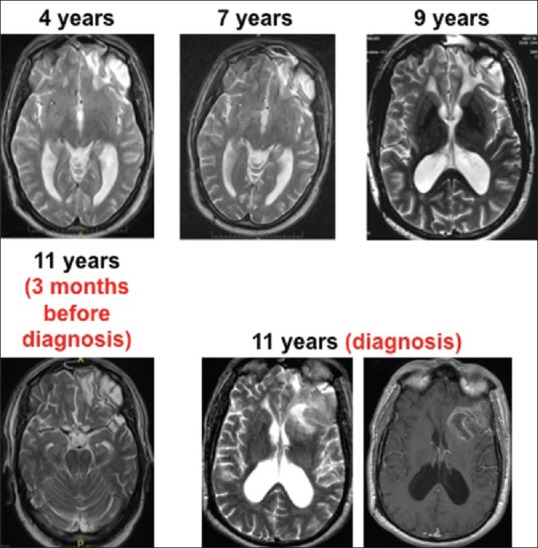
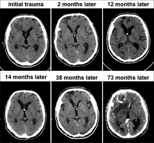
Results: Epidemiological studies are equivocal on a possible link between brain trauma and increased risk of malignant glioma formation. We present two case reports of patients with GBM arising at the site of prior brain injury.
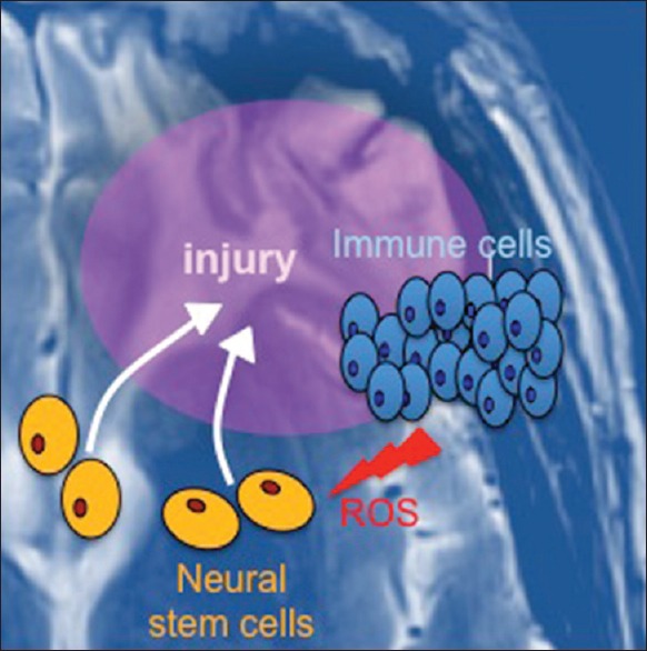
Conclusion: The hypothesis that TBI may predispose to gliomagenesis is disputed by several large-scale epidemiological studies, but supported by some. Radiographic evidence from two cases presented here suggest that GBM formed at the site of brain injury. We propose a putative pathogenesis model that connects post-traumatic inflammation, stem and progenitor cell transformation, and gliomagenesis.
Keywords: Brain tumor; glioblastoma; traumatic brain injury.
Figures
References
-
- Adeberg S, König L, Bostel T, Harrabi S, Welzel T, Debus J, et al. Glioblastoma recurrence patterns after radiation therapy with regard to the subventricular zone. Int J Radiat Oncol Biol Phys. 2014;90:886–93. - PubMed
-
- Amary MF, Damato S, Halai D, Eskandarpour M, Berisha F, Bonar F, et al. Ollier disease and Maffucci syndrome are caused by somatic mosaic mutations of IDH1 and IDH2. Nature Genet. 2011;43:1262–5. - PubMed
-
- Anselmi E, Vallisa D, Bertè R, Vanzo C, Cavanna L. Post-traumatic glioma: Report of two cases. Tumori. 2006;92:175–7. - PubMed
-
- Barnabé-Heider F, Göritz C, Sabelström H, Takebayashi H, Pfrieger FW, Meletis K, et al. Origin of new glial cells in intact and injured adult spinal cord. Cell Stem Cell. 2010;7:470–82. - PubMed
LinkOut - more resources
Full Text Sources
Other Literature Sources
