

Flash photography-induced maculopathy
- PMID: 27625926
- PMCID: PMC5015607
- DOI: 10.3205/oc000004
Flash photography-induced maculopathy
Abstract
Objective: To report a flash photography-induced maculopathy.
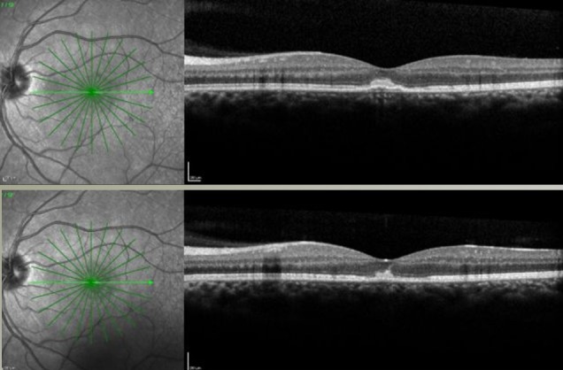
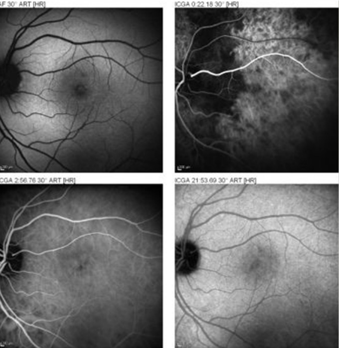
Methods: A professional photographer blinded himself accidentally and he consulted 3 days after the event with a scotoma in his dominant left eye. A unilateral acute light-induced maculopathy with hemorrhage was observed. The lesion was studied with colour photography, fluorescein and indocyanin angiography, autofluorescence imaging and repeated optical coherence tomography (OCT) imaging.
Results: At age 43, this professional photographer was blinded by the flash light of his camera and subsequently realized he had a scotoma in his dominant eye. Three days after the event visual acuity (VA) was 20/70 and an acute light-induced maculopathy was noted. Another three days later, VA was 20/50 and the lesions were less prominent. After one month, the photographer still had problems making sharp pictures, VA was 20/25 and a macular scar was observed. During further follow-up, he regained full vision and experienced no professional problems.
Conclusions: This case illustrates that the light of flash photography can accidentally hit an eye and induce a light-induced maculopathy.
Keywords: acute light-induced maculopathy; flash photography; foveal atrophy; hemorrhage; professional photographer.
Figures


References
-
- Leys A, Swinnen T, Hannon L, Van Wing F. Solar retinopathy and foveal cysts. Bull Soc Belge Ophtalmol. 1978;182:74–81. - PubMed
-
- Stangos A, Petropoulos I, Pournaras JA, et al. Optical coherence tomography and multifocal elecroretinogram findings in chronic solar retinopthy. Am J Ophthalmol. 2007;144:131–134. doi: 10.1016/j.ajo.2007.03.003. Available from: http://dx.doi.org/10.1016/j.ajo.2007.03.003. - DOI - DOI - PubMed
-
- Bechmann M, Ehrt O, Thiel MJ, et al. Optical coherence tomography findings in early solar retinopathy. Br J Ophthalmol. 2000;84:547–548. doi: 10.1136/bjo.84.5.546b. Available from: http://dx.doi.org/10.1136/bjo.84.5.546b. - DOI - DOI - PMC - PubMed
-
- Hope-Ross MW, Mahon GJ, Gardiner TA, et al. Ultrastructural findings in solar retinopathy. Eye. 1993;7:29–33. doi: 10.1038/eye.1993.7. Available from: http://dx.doi.org/10.1038/eye.1993.7. - DOI - DOI - PubMed
-
- Stalmans P, Weckhuysen B, Schoonheydt R, et al. How to protect your eyes from solar retinopathy. Bull Soc Ophthalmol. 1999;272:93–100. - PubMed
Publication types
LinkOut - more resources
Full Text Sources
