. 2016 Aug 26;5(1):1427.
doi: 10.1186/s40064-016-3091-7. eCollection 2016.

Detection of mesenchymal stem cells senescence by prelamin A accumulation at the nuclear level

Affiliations

Detection of mesenchymal stem cells senescence by prelamin A accumulation at the nuclear level

Chiara Bellotti et al. Springerplus. .

Abstract

Background: Human mesenchymal stem cells (MSC), during in vitro expansion, undergo a progressive loss of proliferative potential that leads to the senescent state, associated with a reduction of their "medicinal" properties. This may hampers their efficacy in the treatment of injured tissues. Quality controls on MSC-based cell therapy products should include an assessment of the senescent state. However, a reliable and specific marker is still missing. From studies on lamin-associated disorders, has emerged the correlation between defective lamin A maturation and cellular senescence.

Findings: Primary cultured hMSC lines (n = 3), were analyzed by immunostaining at different life-span stages for the accumulation of prelamin A, along with other markers of cellular senescence. During culture, cells at the last stage of their life span displayed evident signs of senescence consistent with the positivity of SA-β-gal staining. We also observed a significant increase of prelamin A positive cells. Furthermore, we verified that the cells marked by prelamin A were also positive for p21(Waf1) while negative for Ki67.

Conclusions: Overall data support that the detection of prelamin A identifies senescent MSC, providing an easy and reliable tool to be use alone or in combination with known senescence markers to screen MSC before their use in clinical applications.

Keywords: Cell- and tissue-based therapy; Lamin A; Mesenchymal stem cells; Prelamin A; Senescence.


Figures

Fig. 1
Fig. 1
MSC undergo replicative senescence during in vitro expansion. a Cumulative population doublings (CPD) of cell cultures from three different donors. b Passages and PDT intervals corresponding to the early and late stages of each MSC cell line. The end of the late stage matches with replicative senescence of the culture. c Representative brightfield images of MSC at early and late stages. White boxes indicate areas of magnification highlighting the altered shape of senescent cells compared to the well defined spindle-shape of MSC at early stages. Scale bar 200 µm. d Doubling times (DT) calculated from proliferation assay confirm the remarked reduction of proliferation potential of cells according to the progression of life-span stages
Fig. 2
Fig. 2
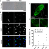
MSC under replicative senescence accumulate prelamin A. a Representative microphotograph of SA-β-gal assay performed on early and late stage cells. Blue staining indicates the presence of β-galactosidase activity in senescent cells. Scale bar 200 μm. b Cells at early and late stages were fixed and immunostained with a specific antibody against prelamin A and counterstained with DAPI. Scale bar 50 µm. c 3D digital rendering of Z-slices confocal images of a representative prelamin A positive cell at late stage. c’ Single slice images of different Z-stacks illustrating the distribution of prelamin A, from surface (left panel) and from middle plane (right panel) point of views. Scale bar 10 µm. d Quantification of β-gal positive and prelamin A positive cells at early and late stages. Blue stained cells and Hoechst stained nuclei were counted in a minimum of six random fields to report the percentage β-gal positive cells at early and late stages. To count prelamin A positive and total cell numbers a minimum of six random fields was checked at early and late stages. Data are expressed as percentage of positive cells respect to the total cell count
Fig. 3
Fig. 3
Cell-cycle arrested cells are positive for prelamin A. Representative images of MSC immunostained for prelamin A (green), Ki67, and p21 (both red in the panels). Cell nuclei were counterstained with DAPI (blue) and merged images are shown in the last column. Prelamin A positive cells (arrows) are negative for Ki67 and positive for p21. Scale bar 10 µm

References

    1. Banfi A, Muraglia A, Dozin B, et al. Proliferation kinetics and differentiation potential of ex vivo expanded human bone marrow stromal cells: implications for their use in cell therapy. Exp Hematol. 2000;28:707–715. doi: 10.1016/S0301-472X(00)00160-0. - DOI - PubMed
    1. Broers J, Ramaekers F. Nuclear lamins: laminopathies and their role in premature ageing. Physiol Rev. 2006;86:967–1008. doi: 10.1152/physrev.00047.2005. - DOI - PubMed
    1. Caron M, Auclair M, Donadille B, et al. Human lipodystrophies linked to mutations in A-type lamins and to HIV protease inhibitor therapy are both associated with prelamin A accumulation, oxidative stress and premature cellular senescence. Cell Death Differ. 2007;14:1759–1767. doi: 10.1038/sj.cdd.4402197. - DOI - PubMed
    1. Coppe J-P, Desprez P-Y, Krtolica A, Campisi J. The senescence-associated secretory phenotype: the dark side of tumor suppression. Annu Rev Pathol. 2010;5:99–118. doi: 10.1146/annurev-pathol-121808-102144. - DOI - PMC - PubMed
    1. Crowe EP, Nacarelli T, Bitto A, et al. Detecting senescence: methods and approaches. In: Noguchi E, Gadaleta MC, et al., editors. Cell cycle control. New York, NY: Springer; 2014. pp. 425–445. - PubMed