. 2016 Sep 14;11(1):125.
doi: 10.1186/s13023-016-0505-0.

The role of small in-frame insertions/deletions in inherited eye disorders and how structural modelling can help estimate their pathogenicity

Affiliations

The role of small in-frame insertions/deletions in inherited eye disorders and how structural modelling can help estimate their pathogenicity

Panagiotis I Sergouniotis et al. Orphanet J Rare Dis. .

Abstract

Background: Although the majority of small in-frame insertions/deletions (indels) has no/little affect on protein function, a small subset of these changes has been causally associated with genetic disorders. Notably, the molecular mechanisms and frequency by which they give rise to disease phenotypes remain largely unknown. The aim of this study is to provide insights into the role of in-frame indels (≤21 nucleotides) in two genetically heterogeneous eye disorders.

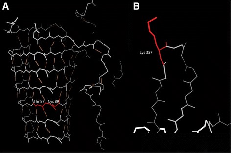
Results: One hundred eighty-one probands with childhood cataracts and 486 probands with retinal dystrophy underwent multigene panel testing in a clinical diagnostic laboratory. In-frame indels were collected and evaluated both clinically and in silico. Variants that could be modeled in the context of protein structure were identified and analysed using integrative structural modeling. Overall, 55 small in-frame indels were detected in 112 of 667 probands (16.8 %); 17 of these changes were novel to this study and 18 variants were reported clinically. A reliable model of the corresponding protein sequence could be generated for 8 variants. Structural modeling indicated a diverse range of molecular mechanisms of disease including disruption of secondary and tertiary protein structure and alteration of protein-DNA binding sites.

Conclusions: In childhood cataract and retinal dystrophy subjects, one small in-frame indel is clinically reported in every ~37 individuals tested. The clinical utility of computational tools evaluating these changes increases when the full complexity of the involved molecular mechanisms is embraced.

Keywords: Childhood cataract; Homology modeling; In-frame insertions/deletions; Inherited eye disease; Retinal dystrophy.


Figures

Fig. 1
Fig. 1
Integrative protein structure modeling for four variants identified in individuals with childhood cataracts. Affected amino acids are highlighted in red. a, b Models of the CRYBA1 c.272_274del, p. (Gly91del) (a) and CRYBA4 c.136_156del, p. (Ser46_Gly52del) (b) variants. The CRYBA1 and CRYBA4 proteins exhibit significant sequence similarity and the template with pdb code 3LWK (human β-crystallin A4) was used on both occasions. The main chain backbone atoms (white/grey lines) and the hydrogen bond network (brown lines) of the affected protein regions are shown. Both sequence alterations involve deleting residues located in β-sheets. c Homology model of the BFSP2 c.697_699del, p. (Glu233del) variant. BFSP2 forms parallel coiled-coil dimers that interact with one another in the form of a symmetrical anti-parallel dimer. The main chain backbone atoms (white/yellow lines) and the side chains that comprise the interaction interface (green) of the affected protein region are shown. The wild-type protein is presented on the left hand-side image. Notably, the affected amino acid is located in an α-helical region (highlighted in red). The right hand-side image shows a model of the mutant protein; the deletion shifts the position of the interacting side chains resulting in disruption of the dimer structure and exposure of the hydrophobic interface residues to the protein surface. d Model of the PITX2 c.429_431del, p. (Arg144del) variant. The main chain backbone atoms of the protein (white/grey lines) complexed with an interacting DNA double helix (brown chain) are shown. The mutated residue (highlighted in red) makes direct contact with the phosphate backbone of DNA, forming a salt bridge
Fig. 2
Fig. 2
Integrative protein structure modeling for two variants identified in individuals with retinal dystrophy. Affected amino acids are highlighted in red. a Model of the RP2 c.260_268del, p. (Thr87_Cys89del) variant. The main chain backbone atoms (white/grey lines) and the hydrogen bond network (brown lines) of the affected protein region are shown. The variant is found in an extended set of β-sheets that form a complex set of hydrogen bonds. b Homology model of the FSCN2 c.1071_1073del, p. (Lys357del) variant. The main chain backbone atoms (white/grey lines) of a small part of the protein is shown. The deletion affects a residue in a surface loop, away from known functional or interaction sites

References

    1. Genomes Project C, Auton A, Brooks LD, et al. A global reference for human genetic variation. Nature. 2015;526:68–74. doi: 10.1038/nature15393. - DOI - PMC - PubMed
    1. Zhang X, Lin H, Zhao H, et al. Impact of human pathogenic micro-insertions and micro-deletions on post-transcriptional regulation. Hum Mol Genet. 2014;23:3024–3034. doi: 10.1093/hmg/ddu019. - DOI - PMC - PubMed
    1. Hu J, Ng PC. SIFT indel: predictions for the functional effects of amino acid insertions/deletions in proteins. PLoS One. 2013;8:e77940. doi: 10.1371/journal.pone.0077940. - DOI - PMC - PubMed
    1. Choi Y, Chan AP. PROVEAN web server: a tool to predict the functional effect of amino acid substitutions and indels. Bioinformatics. 2015;31:2745–2747. doi: 10.1093/bioinformatics/btv195. - DOI - PMC - PubMed
    1. Zhao H, Yang Y, Lin H, et al. DDIG-in: discriminating between disease-associated and neutral non-frameshifting micro-indels. Genome Biol. 2013;14:R23. doi: 10.1186/gb-2013-14-3-r23. - DOI - PMC - PubMed