

Review
doi: 10.1038/nature19948.
Unravelling biological macromolecules with cryo-electron microscopy
Affiliations
- PMID: 27629640
- PMCID: PMC5074357
- DOI: 10.1038/nature19948
Item in Clipboard
Review
Unravelling biological macromolecules with cryo-electron microscopy
Nature.
.
Abstract
Knowledge of the three-dimensional structures of proteins and other biological macromolecules often aids understanding of how they perform complicated tasks in the cell. Because many such tasks involve the cleavage or formation of chemical bonds, structural characterization at the atomic level is most useful. Developments in the electron microscopy of frozen hydrated samples (cryo-electron microscopy) are providing unprecedented opportunities for the structural characterization of biological macromolecules. This is resulting in a wave of information about processes in the cell that were impossible to characterize with existing techniques in structural biology.
Figures
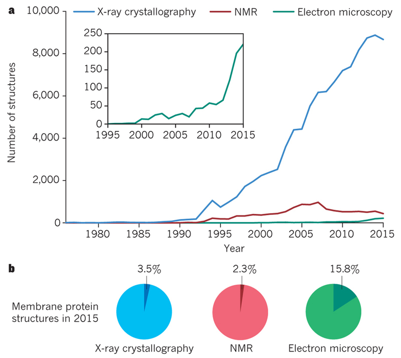
A. Number of structures in the Protein Data Bank as determined by X-ray crystallography (blue), NMR (red) and EM (green). B. Fraction of membrane protein structures in 2015 for each of the three techniques.
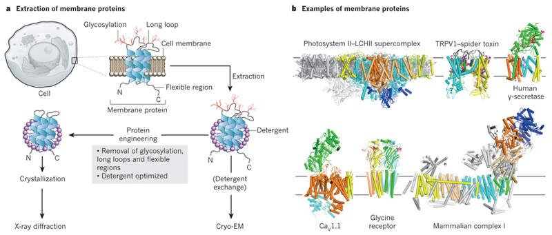
Membrane proteins need to be extracted from their membranes using detergent solubilisation. Membrane protein crystallisation is difficult and often requires protein engineering to remove flexible loops and sugar. Cryo-EM structure determination allows direct imaging of membrane proteins in detergents or in more natural environments like nano-discs. The presence of sugars or very large membrane complexes does not preclude cryo-EM structure determination. The structures of the photosystem II - light harvesting supercomplex, TRPV1 in complex with a spider toxin, gamma-secretase, the voltage-gated calcium channel CaV1.1, the glycine receptor, and bovine complex-I are shown as examples.
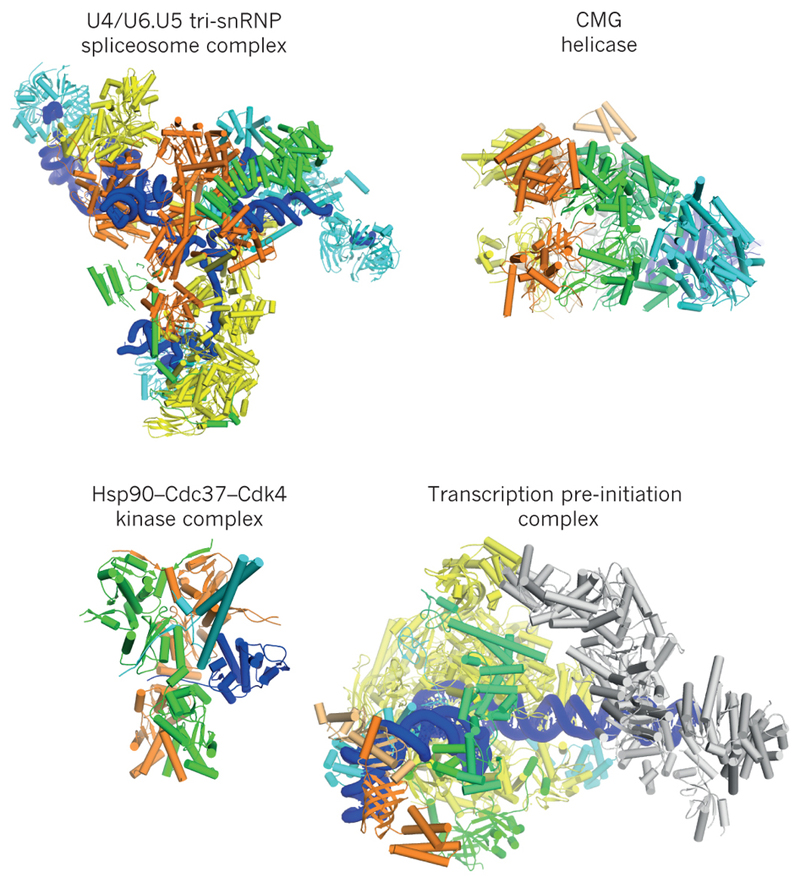
Examples of cryo-EM structures are shown for the U4/U6.U5 tri-snRNP spliceosomal complex from yeast; the eukaryotic replicative CMG helicase; the human Hsp90-Cdc37-Cdk4 kinase complex; and the human transcription pre-initiation complex.
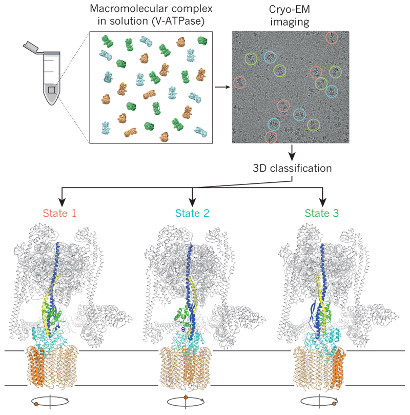
Mixtures of macromolecular complexes in distinct conformational or compositional states may be imaged directly, and image classification may be used to obtain structures for each of the states. Thereby, image classification allows characterization of the functional cycles of dynamic molecular machines from a single experiment. Different functional states of the eukaryotic V-type ATPase are shown as an example.
References
-
- Alberts B. The Cell as a Collection of Protein Machines: Preparing the Next Generation of Molecular Biologists. Cell. 1998;92:291–294. - PubMed
-
- Kuhlbrandt W. The Resolution Revolution. Science. 2014;343:1443–1444. - PubMed
-
- Taylor KA, Glaeser RM. Electron Diffraction of Frozen, Hydrated Protein Crystals. Science. 1974;186:1036–1037. - PubMed
-
- Dubochet J, Chang JJ, Freeman R, Lepault J, McDowall AW. Frozen aqueous suspensions. Ultramicroscopy. 1982;10:55–61.
Publication types
MeSH terms
Substances
Grants and funding
LinkOut - more resources
Full Text Sources
Other Literature Sources
