. 2017 Jan 26;25(2):295-304.
doi: 10.3727/096504016X14732527645922. Epub 2016 Sep 13.

Intratumoral Photodynamic Therapy With Newly Synthesized Pheophorbide a in Murine Oral Cancer

Affiliations

Intratumoral Photodynamic Therapy With Newly Synthesized Pheophorbide a in Murine Oral Cancer

Mee-Young Ahn et al. Oncol Res. .

Abstract

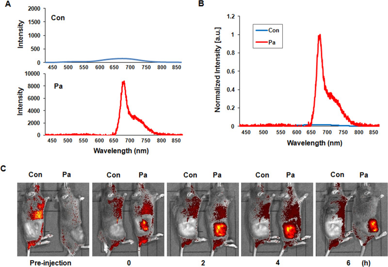
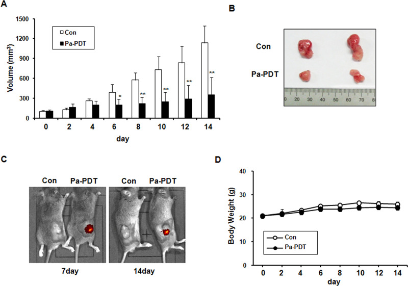
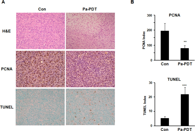
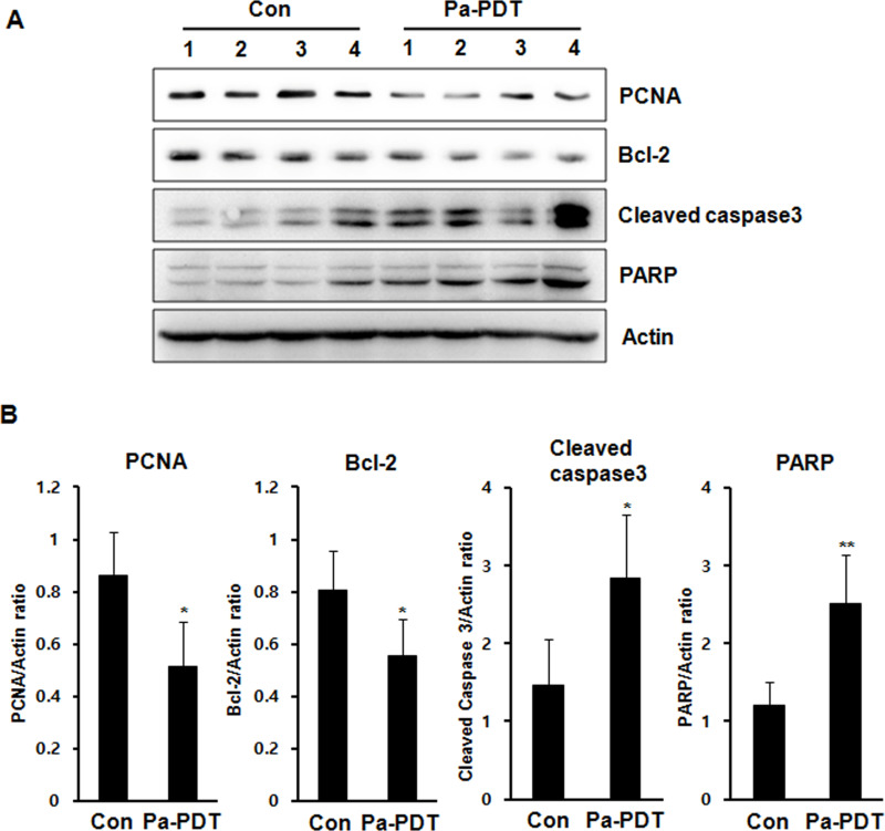
Photodynamic therapy (PDT) is a therapeutic alternative for malignant tumors that uses a photosensitizer. Our group recently synthesized photosensitizer pheophorbide a (Pa) from chlorophyll-a. The present study investigated the therapeutic effect of PDT using intratumoral administration of the synthetic photosensitizer Pa in an in vivo murine oral squamous cell carcinoma (OSCC) animal model. Pa accumulation was measured using the fluorescence spectrum and imaging in living C3H mice. Intratumoral treatment of Pa-PDT (IT Pa-PDT) significantly inhibited the growth of transplanted OSCC cells. Histopathological examination of tumor tissues showed that PCNA expression was significantly decreased, while TUNEL-stained cells were markedly increased in the IT Pa-PDT group compared to controls. IT Pa-PDT-induced apoptosis was confirmed by immunoblot. Reduction of Bcl-2 and cleavage of caspase 3 and PARP were observed in IT Pa-PDT. These data demonstrate that IT Pa-PDT inhibited tumor cell proliferation and induced apoptosis, which is correlated with the anticancer activity of IT Pa-PDT. These potent antitumor activities of IT Pa-PDT were observed in both the immunohistochemistry and Western blot experiments. Our findings suggest the intratumoral therapeutic potential of Pa-PDT on OSCC. Additionally, demonstrated detection of Pa using a fluorescence spectroscopy system or molecular imaging system provides a means for simultaneous diagnosis and treatment of OSCC.


Figures

Figure 1
Figure 1
Chemical structures of chlorophyll-a (A) and synthesized pheophorbide a (B).
Figure 2
Figure 2
Pa accumulation and growth inhibition by Pa-PDT in AT-84 cells. (A) The cells were incubated with 0.25 μM Pa for up to 24 h, and photographs were obtained by fluorescence microscopy (magnification: 200×). (B) The cells were incubated with the indicated concentrations of Pa for 2 h followed by exposure to 4.24 J/cm2 of laser light for 24 h. The levels of cell proliferation were measured using an MTT assay. The percentage of viable cells was calculated as the ratio of treated cells to control cells. The data are reported as the mean ± SD of three independent experiments. *p < 0.05, **p < 0.01 compared to untreated control.
Figure 3
Figure 3
Pa accumulation in the in vivo C3H mice model. Photosensitizer Pa (10 mg/kg) was injected intratumorally into the right flank of C3H mice. (A) The fluorescence spectra were measured from tumor tissues of the control and Pa-treated groups, respectively, using the fluorescence spectroscopy (FS) system. (B) The peak intensity was compared between the control and Pa-treated group. The main fluorescence peaks were observed at ∼672 nm in Pa-treated tumor tissues. (C) In vivo fluorescence imaging was obtained using the Xenogen IVIS-100 Imaging System equipped with a CCD camera system.
Figure 4
Figure 4
Effect of IT Pa-PDT on tumor growth in an in vivo model. (A) Relative tumor volumes in mice inoculated with AT-84 cancer cells. Relative tumor volumes of the mice treated with IT Pa-PDT and the vehicle-treated controls. The tumor volumes were measured and transformed subsequently to the relative tumor volume, as detailed in Materials and Methods. The data are reported as the mean ± SD of five animals. *p < 0.05, **p < 0.01 as determined by a Student’s t-test compared to the control group. (B) In situ appearance of tumors in the control and Pa-PDT-treated mice. (C) In vivo fluorescence imaging of Pa was obtained using the Xenogen IVIS-100 Imaging System equipped with a CCD camera system. (D) An assessment of body weight between the Pa-PDT-treated and control mice during the entire experimental period. The body weight of mice in different groups was recorded every alternate day.
Figure 5
Figure 5
Effect of IT Pa-PDT on the proliferation and apoptosis in vivo model. (A) H&E staining of tumor sections (top), immunohistochemistry for PCNA (middle), and TUNEL assay (bottom) were performed on paraffin sections from the tumor. Photographs were taken under a magnification of 200×. (B) Positive cells for PCNA immunostaining (top) and TUNEL (bottom) were counted, and the results are expressed. The data are reported as the mean ± SD of three independent experiments. **p < 0.01, ***p < 0.001 compared to the control group.
Figure 6
Figure 6
The expression of the apoptosis-related protein levels in the tumor tissues. (A) The protein fraction from tumor tissues and the expression levels of PCNA, Bcl-2, cleaved caspase 3, PARP, and actin were detected using Western blot analysis. The protein levels were normalized by a comparison to the actin levels. (B) The bar graft data are reported as the mean ± SD of three independent experiments. *p < 0.05, **p < 0.01 compared to the control group.

References

    1. Marmur ES, Schmults CD, Goldberg J. A review of laser and photodynamic therapy for the treatment of nonmelanoma skin cancer. Dermatol Surg. 2004;30:264–71. - PubMed
    1. Weishaupt KR, Gomer CJ, Dougherty TJ. Identification of singlet oxygen as the cytotoxic agent in photoinactivation of a murine tumor. Cancer Res. 1976;36:2326–9. - PubMed
    1. Moan J, Berg K. Photochemotherapy of cancer: Experimental research. Photochem Photobiol. 1992;55:931–48. - PubMed
    1. Chiu SM, Oleinick NL. Dissociation of mitochondrial depolarization from cytochrome c release during apoptosis induced by photodynamic therapy. Br J Cancer 2001;84:1099–1106. - PMC - PubMed
    1. Karakullukcu B, Nyst HJ, van Veen RL, Hoebers FJ, Hamming-Vrieze O, Witjes MJ, de Visscher SA, Burlage FR, Levendag PC, Sterenborg HJ, Tan IB. mTHPC mediated interstitial photodynamic therapy of recurrent nonmetastatic base of tongue cancers: Development of a new method. Head Neck 2012;34:1597–1606. - PubMed

MeSH terms