. 2016 Oct 11;134(15):1068-1081.
doi: 10.1161/CIRCULATIONAHA.116.022931. Epub 2016 Sep 14.

Lone Atrial Fibrillation Is Associated With Impaired Left Ventricular Energetics That Persists Despite Successful Catheter Ablation

Affiliations

Lone Atrial Fibrillation Is Associated With Impaired Left Ventricular Energetics That Persists Despite Successful Catheter Ablation

Rohan S Wijesurendra et al. Circulation. .

Abstract

Background: Lone atrial fibrillation (AF) may reflect a subclinical cardiomyopathy that persists after sinus rhythm (SR) restoration, providing a substrate for AF recurrence. To test this hypothesis, we investigated the effect of restoring SR by catheter ablation on left ventricular (LV) function and energetics in patients with AF but no significant comorbidities.

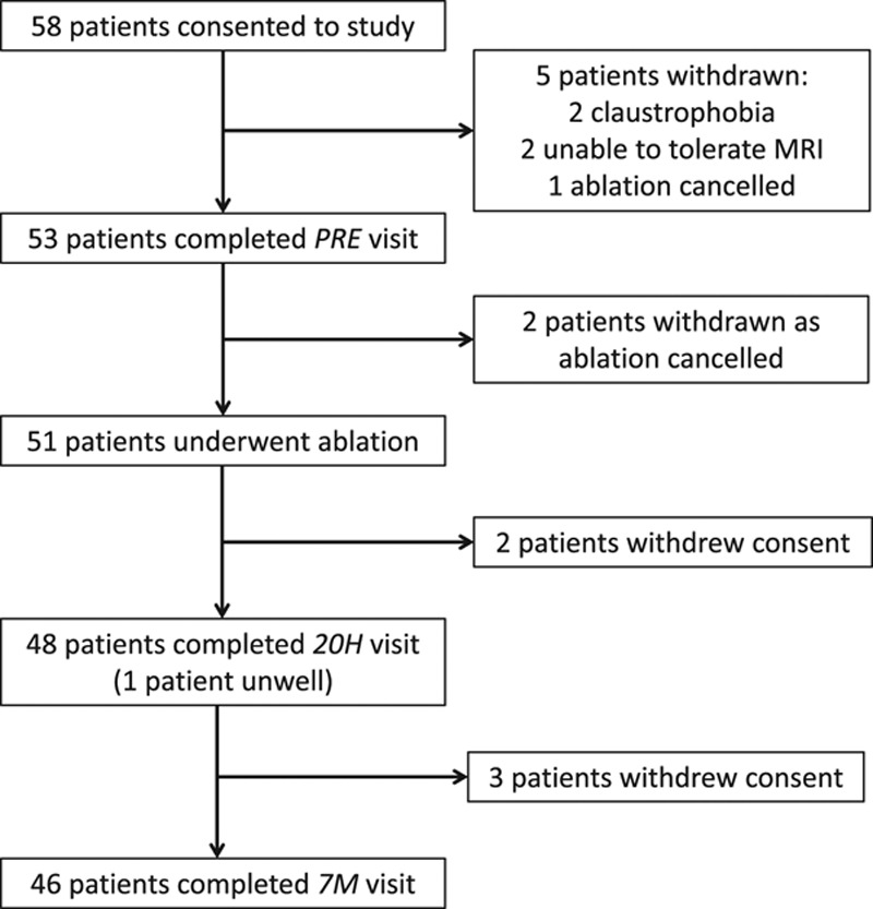
Methods: Fifty-three patients with symptomatic paroxysmal or persistent AF and without significant valvular disease, uncontrolled hypertension, coronary artery disease, uncontrolled thyroid disease, systemic inflammatory disease, diabetes mellitus, or obstructive sleep apnea (ie, lone AF) undergoing ablation and 25 matched control subjects in SR were investigated. Magnetic resonance imaging quantified LV ejection fraction (LVEF), peak systolic circumferential strain (PSCS), and left atrial volumes and function, whereas phosphorus-31 magnetic resonance spectroscopy evaluated ventricular energetics (ratio of phosphocreatine to ATP). AF burden was determined before and after ablation by 7-day Holter monitoring; intermittent ECG event monitoring was also undertaken after ablation to investigate for asymptomatic AF recurrence.

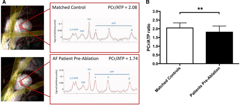
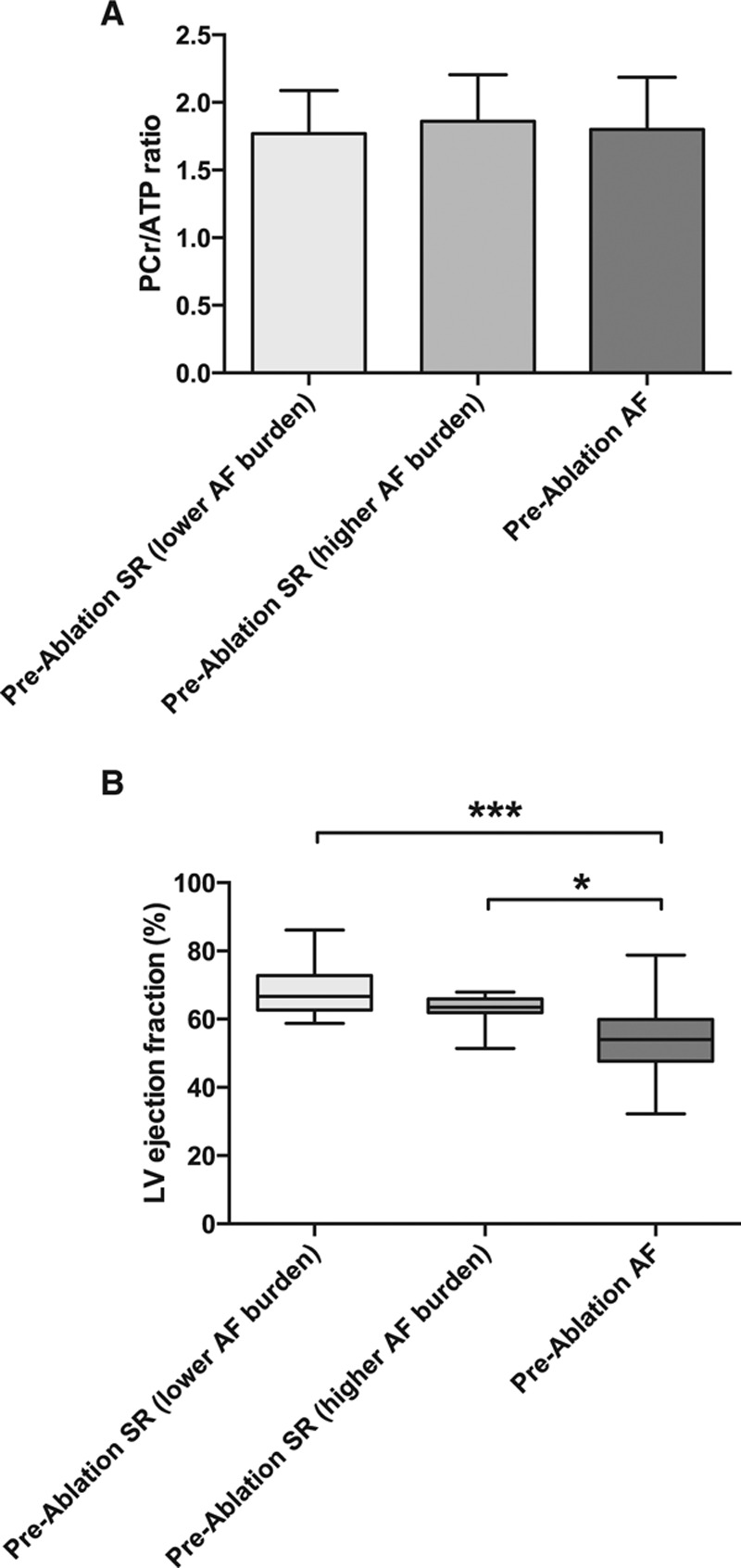
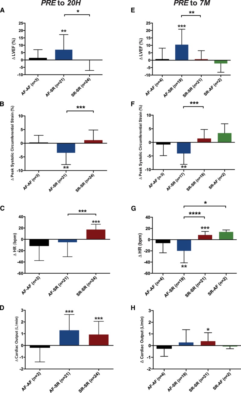
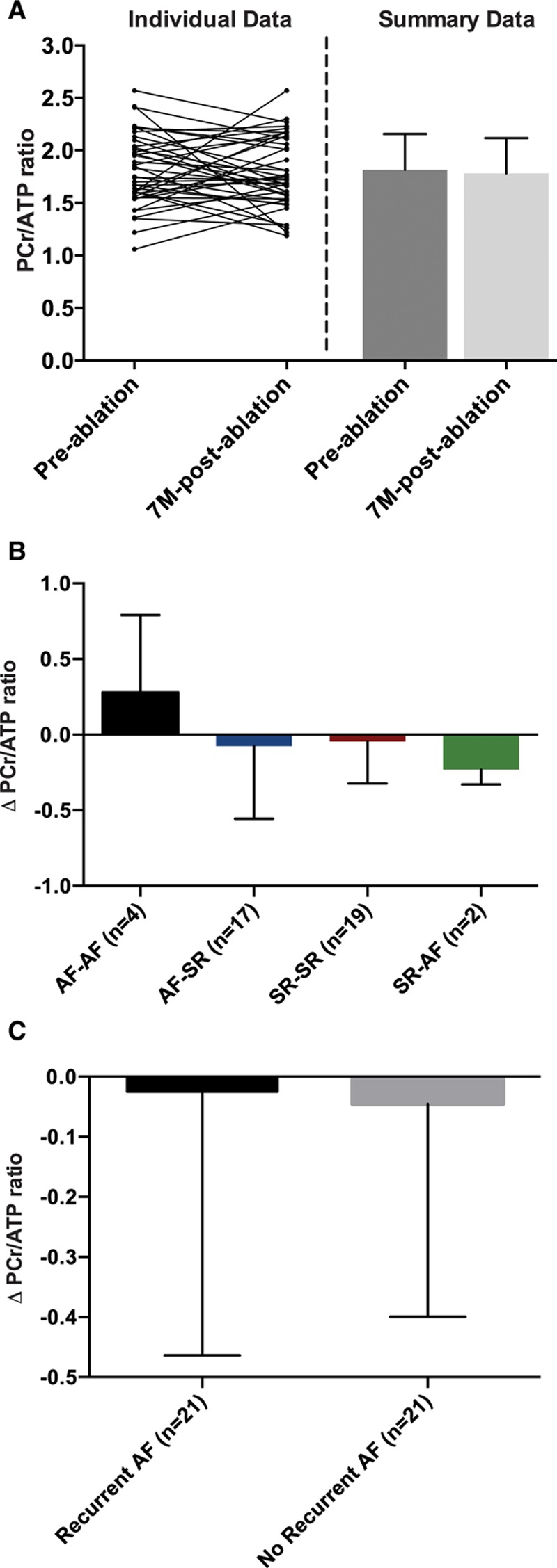
Results: Before ablation, both LV function and energetics were significantly impaired in patients compared with control subjects (LVEF, 61% [interquartile range (IQR), 52%-65%] versus 71% [IQR, 69%-73%], P<0.001; PSCS, -15% [IQR, -11 to -18%] versus -18% [IQR, -17% to -19%], P=0.002; ratio of phosphocreatine to ATP, 1.81±0.35 versus 2.05±0.29, P=0.004). As expected, patients also had dilated and impaired left atria compared with control subjects (all P<0.001). Early after ablation (1-4 days), LVEF and PSCS improved in patients recovering SR from AF (LVEF, 7.0±10%, P=0.005; PSCS, -3.5±4.3%, P=0.001) but were unchanged in those in SR during both assessments (both P=NS). At 6 to 9 months after ablation, AF burden reduced significantly (from 54% [IQR, 1.5%-100%] to 0% [IQR 0%-0.1%]; P<0.001). However, LVEF and PSCS did not improve further (both P=NS) and remained impaired compared with control subjects (P<0.001 and P=0.003, respectively). Similarly, there was no significant improvement in atrial function from before ablation (P=NS), and this remained lower than in control subjects (P<0.001). The ratio of phosphocreatine to ATP was unaffected by heart rhythm during assessment and AF burden before ablation (both P=NS). It was unchanged after ablation (P=0.57), remaining lower than in control subjects regardless of both recovery of SR and freedom from recurrent AF (P=0.006 and P=0.002, respectively).

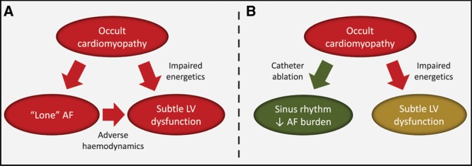
Conclusions: Patients with lone AF have impaired myocardial energetics and subtle LV dysfunction, which do not normalize after ablation. These findings suggest that AF may be the consequence (rather than the cause) of an occult cardiomyopathy, which persists despite a significant reduction in AF burden after ablation.

Keywords: arrhythmias, cardiac; atrial fibrillation; catheter ablation; magnetic resonance imaging; magnetic resonance spectroscopy; ventricular dysfunction, left.


Figures

Figure 1.
Figure 1.
Flowchart of patients with atrial fibrillation through the study. The number of patients consenting to the study, completing the preablation (PRE) visit, undergoing ablation, and completing the early (20H) and late (7M) visits is shown. The number of patients not completing each stage is also indicated, with reasons given. MRI indicates magnetic resonance imaging.
Figure 2.
Figure 2.
Representative phosphorus-31 magnetic resonance(31P-MR) spectra and myocardial energetics in the study groups. A, Images (left) show a midventricular short-axis slice, with the selected voxel positioned on the septum and highlighted (red rectangle). Saturation band positions are also shown (yellow). Spectra (right) show both raw data (black) and fitted line (red); peaks are labeled corresponding to 2,3-diphosphoglycerate (2,3-DPG), phosphodiester (PDE), phosphocreatine (PCr), and γ, α, and β peaks of ATP. Representative 31P-MR spectra from a control subject in SR with a PCr/ATP ratio of 2.08 and a patient with AF before ablation with a PCr/ATP ratio of 1.74. B,PCr/ATP ratio in control subjects and patients (n=25 and n=52, respectively; P=0.004 by unpaired t test).
Figure 3.
Figure 3.
Effect of intrascan rhythm on left ventricular (LV) ejection fraction(EF) and myocardial energetics in patients with atrial fibrillation (AF) before ablation. A, Myocardial energetics determined by the ratio of phosphocreatine to ATP (PCr/ATP) categorized by rhythm during scan and, for patients in sinus rhythm (SR), by Holter-determined AF burden (higher or lower than median). There is no difference between the groups (n=10 for SR with lower AF burden, n=11 for SR with higher AF burden, and n=28 for AF; P=0.84 by 1-way ANOVA). B, LVEF in patients with AF before ablation, similarly categorized. Note the significantly lower EF in those in AF compared with those in SR at the time of the scan, regardless of AF burden (Kruskal-Wallis P=0.0002). Smaller P values stratified by size (**P<0.01, ***P<0.001, ****P<0.0001).
Figure 4.
Figure 4.
Change in left ventricular (LV) ejection fraction(EF), peak systolic circumferential strain (PSCS), heart rate (HR), and cardiac output early and late after ablation, categorized by the intrascan rhythm at each time point. One-sample t tests assessed whether changes within each subgroup are significantly different from zero. Changes between subgroups were compared by use of 1-way ANOVA; P values for subgroup comparisons are Bonferroni corrected for multiple comparisons. A, At 20 hours (20H), LVEF improves only in the atrial fibrillation (AF)–sinus rhythm (SR) subgroup (P=0.005). B, Similarly, PSCS also improves only in the AF-SR subgroup (denoted by a more negative change; P=0.001). C, Only the SR-SR subgroup shows a significant increase in HR early after ablation (P<0.001), and both the AF-SR and SR-SR subgroups show a significant increase in cardiac output, without differences between subgroups (D). At 7 months (7M), the pattern of change in LV function from ≤4 weeks before ablation (PRE) in subgroups is similar to that at 20 hours, with the AF-SR subgroup alone showing significant improvement in LVEF (E; P<0.001) and PSCS (denoted by a more negative change; F; P=0.001). G, The changes in HR from ≤4 weeks before ablationin the AF-SR and SR-SR groups are significantly different (P<0.0001), and there is a significant difference between the AF-SR and SR-AF groups (P=0.03). H, Similar to 20 hours, there are no significant differences between subgroups in the change in cardiac output, with only the SR-SR subgroup demonstrating a small increase (P=0.03). Smaller P values stratified by size (**P<0.01, ***P<0.001, ****P<0.0001).
Figure 5.
Figure 5.
Myocardial energetics before and late after ablation. A, In all patients, there was no change in the ratio of phosphocreatine to ATP (PCr/ATP) from ≤4 weeks before to 7 months (7M) after ablation(n=42, P=0.57, paired t test). There were also no significant differences in the change in PCr/ATP ratio after ablation in any subgroups on the basis of either intrascan rhythm combinations (P=0.37, 1-way ANOVA; B) or the presence or absence of recurrent atrial fibrillation (AF) after ablation (P=0.87, unpaired t test; C). SR indicates sinus rhythm.
Figure 6.
Figure 6.
Proposed schematic representation of the relationships between lone atrial fibrillation (AF), subtle left ventricular (LV) dysfunction, and upstream cardiomyopathy and the effect of ablation. A, Lone AF and subtle LV dysfunction may be tissue-specific manifestations of an upstream occult cardiomyopathy, characterized by impaired energetics. AF further contributes to LV dysfunction via adverse hemodynamics. B, Successful catheter ablation restores sinus rhythm and/or reduces AF burden and leads to a modest increase in LV function via improvement in hemodynamics. However, the underlying cardiomyopathy remains, and myocardial energetics and LV function do not normalize.

Comment in

Similar articles

Cited by

References

    1. Lin HJ, Wolf PA, Kelly-Hayes M, Beiser AS, Kase CS, Benjamin EJ, D’Agostino RB. Stroke severity in atrial fibrillation: the Framingham study. Stroke. 1996;27:1760–1764. - PubMed
    1. Soliman EZ, Safford MM, Muntner P, Khodneva Y, Dawood FZ, Zakai NA, Thacker EL, Judd S, Howard VJ, Howard G, Herrington DM, Cushman M. Atrial fibrillation and the risk of myocardial infarction. JAMA Intern Med. 2014;174:107–114. doi: 10.1001/jamainternmed.2013.11912. - PMC - PubMed
    1. Benjamin EJ, Wolf PA, D’Agostino RB, Silbershatz H, Kannel WB, Levy D. Impact of atrial fibrillation on the risk of death: the Framingham Heart Study. Circulation. 1998;98:946–952. - PubMed
    1. Chugh SS, Havmoeller R, Narayanan K, Singh D, Rienstra M, Benjamin EJ, Gillum RF, Kim YH, McAnulty JH, Jr, Zheng ZJ, Forouzanfar MH, Naghavi M, Mensah GA, Ezzati M, Murray CJ. Worldwide epidemiology of atrial fibrillation: a Global Burden of Disease 2010 Study. Circulation. 2014;129:837–847. doi: 10.1161/CIRCULATIONAHA.113.005119. - PMC - PubMed
    1. Wijffels MC, Kirchhof CJ, Dorland R, Allessie MA. Atrial fibrillation begets atrial fibrillation: a study in awake chronically instrumented goats. Circulation. 1995;92:1954–1968. - PubMed

MeSH terms