. 2016 Dec;30(12):4202-4213.
doi: 10.1096/fj.201600392RR. Epub 2016 Sep 14.

Galectin-3 regulates inflammasome activation in cholestatic liver injury

Affiliations

Galectin-3 regulates inflammasome activation in cholestatic liver injury

Jijing Tian et al. FASEB J. 2016 Dec.

Abstract

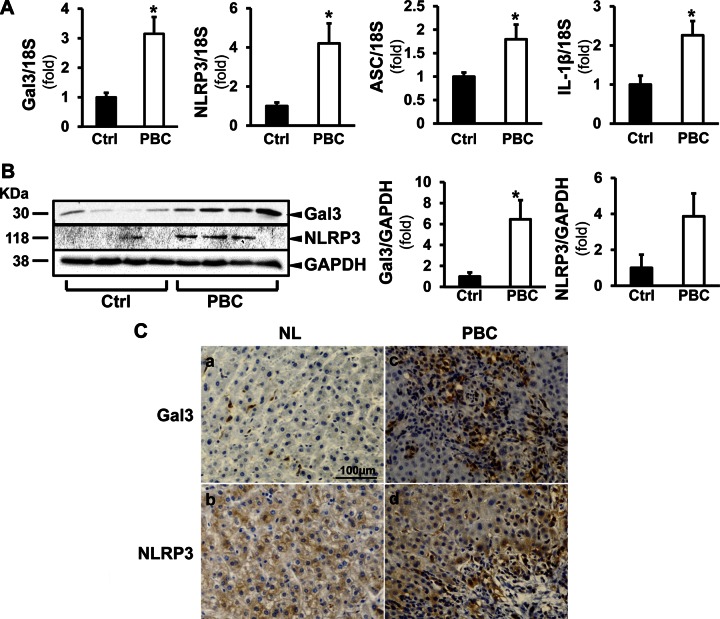
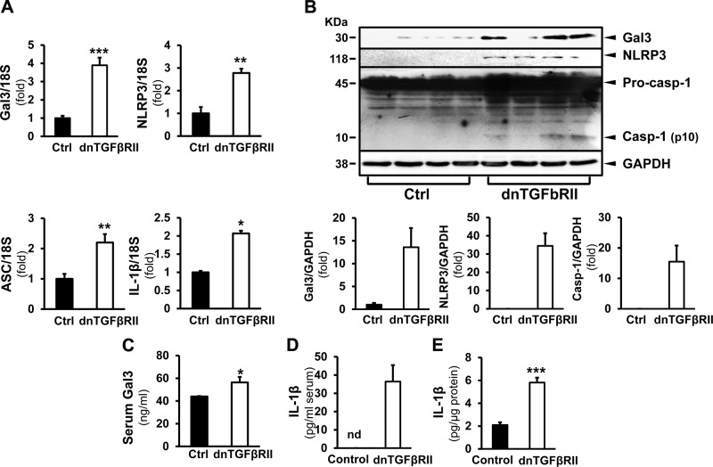
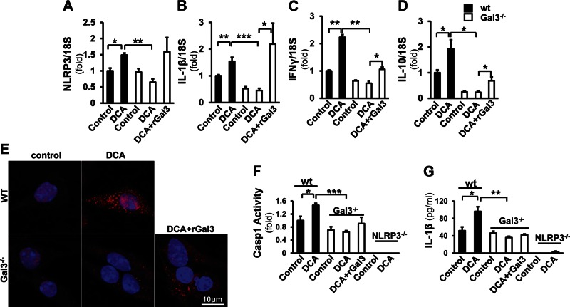
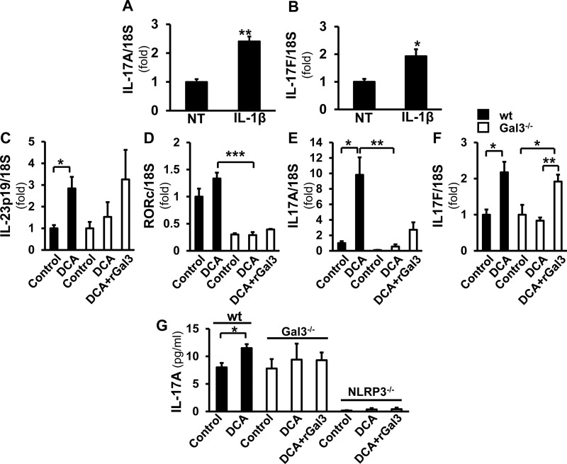
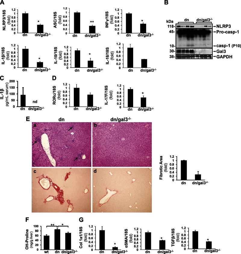
Macrophage activation is an important feature of primary biliary cholangitis (PBC) pathogenesis and other cholestatic liver diseases. Galectin-3 (Gal3), a pleiotropic lectin, is produced by monocytic cells and macrophages. However, its role in PBC has not been addressed. We hypothesized that Gal3 is a key to induce NOD-like receptor family, pyrin domain containing 3 (NLRP3) inflammasome in macrophages and in turn to propagate proinflammatory IL-17 signaling. In liver tissues from patients with PBC and dnTGF-βRII mice, a model of autoimmune cholangitis, the expression of Gal3, NLRP3, and the adaptor protein adaptor apoptosis-associated speck-like protein was induced, with the downstream activation of caspase-1 and IL-1β. In wild-type hepatic macrophages, deoxycholic acid induced the association of Gal3 and NLRP3 with direct activation of the inflammasome, resulting in an increase in IL-1β. Downstream retinoid-related orphan receptor C mRNA, IL-17A, and IL-17F were induced. In Gal3-/- macrophages, no inflammasome activation was detected. To confirm the key role of Gal3 in the pathogenesis of cholestatic liver injury, we generated dnTGF-βRII/galectin-3-/- (dn/Gal3-/-) mice, which showed impaired inflammasome activation along with significantly improved inflammation and fibrosis. Taken together, our data point to a novel role of Gal3 as an initiator of inflammatory signaling in autoimmune cholangitis, mediating the activation of NLRP3 inflammasome and inducing IL-17 proinflammatory cascades. These studies provide a rationale to target Gal3 in autoimmune cholangitis and potentially other cholestatic diseases.-Tian, J., Yang, G., Chen, H.-Y., Hsu, D. K., Tomilov, A., Olson, K. A., Dehnad, A., Fish, S. R., Cortopassi, G., Zhao, B., Liu, F.-T., Gershwin, M. E., Török, N. J., Jiang, J. X. Galectin-3 regulates inflammasome activation in cholestatic liver injury.

Keywords: IL-17; NLRP3; galectin-3; primary biliary cholangitis.


Figures

Figure 1.
Figure 1.
The expression of Gal3 and NLRP3 inflammasome components are induced in patients with PBC. A) Liver tissues from healthy subjects (Ctrl) and patients with PBC were processed for RT-qPCR to evaluate the expression of Gal3, NLRP3, ASC, and IL-1β. A) The mRNA levels of Gal3, NLRP3, the adaptor ASC and the downstream effector IL-1β were all significantly elevated in livers from patients with PBC. *P < 0.05; means ± sem; n = 4. B) Western blot and densitometry analyses showed that the protein levels of Gal3 and NLRP3 were increased in PBC livers. *P < 0.05; means ± sem; n = 4. C) Immunohistochemistry revealed enhanced signals for Gal3 (a, c) and NLRP3 (b, d) in the livers from patients with PBC compared with normal livers (NL) from healthy control subjects.
Figure 2.
Figure 2.
The expression of Gal3 and NLRP3 inflammasome components are induced in dnTGF-βRII mice. A) In transgenic dnTGF-βRII mice that spontaneously develop PBC-like cholangitis, the transcripts of Gal3, NLRP3, ASC, and IL-1β were significantly induced in the livers compared with WT (Ctrl) livers. *P < 0.05, **P < 0.01, ***P < 0.001; means ± sem; n = 4. B) Western blots on liver homogenates depicted increased Gal3 and NLRP3 in the dnTGF-βRII mice and enhanced caspase-1 cleavage. ***P < 0.001; data are representative. C) Serum levels of Gal3 were examined by ELISA. The dnTGF-βRII mice had significantly higher levels of Gal3. *P < 0.05; means ± sem; n = 5, 13. D, E) Sera (D) and the liver homogenates (E) from WT and dnTGF-βRII mice were processed for IL-1β ELISA. Significantly higher levels of IL-1β were seen in both serum and livers from the transgenic mice. ***P < 0.001; means ± sem; n = 6.
Figure 3.
Figure 3.
DCA induces Gal3 and NLRP3 expression and their association in hepatic macrophages. Primary macrophages were isolated from mouse livers and treated with DCA (100 µM for 24 h). A, B) RT-qPCR showed significantly induced Gal3 (A) and NLRP3 (B) expression after DCA treatment (means ± sem; n = 4). *P < 0.05. C) The induction at protein levels was also detected by Western blot analysis. D) Culture medium was collected for ELISA to examine Gal3. The DCA-challenged cells released significantly higher levels of Gal3 (means ± sem; n = 6). *P < 0.05. E) To visualize Gal3 and NLRP3 in macrophages, immunofluorescence studies were done in the DCA-treated macrophages. The signals for Gal3 (green) and NLRP3 (red) were dispersed and weak in nontreated cells. In the DCA-challenged cells, enhanced Gal3 and NLRP3 were seen, and more NLRP3 clusters were observed. F) Immunoprecipitation of Gal3 and NLRP3 Western blot showed their association in DCA-treated macrophages. BLI was conducted to confirm the association. Recombinant NLRP3 loaded sensors were incubated with either recombinant WT Gal3 or N-terminal truncated Gal3 (Gal3-C) (18 μM). The association was recorded for 600 s. G) The sensors were moved to the blank buffer to measure the dissociation. Gal3 showed an obvious association/dissociation pattern. This was not present in Gal3-C. Thus, Gal3 directly interacts with NLRP3, and its N-terminal motif is crucial for this interaction. H) When the NLRP3-loaded sensors were incubated with Gal3 at a series of concentrations, a dose-dependent association was observed.
Figure 4.
Figure 4.
Gal3 is required for the induction of NLRP3 inflammasome signaling by DCA in macrophages. Macrophages from WT and Gal3−/− mice were incubated with DCA (100 μM) and with recombinant Gal3 (rGal3; 1 µM for 24 h) in the knockout cells. AD) Real-time qPCR revealed that the expression of NLRP3 (A), IL-1β (B), IFN-γ (C), and IL-10 (D) was significantly induced in WT cells (means ± sem; n = 4) but not in the Gal3−/− cells. When the knockout cells were exposed to rGal3, the expression of these transcripts was partially reversed (means ± sem; n = 4). *P < 0.05, **P < 0.01. E) The immunofluorescent staining probing ASC (red) revealed that there were no ASC foci in nontreated cells, whereas DCA induced more ASC focus formation in WT cells than in the knockout cells. Application of rGal3 in the knockout cells enhanced the ASC signal and focus formation. F, G) To confirm that the activation of inflammasome by DCA is Gal3 dependent, caspase-1 activity (F) and IL-1β in medium (G) were assessed in WT, Gal3−/−, and NLRP3−/− macrophages. DCA significantly induced caspase-1 activity in WT cells but not in Gal3−/− cells (F). In NLRP3−/− cells, caspase-1 activity could not be detected in either DCA-treated or nontreated groups (means ± sem; n = 4). IL-1β ELISA was conducted with cell culture medium from the above cells. WT cells released more IL-1β upon the challenge of DCA; this was not seen in the Gal3−/− cells (G). A minimal amount of IL-1β was detected in NLRP3−/− cells (means ± sem; n = 4).
Figure 5.
Figure 5.
DCA-induced IL-23/IL-17 signaling is mediated by Gal3. RT-qPCR was performed on macrophages treated with IL-1β (5 ng/ml). A, B) The transcripts of IL-17A (A) and IL-17F (B) were significantly increased (means ± sem; n = 4). *P < 0.05, **P < 0.01. CF) DCA induced IL-23p19 (C), RORc (D), IL-17A (E), and IL-17F (F) in WT cells but not in the knockout cells. This effect was partially reversed by rGal3 (means ± sem; n = 4). *P < 0.05, **P < 0.01, ***P < 0.001. G) IL-17A in medium from DCA-treated WT, Gal3−/−, and NLRP3−/− cells was evaluated by ELISA. The DCA-treated WT cells, but not the Gal3−/− cells, released significantly more IL-17A. Only a trace amount of IL-17A was detected in NLRP3−/− cells, and rGal3 did not change this (means ± sem; n = 4). *P < 0.05.
Figure 6.
Figure 6.
Gal3 deficiency prevents inflammasome activation and reduces collagen deposition in dnTGF-βRII mice. dn/Gal3−/− mice were generated by crossing Gal3−/− and dnTGF-βRII mice. A) RT-qPCR showed that dn/Gal3−/− mice expressed significantly lower levels of NLRP3, ASC, INF-γ, IL-1β, IL-10, and IL-18 compared with dnTGF-βRII (dn) mice (means ± sem; n = 6). *P < 0.05, **P < 0.01. B) Western blots showed less hepatic NLRP3 and caspase-1 cleavage (representative data). C) The dn/Gal3−/− mice also had undetectable serum IL-1β (representative data), suggesting suppressed NLRP3 inflammasome activation. D) The transcripts of RORc and IL-17F were lower in these mice as well (n = 6). *P < 0.05. E) H&E showed inflammatory granulomas in the dnTGF-βRII mice (a, arrows), whereas this was not seen in the dn/Gal3−/− group (b). Picrosirius red staining and image morphometric analysis revealed less fibrosis in the dn/Gal3−/− mice (c, d). F) Liver tissue was subjected to hydroxyproline assay. The dnTGF-βRII mice showed significantly higher amount of hydroxyproline compared with their WT littermates, and this was significantly reduced in dn/Gal3−/− mice (means ± sem; n = 9). *P < 0.05, **P < 0.01. G) Fibrogenic transcripts procollagen Ia1, α-SMA, and TGF-β also significantly decreased in the dn/Gal3−/− mice (means ± sem, n = 6). *P < 0.05.

References

    1. He X. S., Ansari A. A., Ridgway W. M., Coppel R. L., Gershwin M. E. (2006) New insights to the immunopathology and autoimmune responses in primary biliary cirrhosis. Cell. Immunol. 239, 1–13 - PubMed
    1. Chuang Y. H., Lian Z. X., Yang G. X., Shu S. A., Moritoki Y., Ridgway W. M., Ansari A. A., Kronenberg M., Flavell R. A., Gao B., Gershwin M. E. (2008) Natural killer T cells exacerbate liver injury in a transforming growth factor beta receptor II dominant-negative mouse model of primary biliary cirrhosis. Hepatology 47, 571–580 - PubMed
    1. Lleo A., Bowlus C. L., Yang G. X., Invernizzi P., Podda M., Van de Water J., Ansari A. A., Coppel R. L., Worman H. J., Gores G. J., Gershwin M. E. (2010) Biliary apotopes and anti-mitochondrial antibodies activate innate immune responses in primary biliary cirrhosis. Hepatology 52, 987–998 - PMC - PubMed
    1. Jiang J. X., Chen X., Hsu D. K., Baghy K., Serizawa N., Scott F., Takada Y., Takada Y., Fukada H., Chen J., Devaraj S., Adamson R., Liu F.-T., Török N. J. (2012) Galectin-3 modulates phagocytosis-induced stellate cell activation and liver fibrosis in vivo. Am. J. Physiol. Gastrointest. Liver Physiol. 302, G439–G446 - PMC - PubMed
    1. Liu F.-T. (1990) Molecular biology of IgE-binding protein, IgE-binding factors, and IgE receptors. Crit. Rev. Immunol. 10, 289–306 - PubMed

Publication types