. 2017 Feb;101(2):531-542.
doi: 10.1189/jlb.4A0815-365RR. Epub 2016 Sep 14.

Lipoxin A4, a 5-lipoxygenase pathway metabolite, modulates immune response during acute respiratory tularemia

Affiliations

Lipoxin A4, a 5-lipoxygenase pathway metabolite, modulates immune response during acute respiratory tularemia

Anju Singh et al. J Leukoc Biol. 2017 Feb.

Abstract

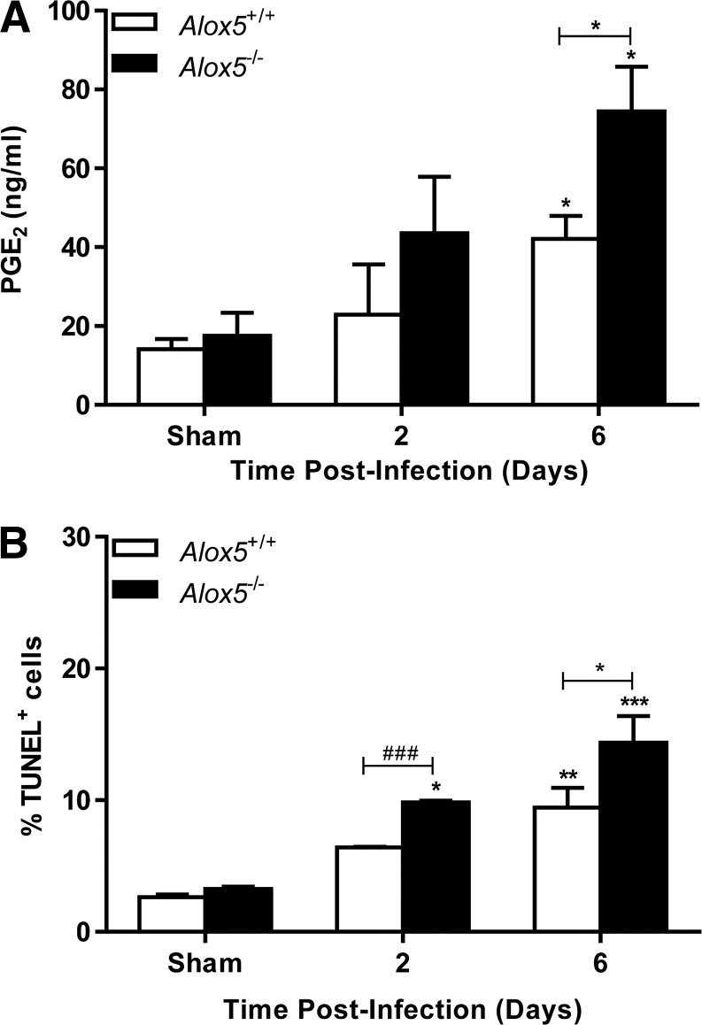
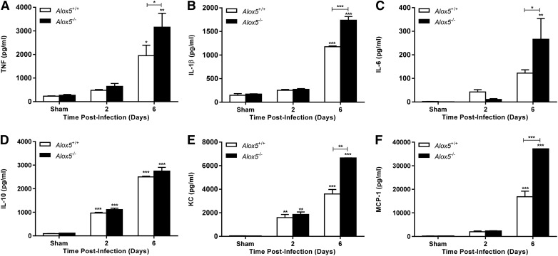
Respiratory infection with Francisella tularensis (Ft) is characterized by a muted, acute host response, followed by sepsis-like syndrome that results in death. Infection with Ft establishes a principally anti-inflammatory environment that subverts host-cell death programs to facilitate pathogen replication. Although the role of cytokines has been explored extensively, the role of eicosanoids in tularemia pathogenesis is not fully understood. Given that lipoxin A4 (LXA4) has anti-inflammatory properties, we investigated whether this lipid mediator affects host responses manifested early during infection. The addition of exogenous LXA4 inhibits PGE2 release by Ft-infected murine monocytes in vitro and diminishes apoptotic cell death. Tularemia pathogenesis was characterized in 5‑lipoxygenase-deficient (Alox5-/-) mice that are incapable of generating LXA4 Increased release of proinflammatory cytokines and chemokines, as well as increased apoptosis, was observed in Alox5-/- mice as compared with their wild-type counterparts. Alox5-/- mice also exhibited elevated recruitment of neutrophils during the early phase of infection and increased resistance to lethal challenge. Conversely, administration of exogenous LXA4 to Alox5-/- mice made them more susceptible to infection thus mimicking wild-type animals. Taken together, our results suggest that 5-LO activity is a critical regulator of immunopathology observed during the acute phase of respiratory tularemia, regulating bacterial burden and neutrophil recruitment and production of proinflammatory modulators and increasing morbidity and mortality. These studies identify a detrimental role for the 5-LO-derived lipid mediator LXA4 in Ft-induced immunopathology. Targeting this pathway may have therapeutic benefit as an adjunct to treatment with antibiotics and conventional antimicrobial peptides, which often have limited efficacy against intracellular bacteria.

Keywords: anti-inflammatory; cytokines; inflammation; lipid mediators.


Figures

Figure 1.
Figure 1.. Lipid mediators PGE2 and LXA4 are reciprocally regulated during the course of Ft LVS infection.
(A) Wild-type C57BL/6 mice were infected with 1000 CFU of Ft LVS, and the BALF cells were isolated from individual mice on d 2 postinfection. The BALF cells were stained with Nile Red (0.1 ng/ml) and cytospun. The cells were fixed in 3.7% formaldehyde and were analyzed by confocal microscopy. The results are representative of 6 individual mice. Levels of PGE2 (B) and LXA4 (C) during the course of Ft infection were measured in lung homogenates by mass spectrometry. Data are presented as the means ± sem from 3 independent experiments (n = 6 mice/group; 18 mice total). *P < 0.05, ***P < 0.001. All results shown were subjected to 1-way ANOVA with Bonferroni’s posttest.
Figure 2.
Figure 2.. Addition of exogenous LXA4 ablates PGE2 secretion by Ft-infected BMDMs.
(A) Wild-type C57BL/6 BMDMs (2.5 × 10⁵ cells/well) were seeded into 24-well plates and infected with Ft LVS at an MOI of 100. Exogenous LXA4 (1 µM) was added at the time of infection to the cells. Supernatants were collected after incubation for 24 h at 37°C and were analyzed for the presence of PGE2. **P < 0.01, ***P < 0.001. (B) Wild-type C57BL/6 BMDMs (1 × 10⁶ cells/well) were infected with Ft LVS at an MOI of 100. Exogenous LXA4 (1 µM) was added to cells at the time of infection, and after incubation for 24 h at 37°C, the cells were stained with TUNEL to quantify the percentage of cells undergoing apoptosis. ***P < 0.001. Data are presented as the means ± sem from 3 independent experiments. All results shown were subjected to 1-way ANOVA with Bonferroni’s posttest.
Figure 3.
Figure 3.. Alox5−/− BMDMs secrete significantly more PGE2 in response to Ft infection.
(A) Wild-type (Alox5+/+) and Alox5−/− C57BL/6 BMDMs (2.5 × 10⁵ cells/well) were seeded into 24-well plates and infected with Ft LVS at an MOI of 100. Cells were incubated at 37°C, and supernatants were collected at various times and analyzed for the presence of PGE2. ***P < 0.001. (B) Wild-type (Alox5+/+) and Alox5−/− C57BL/6 BMDMs (1 × 10⁶ cells/well) were seeded into 6-well plates and infected with Ft LVS at an MOI of 100 and were incubated for 24 h at 37°C. The cells were stained with TUNEL to quantify the percentage of undergoing apoptosis. ***P < 0.001. Data are presented as the means ± sem from 3 independent experiments. All results shown were subjected to One-way ANOVA with Bonferroni’s Posttest. Asterisks immediately above columns indicate a significant difference compared with their corresponding control while asterisks above a bracket indicate a significant difference between 2 experimental groups.
Figure 4.
Figure 4.. Increased proinflammatory cytokine and chemokine production is observed by Ft-infected Alox5−/− BMDMs.
(A-F) Wild-type (Alox5+/+) and Alox5−/− C57BL/6 BMDMs (2.5 × 10⁵ cells/well) were infected with Ft LVS at an MOI of 100. Supernatants were collected 24 h postinfection and were assayed for the presence of various cytokines and chemokines with the Luminex assay. *P < 0.05, **P < 0.01, ***P < 0.001. Data are presented as the means ± sem from 3 independent experiments. All results shown were subjected to 1-way ANOVA with Bonferroni’s posttest. Asterisks immediately above columns indicate a significant difference compared with its corresponding control, whereas asterisks above a bracket indicate a significant difference between 2 experimental groups.
Figure 5.
Figure 5.. Alox5−/− mice secrete significantly higher levels of PGE2 in response to Ft infection.
(A) Wild-type (Alox5+/+) and Alox5−/− C57BL/6 mice were infected with 1000 CFU of Ft LVS, and total lung cells were isolated from individual mice at different times. The lung homogenates were analyzed for the presence of PGE2 by ELISA. *P < 0.05, #P < 0.051 (Student’s t test) (B) Single-cell lung suspensions isolated from wild-type and Alox5−/− C57BL/6 mice were infected with 1000 CFU of Ft LVS and were stained with TUNEL to quantify the percentage of cells undergoing apoptosis. **P < 0.01, ***P < 0.001, ###P < 0.0018 (Student’s t test). Data are presented as the means ± sem from 3 independent experiments. All results shown were subjected to 1-way ANOVA with Bonferroni’s posttest. Asterisks immediately above columns indicate a significant difference compared with its corresponding control, whereas asterisks above a bracket indicate a significant difference between 2 experimental groups.
Figure 6.
Figure 6.. Increased proinflammatory cytokine and chemokine production is observed in Ft-infected Alox5−/− mice.
(A–F) Cell-free lung homogenates from wild-type (Alox5+/+) and Alox5−/− mice were recovered at various times following i.n. administration of 1000 CFUs of Ft LVS. The homogenates were analyzed for the presence of proinflammatory cytokines and chemokines with the Luminex assay. *P < 0.05, **P < 0.01, ***P < 0.001. Data are presented as the means ± sem from 2 independent experiments. All results shown were subjected to 1-way ANOVA with Bonferroni’s posttest. Asterisks immediately above columns indicate a significant difference compared with its corresponding control, whereas asterisks above a bracket indicate a significant difference between 2 experimental groups.
Figure 7.
Figure 7.. Alox5 deficiency is associated with reduced bacterial burdens in the various organs during the course of Ft infection.
Wild-type and Alox5−/− C57BL/6 mice were inoculated i.n. with 1000 CFU of Ft LVS. At the times indicated, mice were sacrificed and homogenates of the lungs, liver, and spleen were plated for determination of bacterial burden. *P < 0.05, **P < 0.01. Results shown are the means ± sem and are representative of 3 independent experiments (n = 4 mice/time point; 12 mice total). Asterisks immediately above columns indicate a significant difference compared with its corresponding control, whereas asterisks above a bracket indicate a significant difference between 2 experimental groups.
Figure 8.
Figure 8.. The inhibitor of the 5-LO pathway, MK-886, results in lower bacterial burden.
(A) Wild-type C57BL/6 mice were infected i.n. with Ft LVS alone or with doses of MK-886 (5 mg/kg) at the time of infection and, thereafter, every 24 h. The infected mice were sacrificed at different times as shown and were analyzed for the presence of LXA4 by ELISA. (B) The lung homogenates were prepared and plated for determination of bacterial burden. *P < 0.05, **P < 0.01. Results shown are the means ± sem and are representative of 2 independent experiments (n = 6 mice/time point; 12 mice total). Asterisks immediately above columns indicate a significant difference compared with its corresponding control, whereas asterisks above a bracket indicate a significant difference between 2 experimental groups.
Figure 9.
Figure 9.. 5-LO deficiency enhances host resistance to Ft infection.
Wild-type (Alox5+/+) and Alox5−/− mice were inoculated i.n. with 250 CFU of Ft LVS (A and B, respectively) and were monitored for morbidity and mortality, and weight loss (A and B, respectively). Results are expressed as Kaplan-Meier curves, and P values were determined using the log-rank test. The results shown are representative of 2 independent experiments (n = 8 mice/group; 16 mice total).
Figure 10.
Figure 10.. Exogenous LXA4 renders the Alox5−/− mice more susceptible to Ft infection.
(A) Wild-type (Alox5+/+) and Alox5−/− mice were infected i.n. with 250 CFU of Ft LVS. Alox5−/− mice were either untreated or received doses of LXA4 (2.5 µg/kg) at the time of infection and, thereafter, every 24 h for 3 d. Following initiation of infection, all mice were monitored for morbidity and mortality over 21 d. (B) Lung homogenates were prepared and plated for determination of bacterial burden. *P < 0.05, **P < 0.01. Results are expressed as Kaplan-Meier curves, and P values were determined using the log-rank test. The results shown are representative of 2 independent experiments (n = 8 mice/group; 16 mice total). Asterisks immediately above columns indicate a significant difference compared with its corresponding control, whereas asterisks above a bracket indicate a significant difference between 2 experimental groups.

Similar articles

Cited by

References

    1. Sharma J., Mares C. A., Li Q., Morris E. G., Teale J. M. (2011) Features of sepsis caused by pulmonary infection with Francisella tularensis Type A strain. Microb. Pathog. 51, 39–47. - PMC - PubMed
    1. Cowley S. C., Elkins K. L. (2011) Immunity to Francisella. Front. Microbiol. 2, 26. - PMC - PubMed
    1. Periasamy S., Singh A., Sahay B., Rahman T., Feustel P. J., Pham G. H., Gosselin E. J., Sellati T. J. (2011) Development of tolerogenic dendritic cells and regulatory T cells favors exponential bacterial growth and survival during early respiratory tularemia. J. Leukoc. Biol. 90, 493–507. - PMC - PubMed
    1. Periasamy S., Avram D., McCabe A., MacNamara K. C., Sellati T. J., Harton J. A. (2016) An immature myeloid/myeloid-suppressor cell response associated with necrotizing inflammation mediates lethal pulmonary tularemia. PLoS Pathog. 12, e1005517. - PMC - PubMed
    1. Chen M., Divangahi M., Gan H., Shin D. S., Hong S., Lee D. M., Serhan C. N., Behar S. M., Remold H. G. (2008) Lipid mediators in innate immunity against tuberculosis: opposing roles of PGE2 and LXA4 in the induction of macrophage death. J. Exp. Med. 205, 2791–2801. - PMC - PubMed

MeSH terms