. 2016 Aug 31:7:1405.
doi: 10.3389/fmicb.2016.01405. eCollection 2016.

Subcutaneous Phaeohyphomycosis Due to Pyrenochaeta romeroi Mimicking a Synovial Cyst

Affiliations

Subcutaneous Phaeohyphomycosis Due to Pyrenochaeta romeroi Mimicking a Synovial Cyst

Aurélien Dinh et al. Front Microbiol. .

Abstract

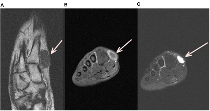
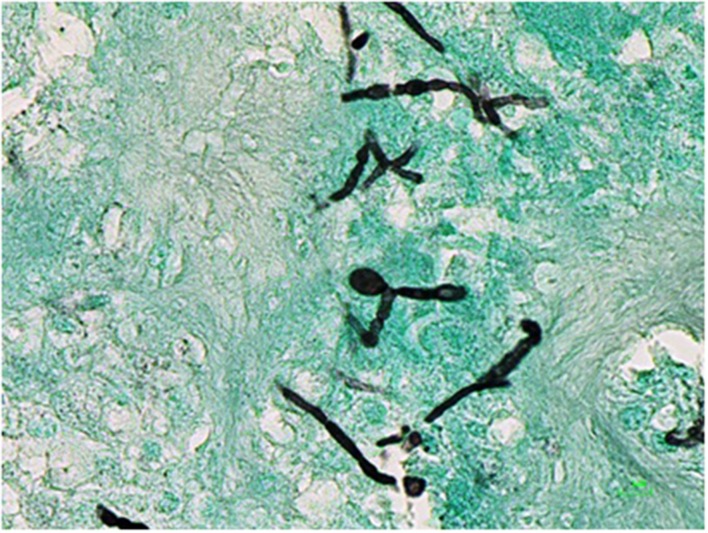
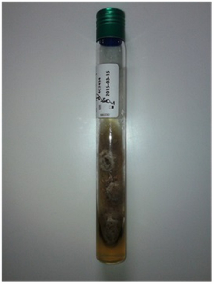
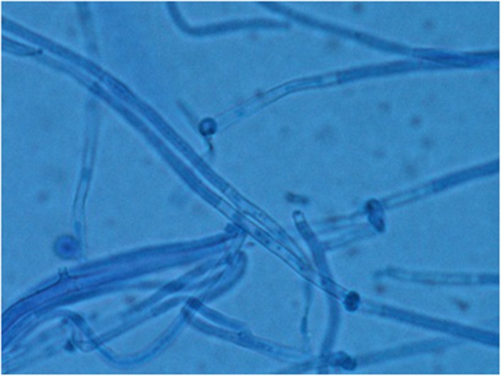
Opportunistic subcutaneous fungal infections are increasing nowadays due to the growing number of medical conditions causing immunosuppression, especially organ transplant. The incidence rate of subcutaneous phaeohyphomycosis is very low. Most studies found are case reports. They showed a wide variation of clinical presentations. Pyrenochaeta romeroi, a fungus from the Dematiaceae group is a saprophyte found in soil and plants and a possible causative agent of phaeohyphomycosis. We present a rare case of subcutaneous phaeohyphomycosis caused by P. romeroi mimicking a synovial cyst in a diabetic patient.

Keywords: Pyrenochaeta; fungal infection; immunosuppression; phaeohyphomycosis; surgical treatment.


Figures

FIGURE 1
FIGURE 1
Left Foot MRI (Magnetic Resonance Image). Axial MRI T1 left foot image (A) shows a subcutaneous ovoid mass of 3cm × 2cm in contact with the first metatarse (white arrow), coronal T1 fat sat with gadolinium (B) and Coronal T2 fat sat (C) show, respectively, an hyposignal T1 and a hyper signal T2 with peripheric enhancement after injection.
FIGURE 2
FIGURE 2
Histological examination of the cyst on Grocott staining.
FIGURE 3
FIGURE 3
Macroscopic aspect of Pyrenochaeta romeroi isolate on Sabouraud Chloramphenicol dextrose agar.
FIGURE 4
FIGURE 4
Microscopic aspect of P. romeroi isolate (Blue cotton, ×40).

References

    1. Badali H., Chander J., Gulati N., Attri A., Chopra R., Najafzadeh M. J., et al. (2010a). Subcutaneous phaeohyphomycotic cyst caused by Pyrenochaeta romeroi. Med. Mycol. 48 763–768. 10.3109/13693780903440383 - DOI - PubMed
    1. Badali H., Najafzadeh M. J., van Esbroeck M., van den Enden E., Tarazooie B., Meis J. F. G. M., et al. (2010b). The clinical spectrum of Exophiala jeanselmei, with a case report and in vitro antifungal susceptibility of the species. Med. Mycol. 48 318–327. 10.1080/13693780903148353 - DOI - PubMed
    1. Ben-Ami R., Lewis R. E., Raad I. I., Kontoyiannis D. P. (2009). Phaeohyphomycosis in a tertiary care cancer center. Clin. Infect. Dis. 48 1033–1041. 10.1086/597400 - DOI - PubMed
    1. Borelli D. (1979). Opportunistic fungi as producers of gray colonies and mycetomata. Dermatologica 159 168–174. 10.1159/000250685 - DOI - PubMed
    1. Chowdhary A., Meis J. F., Guarro J., de Hoog G. S., Kathuria S., Arendrup M. C., et al. (2014). ESCMID and ECMM joint clinical guidelines for the diagnosis and management of systemic phaeohyphomycosis: diseases caused by black fungi. Clin. Microbiol. Infect. 20(Suppl. 3), 47–75. 10.1111/1469-0691.12515 - DOI - PubMed

LinkOut - more resources