

Applications of Chondrocyte-Based Cartilage Engineering: An Overview
- PMID: 27631002
- PMCID: PMC5007317
- DOI: 10.1155/2016/1879837
Applications of Chondrocyte-Based Cartilage Engineering: An Overview
Abstract
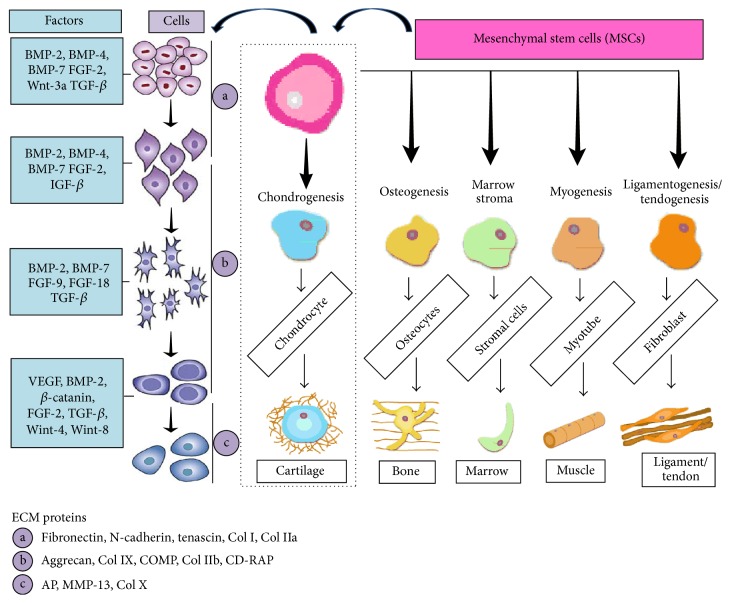
Chondrocytes are the exclusive cells residing in cartilage and maintain the functionality of cartilage tissue. Series of biocomponents such as different growth factors, cytokines, and transcriptional factors regulate the mesenchymal stem cells (MSCs) differentiation to chondrocytes. The number of chondrocytes and dedifferentiation are the key limitations in subsequent clinical application of the chondrocytes. Different culture methods are being developed to overcome such issues. Using tissue engineering and cell based approaches, chondrocytes offer prominent therapeutic option specifically in orthopedics for cartilage repair and to treat ailments such as tracheal defects, facial reconstruction, and urinary incontinence. Matrix-assisted autologous chondrocyte transplantation/implantation is an improved version of traditional autologous chondrocyte transplantation (ACT) method. An increasing number of studies show the clinical significance of this technique for the chondral lesions treatment. Literature survey was carried out to address clinical and functional findings by using various ACT procedures. The current study was conducted to study the pharmacological significance and biomedical application of chondrocytes. Furthermore, it is inferred from the present study that long term follow-up studies are required to evaluate the potential of these methods and specific positive outcomes.
Figures

References
-
- Hall B. K., Miyake T. All for one and one for all: condensations and the initiation of skeletal development. BioEssays. 2000;22(2):138–147. - PubMed
Publication types
MeSH terms
LinkOut - more resources
Full Text Sources
Other Literature Sources
